

The Role of Chondrocyte Hypertrophy and Senescence in Osteoarthritis Initiation and Progression
- PMID: 32235300
- PMCID: PMC7177949
- DOI: 10.3390/ijms21072358
The Role of Chondrocyte Hypertrophy and Senescence in Osteoarthritis Initiation and Progression
Abstract
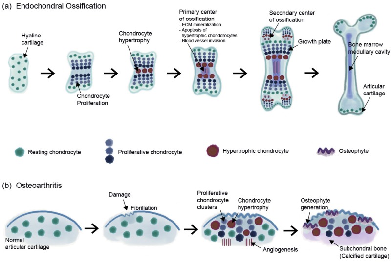
Osteoarthritis (OA) is the most common joint disease that causes pain and disability in the adult population. OA is primarily caused by trauma induced by an external force or by age-related cartilage damage. Chondrocyte hypertrophy or chondrocyte senescence is thought to play a role in the initiation and progression of OA. Although chondrocyte hypertrophy and cell death are both crucial steps during the natural process of endochondral bone formation, the abnormal activation of these two processes after injury or during aging seems to accelerate the progression of OA. However, the exact mechanisms of OA progression and these two processes remain poorly understood. Chondrocyte senescence and hypertrophy during OA share various markers and processes. In this study, we reviewed the changes that occur during chondrocyte hypertrophy or senescence in OA and the attempts that were made to regulate them. Regulation of hypertrophic or senescent chondrocytes might be a potential therapeutic target to slow down or stop OA progression; thus, a better understanding of the processes is required for management.
Keywords: articular cartilage; chondrocyte; hypertrophy; osteoarthritis; senescence.
Conflict of interest statement
The authors declare no conflict of interest.
Figures

Similar articles
-
Chondrocyte hypertrophy and osteoarthritis: role in initiation and progression of cartilage degeneration?Osteoarthritis Cartilage. 2012 Mar;20(3):223-32. doi: 10.1016/j.joca.2011.12.003. Epub 2011 Dec 11. Osteoarthritis Cartilage. 2012. PMID: 22178514 Review.
-
Regulation of α5 and αV Integrin Expression by GDF-5 and BMP-7 in Chondrocyte Differentiation and Osteoarthritis.PLoS One. 2015 May 26;10(5):e0127166. doi: 10.1371/journal.pone.0127166. eCollection 2015. PLoS One. 2015. PMID: 26010756 Free PMC article.
-
The Regulatory Role of Signaling Crosstalk in Hypertrophy of MSCs and Human Articular Chondrocytes.Int J Mol Sci. 2015 Aug 14;16(8):19225-47. doi: 10.3390/ijms160819225. Int J Mol Sci. 2015. PMID: 26287176 Free PMC article. Review.
-
Molecular regulation of articular chondrocyte function and its significance in osteoarthritis.Histol Histopathol. 2011 Mar;26(3):377-94. doi: 10.14670/HH-26.377. Histol Histopathol. 2011. PMID: 21210351 Review.
-
BAPX-1/NKX-3.2 acts as a chondrocyte hypertrophy molecular switch in osteoarthritis.Arthritis Rheumatol. 2015 Nov;67(11):2944-56. doi: 10.1002/art.39293. Arthritis Rheumatol. 2015. PMID: 26245691
Cited by
-
Differential gene co-expression network analyses reveal novel molecules associated with transcriptional dysregulation of key biological processes in osteoarthritis knee cartilage.Osteoarthr Cartil Open. 2022 Oct 23;4(4):100316. doi: 10.1016/j.ocarto.2022.100316. eCollection 2022 Dec. Osteoarthr Cartil Open. 2022. PMID: 36474801 Free PMC article.
-
Spermidine attenuates chondrocyte inflammation and cellular pyroptosis through the AhR/NF-κB axis and the NLRP3/caspase-1/GSDMD pathway.Front Immunol. 2024 Oct 2;15:1462777. doi: 10.3389/fimmu.2024.1462777. eCollection 2024. Front Immunol. 2024. PMID: 39416781 Free PMC article.
-
Changes in Small Noncoding RNA Expression during Chondrocyte Senescence.Cartilage. 2022 Jul-Sep;13(3):19476035221118165. doi: 10.1177/19476035221118165. Cartilage. 2022. PMID: 35993268 Free PMC article.
-
Protocatechuic aldehyde attenuates chondrocyte senescence via the regulation of PTEN-induced kinase 1/Parkin-mediated mitochondrial autophagy.Int J Immunopathol Pharmacol. 2024 Jan-Dec;38:3946320241271724. doi: 10.1177/03946320241271724. Int J Immunopathol Pharmacol. 2024. PMID: 39116410 Free PMC article.
-
In Vitro Characterization of Doxorubicin-Mediated Stress-Induced Premature Senescence in Human Chondrocytes.Cells. 2022 Mar 25;11(7):1106. doi: 10.3390/cells11071106. Cells. 2022. PMID: 35406671 Free PMC article.
References
Publication types
MeSH terms
Substances
Grants and funding
LinkOut - more resources
Full Text Sources
Medical
