

Adipose derived mesenchymal stem cells seeded onto a decellularized nerve allograft enhances angiogenesis in a rat sciatic nerve defect model
- PMID: 32233045
- PMCID: PMC7570204
- DOI: 10.1002/micr.30579
Adipose derived mesenchymal stem cells seeded onto a decellularized nerve allograft enhances angiogenesis in a rat sciatic nerve defect model
Abstract
Purpose: Adipose derived mesenchymal stem cells (MSCs) are hypothesized to supplement tissues with growth factors essential for regeneration and neovascularization. The purpose of this study was to determine the effect of MSCs with respect to neoangiogenesis when seeded onto a decellularized nerve allograft in a rat sciatic nerve defect model.
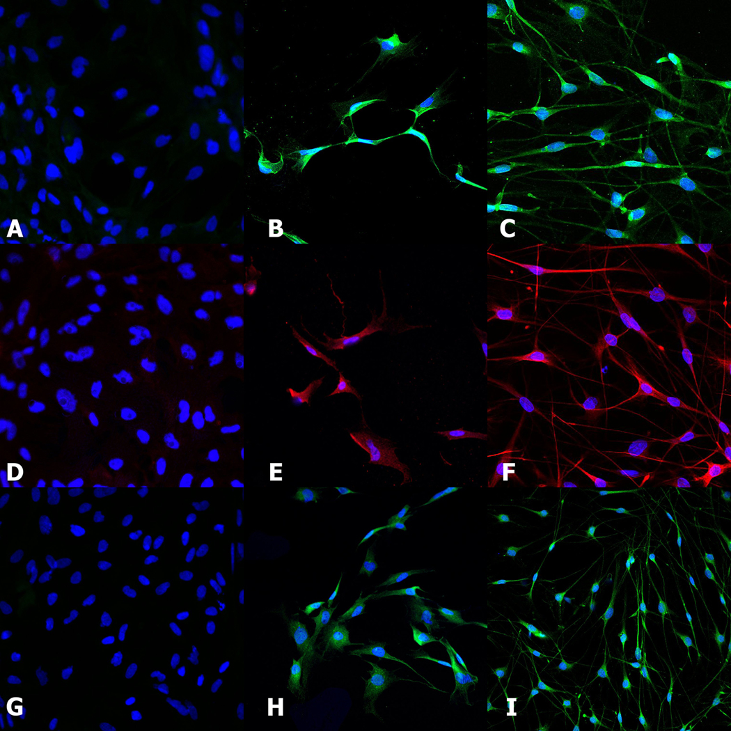
Methods: Allograft nerves were harvested from Sprague-Dawley rats and decellularized. MSCs were obtained from Lewis rats. 10 mm sciatic nerve defects in Lewis rats were reconstructed with reversed autograft nerves, decellularized allografts, decellularized allografts seeded with undifferentiated MSC or decellularized allografts seeded with differentiated MSCs. At 16 weeks, the vascular surface area and volume were evaluated.
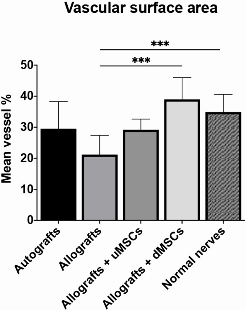
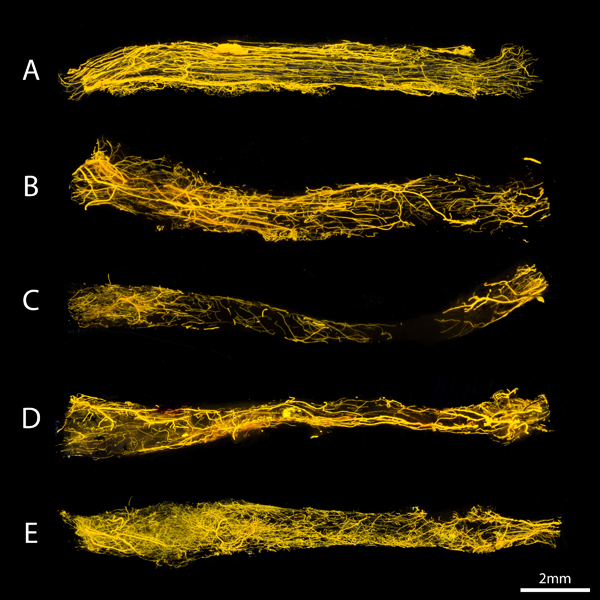
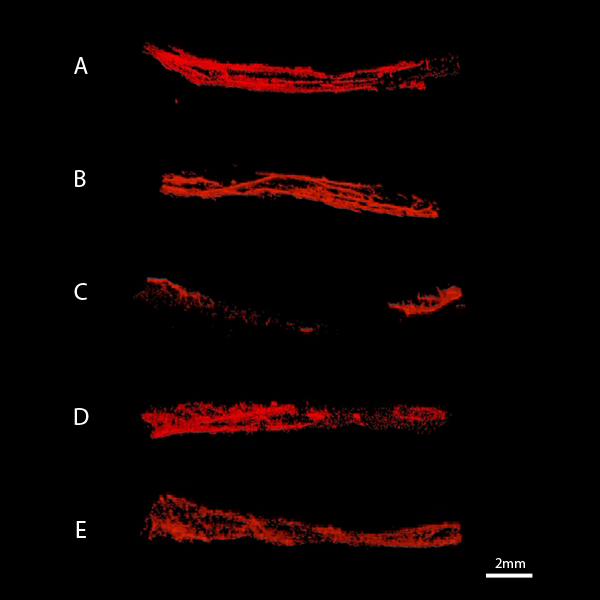
Results: The vascular surface area in normal nerves (34.9 ± 5.7%), autografts (29.5 ± 8.7%), allografts seeded with differentiated (38.9 ± 7.0%) and undifferentiated MSCs (29.2 ± 3.4%) did not significantly differ from each other. Unseeded allografts (21.2 ± 6.2%) had a significantly lower vascular surface area percentage than normal nonoperated nerves (13.7%, p = .001) and allografts seeded with differentiated MSCs (17.8%, p = .001). Although the vascular surface area was significantly correlated to the vascular volume (r = .416; p = .008), no significant differences were found between groups concerning vascular volumes. The vascularization pattern in allografts seeded with MSCs consisted of an extensive nonaligned network of microvessels with a centripetal pattern, while the vessels in autografts and normal nerves were more longitudinally aligned with longitudinal inosculation patterns.
Conclusions: Neoangiogenesis of decellularized allograft nerves was enhanced by stem cell seeding, in particular by differentiated MSCs. The pattern of vascularization was different between decellularized allograft nerves seeded with MSCs compared to autograft nerves.
© 2020 Wiley Periodicals, Inc.
Figures

Similar articles
-
Adhesion, distribution, and migration of differentiated and undifferentiated mesenchymal stem cells (MSCs) seeded on nerve allografts.J Plast Reconstr Aesthet Surg. 2020 Jan;73(1):81-89. doi: 10.1016/j.bjps.2019.05.030. Epub 2019 May 22. J Plast Reconstr Aesthet Surg. 2020. PMID: 31202698 Free PMC article.
-
Functional Outcomes of Nerve Allografts Seeded with Undifferentiated and Differentiated Mesenchymal Stem Cells in a Rat Sciatic Nerve Defect Model.Plast Reconstr Surg. 2021 Aug 1;148(2):354-365. doi: 10.1097/PRS.0000000000008191. Plast Reconstr Surg. 2021. PMID: 34153019 Free PMC article.
-
Gene expression profiles of differentiated and undifferentiated adipose derived mesenchymal stem cells dynamically seeded onto a processed nerve allograft.Gene. 2020 Jan 15;724:144151. doi: 10.1016/j.gene.2019.144151. Epub 2019 Oct 15. Gene. 2020. PMID: 31626959
-
Functional and immunological peculiarities of peripheral nerve allografts.Neural Regen Res. 2022 Apr;17(4):721-727. doi: 10.4103/1673-5374.322445. Neural Regen Res. 2022. PMID: 34472457 Free PMC article. Review.
-
Nerve Autografts Versus Allografts for Mixed Motor/Sensory Nerve Reconstruction.J Hand Surg Glob Online. 2024 Apr 20;6(5):694-699. doi: 10.1016/j.jhsg.2024.01.025. eCollection 2024 Sep. J Hand Surg Glob Online. 2024. PMID: 39381403 Free PMC article. Review.
Cited by
-
The interaction of stem cells and vascularity in peripheral nerve regeneration.Neural Regen Res. 2021 Aug;16(8):1510-1517. doi: 10.4103/1673-5374.303009. Neural Regen Res. 2021. PMID: 33433464 Free PMC article.
-
Advances in nerve guidance conduits for peripheral nerve repair and regeneration.Am J Stem Cells. 2023 Dec 15;12(5):112-123. eCollection 2023. Am J Stem Cells. 2023. PMID: 38213640 Free PMC article. Review.
-
ESWT Diminishes Axonal Regeneration following Repair of the Rat Median Nerve with Muscle-In-Vein Conduits but Not after Autologous Nerve Grafting.Biomedicines. 2022 Jul 22;10(8):1777. doi: 10.3390/biomedicines10081777. Biomedicines. 2022. PMID: 35892677 Free PMC article.
-
Optimized design of a hyperflexible sieve electrode to enhance neurovascular regeneration for a peripheral neural interface.Biomaterials. 2021 Aug;275:120924. doi: 10.1016/j.biomaterials.2021.120924. Epub 2021 Jun 8. Biomaterials. 2021. PMID: 34147716 Free PMC article.
-
Biological nerve conduit model with de-epithelialized human amniotic membrane and adipose-derived mesenchymal stem cell sheet for repair of peripheral nerve defects.Cell Tissue Res. 2023 Mar;391(3):505-522. doi: 10.1007/s00441-022-03732-8. Epub 2022 Dec 23. Cell Tissue Res. 2023. PMID: 36562866
References
-
- Zhu Z, Huang Y, Zou X, Zheng C, Liu J, Qiu L, et al. The vascularization pattern of acellular nerve allografts after nerve repair in Sprague-Dawley rats. Neurolog Res 2017;39:1014–1021. - PubMed
-
- Niu X, Liu X, Hu J, Jiang L. [Experimental research on revascularization of chemically extracted acellular allogenous nerve graft]. Zhongguo xiu fu chong jian wai ke za zhi = Zhongguo xiufu chongjian waike zazhi = Chin J Repar Reconstr Surg 2009;23:235–238. - PubMed
-
- Donzelli R, Capone C, Sgulo FG, Mariniello G, Maiuri F. Vascularized nerve grafts: an experimental study. Neurolog Res 2016;38:669–677. - PubMed
-
- Ferretti A, Boschi E, Stefani A, Spiga S, Romanelli M, Lemmi M, et al. Angiogenesis and nerve regeneration in a model of human skin equivalent transplant. Life Sci 2003;73:1985–1994. - PubMed
MeSH terms
Grants and funding
LinkOut - more resources
Full Text Sources
