. 2020 Mar 13:8:117.
doi: 10.3389/fcell.2020.00117. eCollection 2020.

A Positive Feedback Loop of Hippo- and c-Jun-Amino-Terminal Kinase Signaling Pathways Regulates Amyloid-Beta-Mediated Neurodegeneration

Affiliations

A Positive Feedback Loop of Hippo- and c-Jun-Amino-Terminal Kinase Signaling Pathways Regulates Amyloid-Beta-Mediated Neurodegeneration

Madison Irwin et al. Front Cell Dev Biol. .

Abstract

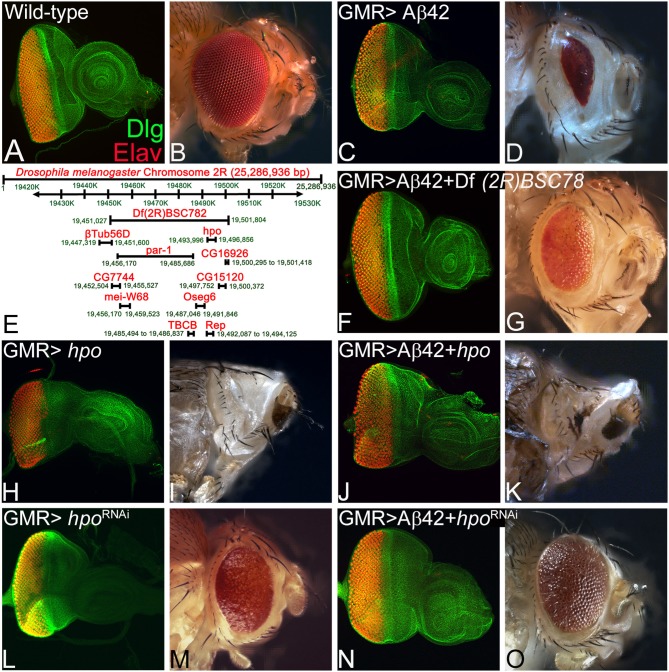
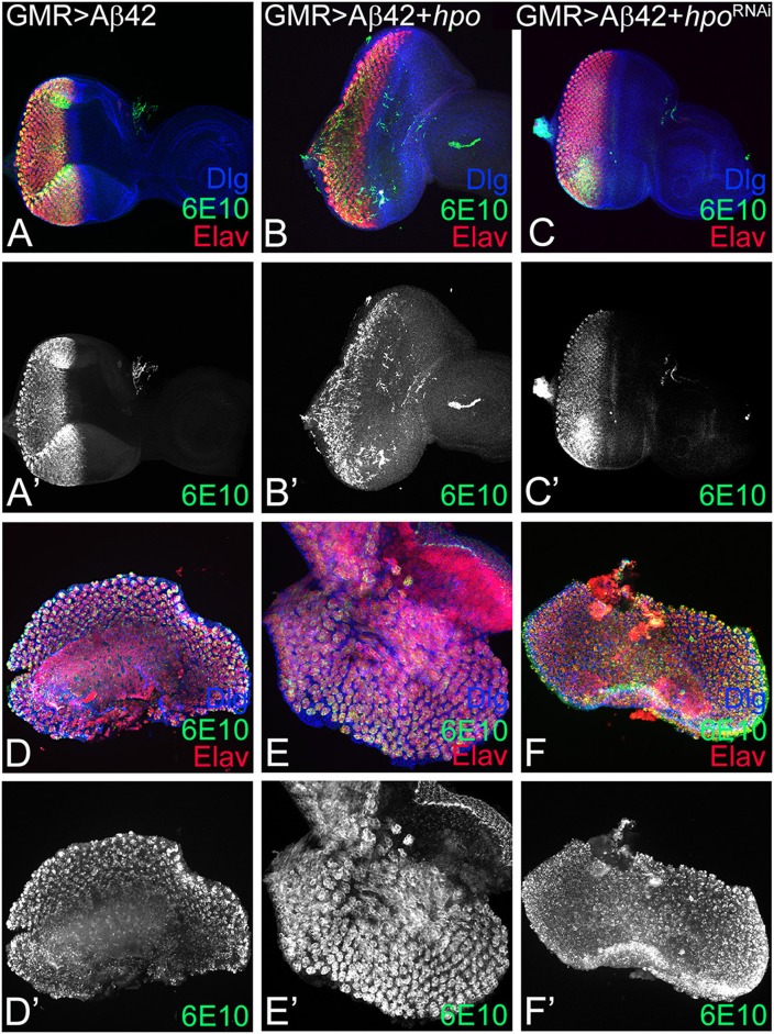
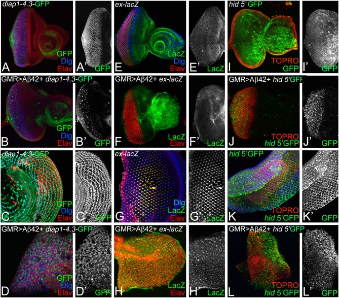
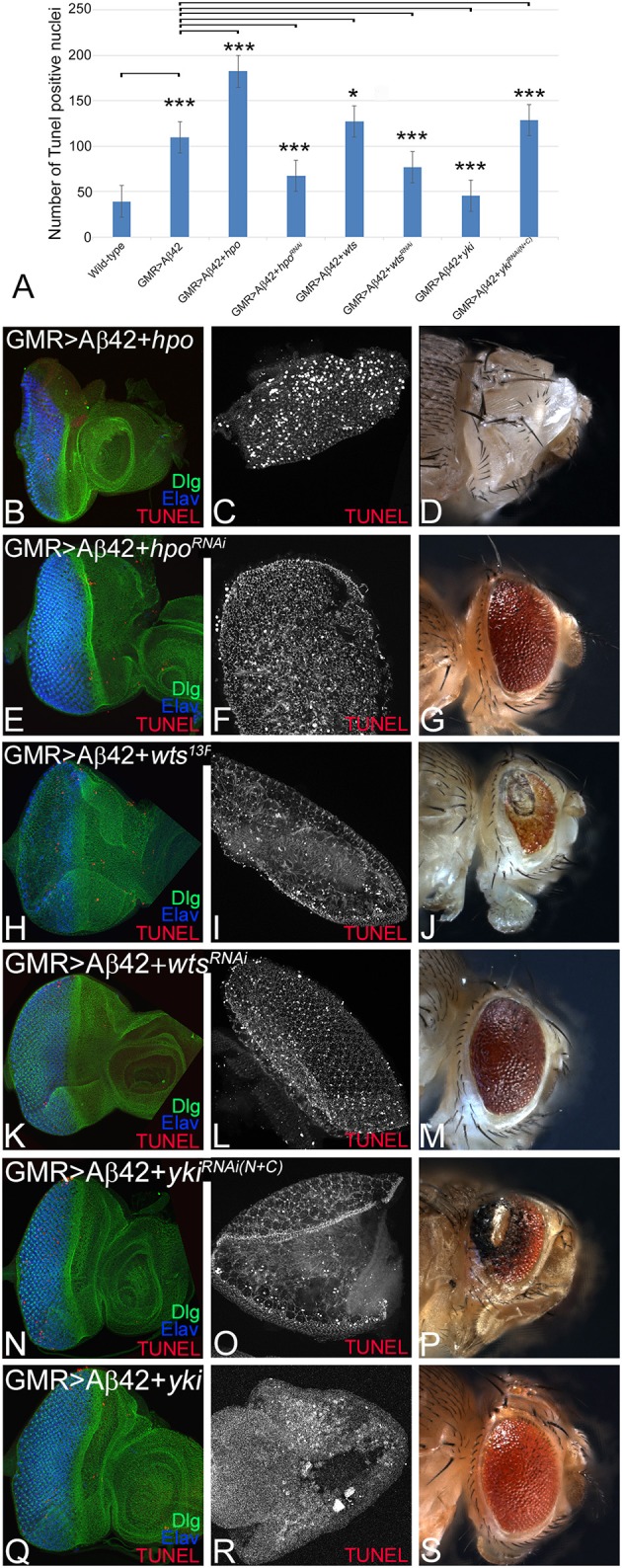
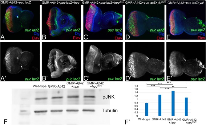
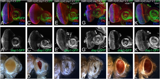
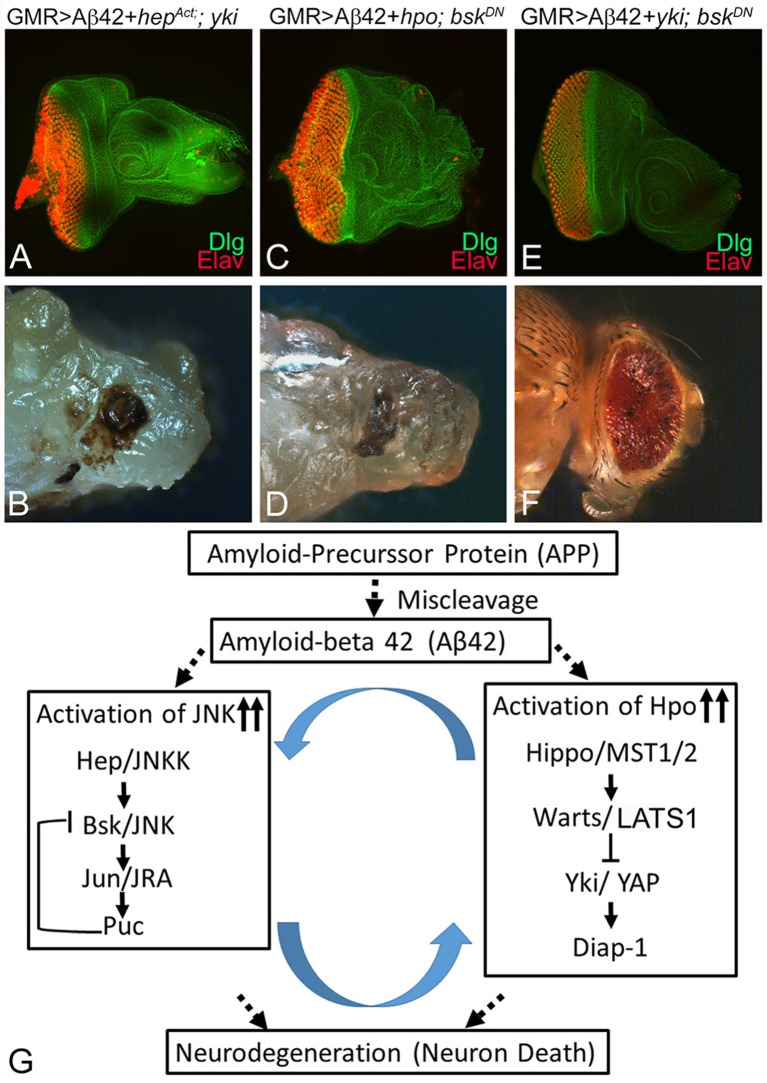
Alzheimer's disease (AD, OMIM: 104300) is an age-related disorder that affects millions of people. One of the underlying causes of AD is generation of hydrophobic amyloid-beta 42 (Aβ42) peptides that accumulate to form amyloid plaques. These plaques induce oxidative stress and aberrant signaling, which result in the death of neurons and other pathologies linked to neurodegeneration. We have developed a Drosophila eye model of AD by targeted misexpression of human Aβ42 in the differentiating retinal neurons, where an accumulation of Aβ42 triggers a characteristic neurodegenerative phenotype. In a forward deficiency screen to look for genetic modifiers, we identified a molecularly defined deficiency, which suppresses Aβ42-mediated neurodegeneration. This deficiency uncovers hippo (hpo) gene, a member of evolutionarily conserved Hippo signaling pathway that regulates growth. Activation of Hippo signaling causes cell death, whereas downregulation of Hippo signaling triggers cell proliferation. We found that Hippo signaling is activated in Aβ42-mediated neurodegeneration. Downregulation of Hippo signaling rescues the Aβ42-mediated neurodegeneration, whereas upregulation of Hippo signaling enhances the Aβ42-mediated neurodegeneration phenotypes. It is known that c-Jun-amino-terminal kinase (JNK) signaling pathway is upregulated in AD. We found that activation of JNK signaling enhances the Aβ42-mediated neurodegeneration, whereas downregulation of JNK signaling rescues the Aβ42-mediated neurodegeneration. We tested the nature of interactions between Hippo signaling and JNK signaling in Aβ42-mediated neurodegeneration using genetic epistasis approach. Our data suggest that Hippo signaling and JNK signaling, two independent signaling pathways, act synergistically upon accumulation of Aβ42 plaques to trigger cell death. Our studies demonstrate a novel role of Hippo signaling pathway in Aβ42-mediated neurodegeneration.

Keywords: Alzheimer's disease; Drosophila eye; Hippo signaling; amyloid-beta 42; c-Jun-amino-terminal kinase (JNK) signaling; cell death; growth regulation; neurodegeneration.


Figures

Figure 1
Figure 1
hippo is a genetic modifier of amyloid-beta 42 (Aβ42)-mediated neurodegeneration in the Drosophila eye. Panels show images of eye imaginal discs stained for the proneural marker embryonic lethal abnormal vision (ELAV; red) and a membrane-specific marker discs large (Dlg; green), and the resulting eye phenotype in the adult from (A,B) wild-type and (C,D) glass multiple repeat (GMR)> Aβ42. (D) Note that the GMR> Aβ42 adult eyes are highly reduced and have glazed morphology with black necrotic spots. (E) A map showing the deficiency BSC782 identified in the forward genetic screen and position of hpo and other genes within this deficiency is depicted. (F–O) Panels show the eye disc stained with Dlg (green) and ELAV (red) and accompanying adult eye phenotypes from (F,G) GMR> Aβ42 + Df(2R)BSC782/+, (H,I) GMR> hpo, (J,K) GMR> Aβ42 + hpo, (L,M) GMR> hpoRNAi, and (N,O) GMR> Aβ42 + hpoRNAi. Note that downregulation of Hippo signaling (N,O) GMR> Aβ42 + hpoRNAi significantly rescues the GMR> Aβ42 neurodegenerative phenotype, whereas activation of Hippo signaling (J,K) GMR> Aβ42 + hpo enhances the GMR> Aβ42 neurodegenerative phenotype. The orientation of all imaginal discs is identical with posterior to the left and dorsal up. Magnification of the eye disc or adult eye images is the same across all panels.
Figure 2
Figure 2
hpo does not affect the amyloid-beta 42 (Aβ42) accumulation. Panels show eye imaginal discs and pupal retinae stained with the proneural marker embryonic lethal abnormal vision (ELAV; shown in red); 6E10, an anti-Aβ42 antibody (green or gray), and the membrane-specific marker discs large (Dlg; blue). (A–C) shows confocal images of eye discs for all markers (Dlg, 6E10, ELAV), whereas the 6E10 expression alone (gray) is shown in panels (A'–C'). Eye discs (A–C) and pupal retinae (D–F) of the following genotypes were compared: (A,A',D,D') glass multiple repeat (GMR)> Aβ42, (B,B',E,E') GMR> Aβ42 + hpo, and (C,C',F,F') GMR> Aβ42 + hpoRNAi. Note that activation (GMR> Aβ42 + hpo) or downregulation (GMR> Aβ42 + hpoRNAi) of Hippo signaling in GMR> Aβ42 background does not affect the accumulation of Aβ42 plaques.
Figure 3
Figure 3
Accumulation of amyloid-beta 42 (Aβ42) activates Hippo signaling. Expression levels of Hippo pathway reporters were tested in eye discs and pupal retinae. Expression of diap1 4.3-green fluorescent protein (GFP) (green, gray) is shown for eye discs and pupal retinae from (A,A',C,C') wild-type, (B,B',D,D') glass multiple repeat (GMR)> Aβ42, respectively. diap1-4.3-GFP expression is shown in a split channel in gray in (C',D') panels. Changes in ex-lacZ (green, gray) levels is shown in eye discs and pupal retinae from (E,E',F,F') wild type, (G,G',H,H') GMR> Aβ42, respectively. ex-lacZ expression is shown in a split channel in gray in (E'–H'). (I–L) shows the expression of hid-5' GFP (green), a reporter for cell death in wild-type (I,I') eye imaginal disc and (K,K') pupal retina and GMR> Aβ42 (J,J') eye imaginal disc and the (L,L') pupal retina. Gray panels (I'–L') show hid-5' GFP expression in indicated genotypes. In (I–L), nuclei are marked by the nuclear dye TOPRO (red).
Figure 4
Figure 4
Hippo activation triggers cell death in glass multiple repeat (GMR)> amyloid-beta 42 (Aβ42) background. For each genotype, we counted terminal deoxynucleotidyl transferase dUTP nick end labeling (TUNEL)-positive nuclei from five (n = 5) eye imaginal discs to determine dying cell population. (A) Quantification of TUNEL-positive nuclei in indicated genotypes is shown (n = 5, p ≤ 0.05). The p-values obtained from Student's t-test between wild-type and GMR> Aβ42 was significant (p < 0.001; ***), GMR> Aβ42 and GMR>Aβ42+hpo (gain-of-function) was significant (p < 0.001; ***), and between GMR> Aβ42 and GMR>Aβ42+hpoRNAi (loss-of-function) was significant (p < 0.001; ***). The p-value obtained from Student's t-test between GMR>Aβ42 and GMR> Aβ42+wts was significant (p < 0.05; *), between GMR> Aβ42 and GMR> Aβ42+wtsRNAi was significant (p < 0.001; ***), between GMR> Aβ42 and GMR> Aβ42+yki was significant (p < 0.001; ***), and between GMR> Aβ42 and GMR> Aβ42+ykiRNAi was significant (p < 0.001; ***). (B–S) shows the extent of cell death based on TUNEL assays (red, gray) in indicated genotypes in imaginal discs, pupal retinae, and adult eyes. The eye discs (B,E,H,K,N,Q) were assessed for dying cells using TUNEL assay (red) and stained with Dlg (green) and embryonic lethal abnormal vision (ELAV; blue). The pupal retinae (C,F,I,L,O,R) assessed for dying cells using TUNEL assays (gray). Adult eye phenotypes are shown in panels (D,G,J,M,P,S). Panels show the extent of cell death in (B–D) GMR> Aβ42+hpo, (E–G) GMR> Aβ42+hpo RNAi, (H–J) GMR> Aβ42 +wts, (K–M) GMR> Aβ42 +wtsRNAi, (N–P) GMR> Aβ42 + ykiRNAi, and (Q–S) GMR> Aβ42 + yki. The orientation of all imaginal discs is identical with posterior to the left and dorsal up. Magnification of all eye imaginal discs is 20×.
Figure 5
Figure 5
Activation of Hippo signaling upon amyloid-beta 42 (Aβ42) accumulation also activates c-Jun-amino-terminal kinase (JNK) signaling. (A–E) shows the expression of embryonic lethal abnormal vision (ELAV; red), JNK signaling pathway reporter puc-lacZ (green, gray), and discs large (Dlg; blue) in eye discs from (A,A') glass multiple repeat (GMR)> Aβ42, (B,B') GMR> Aβ42 + hpo, (C,C') GMR> Aβ42 + hpoRNAi, (D,D') GMR> Aβ42 + ykiRNAi, and (E,E') GMR> Aβ42 + yki. For comparison between genotypes, (A'–E') shows puc-lacZ (Gray) levels. (F) A semiquantitative Western blot is presented to show phospho-JNK (p-JNK) levels in the wild-type, GMR> Aβ42, GMR> Aβ42 + hpo, and GMR> Aβ42 + hpoRNAi background. The samples were loaded in the following sequence: Lane 1-Molecular weight marker, Lane 2-Wild-type (Canton-S), Lane 3-GMR> Aβ42, Lane 4-GMR> Aβ42+hpo (gain-of-function), Lane 5-GMR> Aβ42+hpoRNAi (loss-of-function). Alpha-tubulin is used as a loading control, and (F') graph shows the quantification of p-JNK levels, which were calculated from a set of three (n = 3) in wild-type and other indicated genotypes from the Western blot (F). The p-values for estimation of p-JNK levels in all combination in a semiquantitative Western blot was calculated in a set of three (n = 3) using Student's t-test in Microsoft Excel software. The p-value between wild-type and GMR> Aβ42 was significant (p < 0.001; ***), wild-type and GMR> Aβ42+hpo was significant (p < 0.001; ***), and between wild-type (Canton-S) and GMR> Aβ42+hpoRNAi (loss-of-function) was significant (p <0.001; ***). The p-value between GMR> Aβ42 and GMR> Aβ42+hpo was significant (p < 0.001; ***) and between GMR> Aβ42 and GMR> Aβ42+hpoRNAi was significant (p < 0.01; **).
Figure 6
Figure 6
Activation of c-Jun-amino-terminal kinase (JNK) signaling pathway in amyloid-beta 42 (Aβ42) background activates Hippo pathway. Eye imaginal discs from larvae of indicated genotypes assessed for changes in expression of diap1-4.3-green fluorescent protein (GFP; green), a reporter for Hippo pathway, are shown. All discs show expression of discs large (Dlg; red), diap1-4.3-GFP (green), and embryonic lethal abnormal vision (ELAV; blue). (A,A') show wild-type control glass multiple repeat (GMR)> diap1-4.3-GFP eyes discs and (B) wild-type adult eye phenotypes; and (C,C') GMR> Aβ42 eye discs and (D) adult eye phenotype. (E–K) shows effects of modulating JNK activity on Hippo pathway reporter diap1-4.3-GFP in eye-antennal imaginal disc of (E,E') GMR> Aβ42 + junaspv7, (G,G') GMR> Aβ42 + hepAct, (I,I') GMR> Aβ42 + bskDN, (K,K') GMR> Aβ42 + puc backgrounds. Note that (E,E',G,G') diap1-4.3-GFP exhibits robust induction when JNK signaling is activated, whereas (I,I',K,K') diap1-4.3-GFP levels are significantly reduced when JNK signaling is downregulated in GMR> Aβ42 background. The adult eye phenotypes associated with (D) GMR> Aβ42, (F) GMR> Aβ42+ junaspv7, (H) GMR> Aβ42+ hepAct, (J) GMR> Aβ42+ bskDN, and (L) GMR> Aβ42+ puc. Note that (J,L) downregulation of JNK signaling showed significant rescues in the adult eye phenotypes, whereas (F,H) activation of JNK signaling enhanced the neurodegenerative phenotype of (D) GMR> Aβ42.
Figure 7
Figure 7
Hippo and c-Jun-amino-terminal kinase (JNK) signaling act synergistically to induce amyloid-beta 42 (Aβ42)-mediated neurodegeneration. Panels show eye discs stained with discs large (Dlg; green) and embryonic lethal abnormal vision (ELAV; red) and the resulting adult eye phenotypes when JNK or Hippo pathway is blocked. (A,B) Activation of JNK signaling along with inactivation of Hippo signaling in the glass multiple repeat (GMR)> Aβ42 background (GMR> Aβ42+hepAct+yki) does not rescue the neurodegenerative phenotype. (C,D) Activation of Hippo signaling along with inactivation of JNK signaling in the GMR> Aβ42 background (GMR> Aβ42+hpo+BskDN) does not rescue the neurodegenerative phenotype. (E,F) Inactivation of both JNK signaling and Hippo signaling (GMR> Aβ42+ yki + BskDN) together in the GMR> Aβ42 background exhibits significant rescue of the neurodegenerative phenotype. (G) A model that reconciles the data from this study, which shows that Hippo signaling and JNK signaling act synergistically via a positive feedback loop to induce Aβ42-mediated neurodegeneration.

Similar articles

Cited by

References

    1. Adachi-Yamada T. (2002). Puckered-GAL4 driving in JNK-active cells. Genesis 34, 19–22. 10.1002/gene.10110 - DOI - PubMed
    1. Adachi-Yamada T., Fujimura-Kamada K., Nishida Y., Matsumoto K. (1999). Distortion of proximodistal information causes JNK-dependent apoptosis in Drosophila wing. Nature 400, 166–169. 10.1038/22112 - DOI - PubMed
    1. Adachi-Yamada T., O'connor M. B. (2004). Mechanisms for removal of developmentally abnormal cells: cell competition and morphogenetic apoptosis. J. Biochem. 136, 13–17. 10.1093/jb/mvh099 - DOI - PubMed
    1. Barnes D. E., Yaffe K. (2011). Accuracy of summary risk score for prediction of Alzheimer disease: better than demographics alone? Arch. Neurol. 68, 268–270. 10.1001/archneurol.2011.4 - DOI - PubMed
    1. Battaglia C., Venturin M., Sojic A., Jesuthasan N., Orro A., Spinelli R., et al. . (2019). Candidate genes and MiRNAs linked to the inverse relationship between cancer and Alzheimer's disease: insights from data mining and enrichment analysis. Front. Genet. 10:846. 10.3389/fgene.2019.00846 - DOI - PMC - PubMed

LinkOut - more resources