

Intestinal resident macrophages: Multitaskers of the gut
- PMID: 32222060
- PMCID: PMC7757264
- DOI: 10.1111/nmo.13843
Intestinal resident macrophages: Multitaskers of the gut
Abstract
Background: Intestinal resident macrophages play a crucial role in homeostasis and have been implicated in numerous gastrointestinal diseases. While historically believed to be largely of hematopoietic origin, recent advances in fate-mapping technology have unveiled the existence of long-lived, self-maintaining populations located in specific niches throughout the gut wall. Furthermore, the advent of single-cell technology has enabled an unprecedented characterization of the functional specialization of tissue-resident macrophages throughout the gastrointestinal tract.
Purpose: The purpose of this review was to provide a panorama on intestinal resident macrophages, with particular focus to the recent advances in the field. Here, we discuss the functions and phenotype of intestinal resident macrophages and, where possible, the functional specialization of these cells in response to the niche they occupy. Furthermore, we will discuss their role in gastrointestinal diseases.
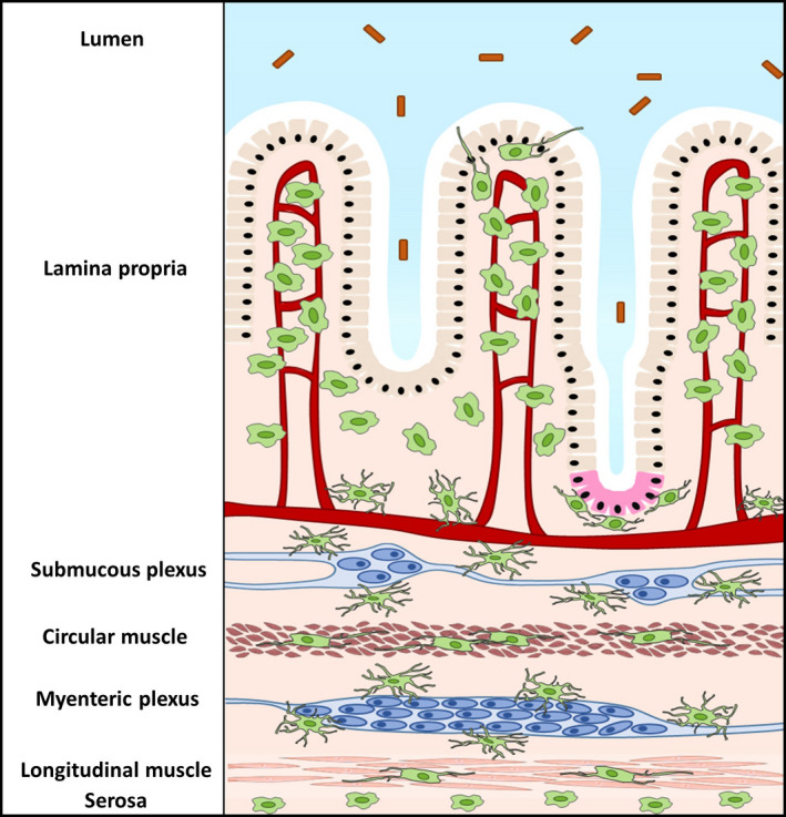
Keywords: enteric nervous system; gastrointestinal tract; lamina propria; muscularis externa; neuroimmune interaction; resident macrophages.
© 2020 The Authors. Neurogastroenterology & Motility published by John Wiley & Sons Ltd.
Conflict of interest statement
The authors declare no conflict of interest.
Figures
Similar articles
-
Niche-specific functional heterogeneity of intestinal resident macrophages.Gut. 2021 Jul;70(7):1383-1395. doi: 10.1136/gutjnl-2020-323121. Epub 2020 Dec 31. Gut. 2021. PMID: 33384336 Free PMC article. Review.
-
Resident macrophages in the healthy and inflamed intestinal muscularis externa.Pflugers Arch. 2017 Apr;469(3-4):541-552. doi: 10.1007/s00424-017-1948-4. Epub 2017 Feb 24. Pflugers Arch. 2017. PMID: 28236119 Review.
-
Beyond Immunity: Underappreciated Functions of Intestinal Macrophages.Front Immunol. 2021 Sep 28;12:749708. doi: 10.3389/fimmu.2021.749708. eCollection 2021. Front Immunol. 2021. PMID: 34650568 Free PMC article. Review.
-
Muscularis macrophages: Key players in intestinal homeostasis and disease.Cell Immunol. 2018 Aug;330:142-150. doi: 10.1016/j.cellimm.2017.12.009. Epub 2017 Dec 26. Cell Immunol. 2018. PMID: 29291892 Free PMC article. Review.
-
New insights into muscularis macrophages in the gut: from their origin to therapeutic targeting.Immunol Res. 2023 Dec;71(6):785-799. doi: 10.1007/s12026-023-09397-x. Epub 2023 May 23. Immunol Res. 2023. PMID: 37219708 Review.
Cited by
-
Gut Innate Immunity and HIV Pathogenesis.Curr HIV/AIDS Rep. 2021 Apr;18(2):128-138. doi: 10.1007/s11904-021-00544-3. Epub 2021 Mar 9. Curr HIV/AIDS Rep. 2021. PMID: 33687703 Free PMC article. Review.
-
Possible Role of Pineal and Extra-Pineal Melatonin in Surveillance, Immunity, and First-Line Defense.Int J Mol Sci. 2021 Nov 10;22(22):12143. doi: 10.3390/ijms222212143. Int J Mol Sci. 2021. PMID: 34830026 Free PMC article. Review.
-
The Interplay between Nutrition, Innate Immunity, and the Commensal Microbiota in Adaptive Intestinal Morphogenesis.Nutrients. 2021 Jun 26;13(7):2198. doi: 10.3390/nu13072198. Nutrients. 2021. PMID: 34206809 Free PMC article. Review.
-
Efficient enzyme-free method to assess the development and maturation of the innate and adaptive immune systems in the mouse colon.Sci Rep. 2024 May 14;14(1):11063. doi: 10.1038/s41598-024-61834-5. Sci Rep. 2024. PMID: 38744932 Free PMC article.
-
Intestinal Macrophage Autophagy and its Pharmacological Application in Inflammatory Bowel Disease.Front Pharmacol. 2021 Nov 24;12:803686. doi: 10.3389/fphar.2021.803686. eCollection 2021. Front Pharmacol. 2021. PMID: 34899362 Free PMC article. Review.
References
-
- Hussell T, Bell TJ. Alveolar macrophages: plasticity in a tissue‐specific context. Nat Rev Immunol. 2014;14:81. - PubMed
Publication types
MeSH terms
Grants and funding
LinkOut - more resources
Full Text Sources
