. 2020 May 18;94(11):e00017-20.
doi: 10.1128/JVI.00017-20. Print 2020 May 18.

Double-Stranded-RNA-Binding Protein 2 Participates in Antiviral Defense

Affiliations

Double-Stranded-RNA-Binding Protein 2 Participates in Antiviral Defense

Károly Fátyol et al. J Virol. .

Abstract

Double-stranded RNA (dsRNA) is a common pattern formed during the replication of both RNA and DNA viruses. Perception of virus-derived dsRNAs by specialized receptor molecules leads to the activation of various antiviral measures. In plants, these defensive processes include the adaptive RNA interference (RNAi) pathway and innate pattern-triggered immune (PTI) responses. While details of the former process have been well established in recent years, the latter are still only partially understood at the molecular level. Nonetheless, emerging data suggest extensive cross talk between the different antiviral mechanisms. Here, we demonstrate that dsRNA-binding protein 2 (DRB2) of Nicotiana benthamiana plays a direct role in potato virus X (PVX)-elicited systemic necrosis. These results establish that DRB2, a known component of RNAi, is also involved in a virus-induced PTI response. In addition, our findings suggest that RNA-dependent polymerase 6 (RDR6)-dependent dsRNAs play an important role in the triggering of PVX-induced systemic necrosis. Based on our data, a model is formulated whereby competition between different DRB proteins for virus-derived dsRNAs helps establish the dominant antiviral pathways that are activated in response to virus infection.IMPORTANCE Plants employ multiple defense mechanisms to restrict viral infections, among which RNA interference is the best understood. The activation of innate immunity often leads to both local and systemic necrotic responses, which confine the virus to the infected cells and can also provide resistance to distal, noninfected parts of the organism. Systemic necrosis, which is regarded as a special form of the local hypersensitive response, results in necrosis of the apical stem region, usually causing the death of the plant. Here, we provide evidence that the dsRNA-binding protein 2 of Nicotiana benthamiana plays an important role in virus-induced systemic necrosis. Our findings are not only compatible with the recent hypothesis that DRB proteins act as viral invasion sensors but also extends it by proposing that DRBs play a critical role in establishing the dominant antiviral measures that are triggered during virus infection.

Keywords: DRB2; PAMP-triggered immunity; RDR6; antiviral RNA interference; double-stranded RNA.


Figures

FIG 1
FIG 1
(A) Multiple alignment of the cloned N. benthamiana (Nb) DRB proteins. Positions of the two conserved N-terminal dsRBD domains are indicated. (B) Phylogenetic tree of the N. benthamiana DRB proteins. Calculated distance values according to the neighbor joining method are given in parentheses.
FIG 2
FIG 2
Analysis of the effects of DRB proteins on PVX replication. (A) An agroinfiltration-based transient virus replication assay was used to evaluate the effects of ectopic expression of DRB proteins on PVX replication. Suspensions of agrobacteria carrying either PVX-GFP- or PVXΔTGB-GFP-encoding binary vectors were infiltrated into leaves of wild-type (wt) or ago2 N. benthamiana. As a control a CaMV 35S promoter-driven GFP expression vector was used. Along with the reporter constructs, agrobacteria carrying binary expression vectors for DRB proteins were also codelivered. As a negative control, a coinfiltrated suspension of empty agrobacteria (∅) was employed. The bacterial suspensions (optical density at 600 nm of 1) were mixed at a 1:1 ratio. GFP expression in the infiltrated leaf patches was monitored using an appropriate UV light source at 3 and 7 dpi. Pictures of representative leaves are shown. At 7 dpi, PVX- and DRB2-coinfiltrated leaf patches frequently exhibited necrosis, which resulted in the apparent fading of the GFP signal. These necrotic areas are circled in red. Experiments were repeated three times. (B) Expression of PVXΔTGB-GFP-encoded GFP was monitored by Western blotting (WB). Leaves of wild-type N. benthamiana were agroinfiltrated with a PVXΔTGB-GFP reporter either alone (right side of leaves) or combined with an expression vector for one of the indicated DRB proteins (left side of leaves). At 5 dpi samples were collected and pooled from three identically infiltrated leaves. Protein lysates were prepared from the pooled samples and analyzed by GFP antibody. To verify the expression of the DRB proteins, the same filter was probed with HA antibody. As a loading control, the Ponceau-stained filter is shown. From the above-described infiltrated leaves, total RNA samples were also prepared, and viral RNA levels were monitored by Northern blotting (right panel). The Northern blot was probed with a radioactively labeled GFP DNA fragment. The ethidium bromide (EtBr)-stained gel is shown as a loading control. Protein lysate or total RNA prepared from PVXΔTGB-GFP and empty agrobacteria (∅)-infiltrated leaves were used as negative controls in the Western and Northern blots.
FIG 3
FIG 3
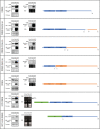
Mapping of the domain of DRB2 necessary for the inhibition of antiviral RNAi. A PVXΔTGB-GFP binary vector was agroinfiltrated either alone or along with binary constructs expressing the depicted mutant DRB proteins into leaves of wild-type N. benthamiana (left or right sides of the same leaves, respectively). At 5 dpi samples were collected and pooled from three identically infiltrated leaves. From the collected samples, protein lysates and total RNA were prepared. GFP and the DRB mutant protein expression levels were monitored by Western blotting (WB) using GFP and HA antibodies, respectively (left panels). PVX gRNA levels were detected by Northern blotting using radioactively labeled GFP probe (right panels). In the case of the bottom two constructs, a GFP tag was used instead of an HA tag (the GFP tag was employed because the HA tag could not stabilize the N-terminally deleted mutant of DRB2). Accordingly, GFP antibody was used to detect DRB expression as well. In these cases, no Northern blots were performed; instead, images of ethidium bromide (EtBr)-stained gels are shown in which the position of the PVX gRNA is indicated by an arrowhead. DRB2B- and DRB3A-derived segments of the chimeric DRB molecules are indicated by blue and orange colors, respectively. The green box represents GFP. The corresponding Ponceau-stained protein filters and EtBr-stained RNA gels are shown as loading controls for the Western and Northern blots, respectively.
FIG 4
FIG 4
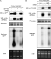
Inhibition of RNAi by DRB2 does not depend on RDR6. (A) Inverted-repeat-initiated posttranscriptional gene silencing is inhibited by DRB2. The indicated binary expression constructs were agroinfiltrated into leaves of wild-type N. benthamiana. At 5 dpi, samples were collected and pooled from three identically infiltrated leaves. From the collected samples protein lysates and total RNA were prepared. Viral protein synthesis and DRB2 protein levels were monitored by Western blotting using GFP and HA antibodies, respectively. Note that the HA antibody detects nonspecific bands in the first and third lanes at positions comparable to those of DRB2. The PVXΔTGB-GFP virus level was followed in Northern blotting using a radioactively labeled GFP probe. (B) DRB2 inhibits RNAi in rdr6 mutant N. benthamiana. Leaves of wild-type (wt) and rdr6 N. benthamiana were agroinfiltrated with a PVXΔTGB-GFP reporter either alone (left side of leaves) or combined with a DRB2 expression vector (right side of leaves). At 5 dpi, samples were collected and pooled from three identically infiltrated leaves. From the collected samples protein lysates and total RNA were prepared. Viral protein synthesis and DRB2 protein levels were monitored by Western blotting using PVX-CP and HA antibodies, respectively. PVXΔTGB-GFP virus level was followed by Northern blotting using a radioactively labeled GFP probe. The corresponding Ponceau-stained protein filters and ethidium bromide (EtBr)-stained RNA gels are shown as loading controls for the Western and Northern blots, respectively.
FIG 5
FIG 5
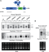
DRB2 inhibits the activity of all DCLs involved in antiviral RNAi. (A) The RNA samples analyzed in the experiment shown in Fig. 1 were used for small RNA Northern blots to examine the vsiRNA production from different regions of PVXΔTGB-GFP in the presence or absence of DRB proteins. Two different probes were used to monitor vsiRNA levels. The positions of the probes are indicated at the top of the cartoon. The filter was first probed with a radioactively labeled PVX-RdRp fragment and subsequently with a PVX-CP fragment (after stripping). The ethidium bromide (EtBr)-stained gel is shown as a loading control. SGP, subgenomic promoter; (A)n, poly(A) tail. (B) DRB2 overexpression inhibits the activity of all DCLs involved in antiviral RNAi in rdr6 N. benthamiana. Leaves of rdr6 N. benthamiana were agroinfiltrated with PVX:ΔTGB-GFP either alone (left side of leaves) or together with a DRB2 binary expression construct (right side of leaves). At 5 dpi samples were collected and pooled from three infiltrated leaves, and total RNAs were prepared. RNAs were analyzed on a small RNA blot as described above. The filter was probed with radioactively labeled PVX-CP probe. The EtBr-stained gel is shown as a loading control.
FIG 6
FIG 6
DRB2 induces HR-like local necrosis in N. benthamiana leaves. (A) PVX-GFP was agroinfiltrated into leaves of wild-type N. benthamiana either alone or in combination with the indicated DRB binary expression vectors. At 7 dpi, infiltrated leaves were photographed under visible (left panel) and UV (right panel) light. The DRB2-PVX-GFP-coinfiltrated necrotic patch is circled. (B) Replication of PVX is necessary for DRB2-dependent necrosis. Wild-type N. benthamiana leaves were agroinfiltrated with PVX-GFP and PVXΔRdRp-GFP (a PVX mutant carrying a frameshift mutation leading to a premature stop codon in the RdRp gene). The viral reporters were infiltrated either alone (left side of leaves) or along with a DRB2 binary expression vector (right side of leaves). Leaves were photographed as described above at 7 dpi. Infiltrated leaf patches are circled (red circle indicates the DRB2-PVX-GFP-coinfiltrated necrotic area). (C) Mapping of domains of DRB2 required for PVX-dependent necrosis. Leaves of wild-type N. benthamiana were agroinfiltrated with PVX-GFP either alone (left side of leaves) or combined with the indicated DRB binary expression vectors (right side of leaves). Photographs of leaves were taken under visible and UV light at 7 dpi. Every experiment was repeated at least twice. In each experiment, a total of six leaves were infiltrated in the same manner (two plants, with three leaves each). Pictures of representative leaves are shown. The necrotic areas of leaves are circled in red. DRB2B- and DRB3A-derived segments of the chimeric DRB molecules are indicated by blue and orange colors, respectively.
FIG 7
FIG 7
DRB2 overexpressing PVX induces necrosis. (A) Infections of wild-type N. benthamiana with the indicated recombinant PVX viruses were initiated by agroinfiltration. Pictures of infected plants were taken at 14 dpi. (B) At 7 dpi protein lysates and total RNA were prepared from systemically infected leaves of N. benthamiana. Presence of PVX RNA was monitored by Northern blotting using radioactively labeled PVX-CP probe. The ethidium bromide (EtBr)-stained gel is shown as a loading control. Viral CP and DRB protein expression levels were analyzed by Western blotting using the indicated antibodies. The Ponceau-stained filter is shown as a loading control. PVX vsiRNA levels were analyzed by small-RNA Northern blotting. The filter was probed with radioactively labeled PVX-CP probe. The ethidium bromide-stained gel is shown as a loading control.
FIG 8
FIG 8
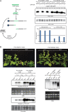
Effects of knockdown of DRB proteins on PVX infection. (A) Wild-type and ago2 N. benthamiana plants were agroinfiltrated with the indicated PVX-DRB-VIGS recombinant constructs. Pictures of mock- and virus-infected plants were taken at 21 dpi. The cartoon at the top depicts the structure of the VIGS constructs. DRB fr., DRB VIGS fragment. (B) Downregulation of DRB2 rescues PVX-induced systemic necrosis of ago2 N. benthamiana. Pictures of PVX-DRB2-VIGS-infected wild-type and ago2 N. benthamiana plants were taken at 21 dpi (top panels). Downregulation of endogenous DRB2 mRNA level was verified by semiquantitative reverse transcription-PCR (bottom left panels). As a control, a fragment of the actin mRNA was amplified. Cleavage-independent translational repression can also contribute to the effects of VIGS. To take this into account, the ability of PVX-DRB2-VIGS to knock down the DRB2 protein level was also assessed (bottom right panels). N. benthamiana plants were infected with PVX-DRB2-VIGS and with empty PVX (as control). At 14 dpi, apical systemically infected leaves of the plants were coinfiltrated with suspensions of agrobacteria expressing epitope-tagged DRB2 and GFP (as negative control). Three days later, the expression levels of HA-DRB2 and GFP were monitored in the infiltrated leaves using Western blotting. The Ponceau-stained filter is shown as a loading control. M, marker lane. (C) Replication of PVX-DRB2-VIGS was verified by Northern blotting (using radioactive PVX-CP probe) and Western blotting (using PVX-CP specific antibody) in systemically infected leaves (left panels). The PR-1a necrotic marker gene expression level was analyzed by Northern blotting (right panel). As loading controls for Northern and Western blots, the corresponding ethidium bromide (EtBr)-stained gel and Ponceau-stained filter are shown, respectively. Each experiment was repeated at least three times with similar results. w, week.
FIG 9
FIG 9
Knockdown of DRB2 and RDR6 inhibits PPV-PVX synergism-induced SN. Wild-type N. benthamiana plants were infected by agroinfiltration using binary plasmid constructs expressing the indicated viruses. Pictures of the infected plants were taken at 14 dpi. Total RNAs were prepared from systemically infected plants at 7 dpi. Samples collected from three identically infected plants were pooled. Viral RNA levels and necrotic marker gene expression levels were analyzed by Northern blotting using the indicated radioactively labeled virus-specific and PR-1a-specific probes. The ethidium bromide (EtBr)-stained gel is shown as a loading control. The experiment was repeated three times with the same outcomes.
FIG 10
FIG 10
Downregulation of RDR6 rescues PVX-induced SN of ago2 N. benthamiana. (A) VIGS-mediated downregulation of RDR6 activity was verified by a functional gene silencing assay. The assay is based on the observation that the inverted repeat (IR)-initiated posttranscriptional gene silencing is significantly enhanced by RDR6-dependent secondary siRNAs. Wild-type N. benthamiana plants were infected with either PVX-RDR6-VIGS or PVX-RDR1-VIGS recombinant PVX viruses (three plants each). Infections were initiated by agroinfiltration of a binary vector expressing the appropriate virus. At 14 dpi, systemically infected leaves of the plants were infiltrated with a 35S CaMV-GFP binary vector either alone (left side of leaves) or together with a GFP-IR binary construct (right side of leaves). Three days later, samples were collected from the leaves, and total protein lysates were prepared. GFP levels were analyzed by quantitative Western blotting using GFP antibody. GFP-IR-induced repression (plotted in the chart at the bottom) was obtained by dividing the GFP band intensities measured in the absence of GFP-IR by the intensities detected in its presence (lane pairs are indicated on the x axis). GFP band intensities were normalized for actin signals. Errors are given as standard deviations. Note that in PVX-RDR6-VIGS-infected plants, efficiency of IR posttranscriptional gene silencing decreased by approximately 1 order of magnitude compared to that of the control PVX-RDR1-VIGS-infected plants, indicating efficient downregulation of RDR6 activity. (B) PVX-RDR6-VIGS and PVX-RDR1-VIGS (negative control) virus infections were initiated in wild-type and ago2 N. benthamiana plants using agroinfiltration. Pictures of infected plants were taken at 21 dpi. Virus levels were monitored by Northern and Western blotting in systemically infected leaves (bottom left panels). The Northern blot was hybridized with radioactively labeled PVX-CP probe, while the Western blot was probed with PVX-CP antibody. As loading controls for Northern and Western blots, the corresponding ethidium bromide-stained gel and Ponceau-stained filter are shown, respectively. Necrotic marker gene expression was analyzed by Northern blotting using radioactively labeled PR-1a-specific probe (bottom right panels). The ethidium bromide-stained gel is shown as a loading control. The experiment was repeated three times with the same outcomes. w, week.
FIG 11
FIG 11
Model for the role of DRB2 in virus infection. In a well-established host-virus interaction, the robust antiviral RNAi response can efficiently limit virus amplification and prevent the buildup of significant amounts of viral dsRNA (left panel). During a mixed virus infection, due to the hampered antiviral RNAi response, virus infection gets out of control, resulting in the accumulation of virus-derived dsRNAs. The excess viral dsRNA interacts with DRB2, triggering necrotic responses (right panel). See further details in the text. RF, replicative form RNA.

Similar articles

Cited by

References

    1. Mandadi KK, Scholthof KB. 2013. Plant immune responses against viruses: how does a virus cause disease? Plant Cell 25:1489–1505. doi:10.1105/tpc.113.111658. - DOI - PMC - PubMed
    1. Paudel DB, Sanfaçon H. 2018. Exploring the diversity of mechanisms associated with plant tolerance to virus infection. Front Plant Sci 9:1575. doi:10.3389/fpls.2018.01575. - DOI - PMC - PubMed
    1. Hornung V. 2014. SnapShot: nucleic acid immune sensors, part 1. Immunity 41:868–868.e. doi:10.1016/j.immuni.2014.10.005. - DOI - PubMed
    1. Hornung V. 2014. SnapShot: nucleic acid immune sensors, part 2. Immunity 41:1066–1066.e1. doi:10.1016/j.immuni.2014.10.006. - DOI - PubMed
    1. Niehl A, Wyrsch I, Boller T, Heinlein M. 2016. Double-stranded RNAs induce a pattern-triggered immune signaling pathway in plants. New Phytol 211:1008–1019. doi:10.1111/nph.13944. - DOI - PubMed

Publication types

LinkOut - more resources