. 2020 May 1;295(18):6053-6063.
doi: 10.1074/jbc.RA119.012307. Epub 2020 Mar 24.

The hibernating 100S complex is a target of ribosome-recycling factor and elongation factor G in Staphylococcus aureus

Affiliations

The hibernating 100S complex is a target of ribosome-recycling factor and elongation factor G in Staphylococcus aureus

Arnab Basu et al. J Biol Chem. .

Abstract

The formation of translationally inactive 70S dimers (called 100S ribosomes) by hibernation-promoting factor is a widespread survival strategy among bacteria. Ribosome dimerization is thought to be reversible, with the dissociation of the 100S complexes enabling ribosome recycling for participation in new rounds of translation. The precise pathway of 100S ribosome recycling has been unclear. We previously found that the heat-shock GTPase HflX in the human pathogen Staphylococcus aureus is a minor disassembly factor. Cells lacking hflX do not accumulate 100S ribosomes unless they are subjected to heat exposure, suggesting the existence of an alternative pathway during nonstressed conditions. Here, we provide biochemical and genetic evidence that two essential translation factors, ribosome-recycling factor (RRF) and GTPase elongation factor G (EF-G), synergistically split 100S ribosomes in a GTP-dependent but tRNA translocation-independent manner. We found that although HflX and the RRF/EF-G pair are functionally interchangeable, HflX is expressed at low levels and is dispensable under normal growth conditions. The bacterial RRF/EF-G pair was previously known to target only the post-termination 70S complexes; our results reveal a new role in the reversal of ribosome hibernation that is intimately linked to bacterial pathogenesis, persister formation, stress responses, and ribosome integrity.

Keywords: 100S ribosome; GTPase; Staphylococcus aureus (S. aureus); bacterial persistence; elongation factor G (EF-G); hibernation; ribosome; ribosome-recycling factor (RRF); translation elongation factor; translation regulation.


Conflict of interest statement

The authors declare that they have no conflicts of interest with the contents of this article

Figures
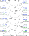
Figure 1.
Figure 1.
In vitro dissociation of the 100S ribosome by the RRF/EF-G pair and HflX in the presence and absence of guanosine analogs. Reactions were programmed with 0.2 μm ribosomes, 2 μm proteins, and 2 mm GTP analogs and incubated at 37 °C for 30 min. The samples were centrifuged in a 5–20% sucrose gradient, and ribosome profiles were monitored from the absorbance at 254 nm (y axis). Quantification of the 100S to 70S ratios were obtained from three technical replicates (of two independently prepared ribosomes and recombinant proteins); mean ± S.D.
Figure 2.
Figure 2.
The effects of RRF and EF-G mutations in 100S ribosome disassembly. A, model of the RRF, EF-G and ribosome co-complex. A structural model was constructed from PDB accession numbers 4V54, 5OT7, and 4WPO. The mutations included in this study are indicated. R59 (red) was mapped to an unstructured region of EF-G in the crystal. B, the dissociation of 100S ribosomes is reduced in GTP-hydrolysis mutants (R29A and R59A) of EF-G but is unaffected by a mutation that compromises tRNA translocation (H572K). The dissociation reactions comprised 0.2 μm ribosomes, 2 μm proteins, and 2 mm GTP analogs and were incubated at 37 °C for 30 min. Ribosome profiles were analyzed via 5–20% sucrose density sedimentation, and the ribosomal species were monitored according to the absorbance at 254 nm. 100S to 70S ratios were obtained from three technical replicates (of two independently prepared ribosomes and recombinant proteins); mean ± S.D. C, malachite green GTPase assay showing the reduction of GTPase hydrolysis in the R29A and R59A mutants. The known translocation inactive H572K mutant does not present impaired GTPase activity. Error bars represent the S.E. obtained from three independent experiments using two different batches of purified proteins. p values were calculated by Student's unpaired t test, ****, p < 0.0001; NS, not significant.
Figure 3.
Figure 3.
In vivo phenotypes of the RRF-depleted strain. A, construction of the RRF-depleted strain. His6-tagged frr was first placed under the control of a tetracycline (Tet)-inducible promoter on a cadmium chloride (CdCl2)-resistant integration plasmid. The plasmid was integrated into the chromosomal attC site, resulting in an S. aureus strain carrying an inducible frr (PTetfrr) and a native copy of frr (frr+). The WT copy of frr was deleted (Δfrr::Km, confers kanamycin resistance) from the frr+,PTetfrr strain by homologous recombination in the presence of 400 ng/ml of aTc. TT, transcriptional terminator. B, relative expression levels of native RRF and aTc-inducible His6-RRF. The expression of RRF was analyzed by immunoblotting with anti-RRF (1:4,000 dilutions). RRF was expressed from the native locus at a much higher level than the His6-RRF. His6-RRF migrated slightly more slowly than the native RRF. C, growth defects of RRF depletion in liquid media. S. aureus cells were grown in TSB at 37 °C with and without aTc. Real-time cell density was monitored on a TECAN plate reader according to the absorbance at 600 nm. The addition of aTc slightly suppressed bacterial growth in all tested strains, but it restored the growth of the RRF-depleted strain to levels comparable with those in other strains with aTc supplementation. Error bars are S.E. from three independent experiments, and each experiment included five replicates. D, the growth defects of RRF depletion were more significant on solid agar plates. Single colonies of different S. aureus strains were streaked on BactoTM agar plates containing TSB base, and bacterial growth was recorded after 16 h of incubation at 37 °C. The RRF-depleted strain (Δfrr,PTetfrr) and its derivative carrying the empty pLI50 plasmid only formed a few microcolonies in the absence of aTc. WT frr and frr(V116D) expressed in the pLI50 plasmid fully rescued the growth defects of Δfrr,PTetfrr.
Figure 4.
Figure 4.
HflX complements the deficiency of RRF. A, spotting assay of the RRF-depleted strain and the hflX knockout harboring the indicated empty and complementation plasmids. Data are representative of three independent experiments. Cells were adjusted to an A600 of ∼0.2 and were serially diluted and spotted (3 μl/spot) on TSB-agar plates, then grown overnight at 37 °C to determine cell viability. The expression of hflX and frr in trans restored the growth of the RRF-depleted strain in the absence of anhydrotetracyline (aTc). B, expression of RRF in S. aureus TSB cultures with and without aTc. Western blots were performed using anti-RRF at 1:4,000 dilutions.
Figure 5.
Figure 5.
Effects of RRF depletion on ribosome pools and stop codon read-through. A, top panels, accumulation of unrecycled ribosomes and queuing of ribosomes in the RRF-depleted strain. Crude ribosomes were isolated from TSB cultures at A600∼ 1.4 (without the aTc inducer) grown at 37 °C and analyzed by 10–40% sucrose gradient sedimentation. No translational elongation inhibitor (e.g. chloramphenicol) was added throughout the experiments; thus, minimal polysomes were preserved in the WT strain. By contrast, unusually high levels of mixed polysomes and stalled ribosomes accumulated upon RRF depletion, which was also observed to a lesser extent in the hflX knockout mutant. Lower panels, conversion of polysomes into lower order ribosomal complexes by RNase A treatment. Excess RNase A (20 μg/ml) collapsed 100S ribosomes and the WT and ΔhflX polysomes, whereas a fraction of disome-like complexes in the RRF-depleted cells are resistant to RNase A. B, possible outcome of RRF depletion. RRF deficiency prevented the recycling of post-termination complexes, which in turn reduced the ribosome pools available for the formation of 100S ribosomes. C, analysis of RRF and His6-RRF production in the RRF-depleted strain and the hflX knockout mutant used in A. The deletion of hflX did not influence RRF expression. D, RRF deficiency reduced the translational capacity and promoted stop codon read-through. The fluorescence intensity of the GFP reporter plasmid (pDM4) was normalized according to the cell density and compared between the WT and RRF-depleted strains (Δfrr::Km,PTetfrr). RRF depletion reduced the expression of the WT GFP reporter. A stop codon was introduced at the Lys-54 position of the GFP to evaluate the degree of translational read-through. In the absence of the aTc inducer, ribosomes bypassed the Lys-54 stop codon, producing low levels of GFP. The addition of aTc to the Δfrr::Km,PTet-frr strain suppressed read-through. Error bars are S.D. from five biological replicates. p values were calculated by Student's unpaired t test (n = 5, ***, p < 0.001; ****, p < 0.0001; NS, not significant).
Figure 6.
Figure 6.
Disassembly pathways of S. aureus 100S ribosomes. RRF and EF-G act in concert to split the PoTc in a GTP hydrolysis-dependent manner. The hibernating 100S ribosome is the second substrate of RRF/EF-G. Thermal stress induces the expression of hflX. HflX recycles 100S ribosomes and rescues stalled 70S ribosomes. A question mark indicates an unknown factor responsible for the hydrolysis of peptidyl-tRNA. HPF is colored in purple.

Similar articles

Cited by

References

    1. Li Y., Sharma M. R., Koripella R. K., Yang Y., Kaushal P. S., Lin Q., Wade J. T., Gray T. A., Derbyshire K. M., Agrawal R. K., and Ojha A. K. (2018) Zinc depletion induces ribosome hibernation in mycobacteria. Proc. Natl. Acad. Sci. U.S.A. 115, 8191–8196 10.1073/pnas.1804555115 - DOI - PMC - PubMed
    1. Polikanov Y. S., Blaha G. M., and Steitz T. A. (2012) How hibernation factors RMF, HPF, and YfiA turn off protein synthesis. Science 336, 915–918 10.1126/science.1218538 - DOI - PMC - PubMed
    1. Akiyama T., Williamson K. S., Schaefer R., Pratt S., Chang C. B., and Franklin M. J. (2017) Resuscitation of Pseudomonas aeruginosa from dormancy requires hibernation promoting factor (PA4463) for ribosome preservation. Proc. Natl. Acad. Sci. U.S.A. 114, 3204–3209 10.1073/pnas.1700695114 - DOI - PMC - PubMed
    1. Basu A., and Yap M. N. (2016) Ribosome hibernation factor promotes Staphylococcal survival and differentially represses translation. Nucleic Acids Res. 44, 4881–4893 10.1093/nar/gkw180 - DOI - PMC - PubMed
    1. Shcherbakova K., Nakayama H., and Shimamoto N. (2015) Role of 100S ribosomes in bacterial decay period. Genes Cells 20, 789–801 10.1111/gtc.12273 - DOI - PubMed

Publication types

MeSH terms

LinkOut - more resources