Parkinson's Disease: A Comprehensive Analysis of Fungi and Bacteria in Brain Tissue
- PMID: 32174790
- PMCID: PMC7053320
- DOI: 10.7150/ijbs.42257
Parkinson's Disease: A Comprehensive Analysis of Fungi and Bacteria in Brain Tissue
Abstract
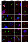
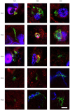
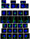
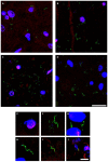
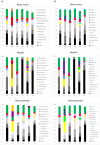
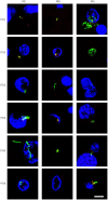
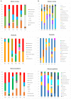
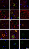
Parkinson's disease (PD) is characterized by motor disorders and the destruction of dopaminergic neurons in the substantia nigra pars compacta. In addition to motor disability, many patients with PD present a spectrum of clinical symptoms, including cognitive decline, psychiatric alterations, loss of smell and bladder dysfunction, among others. Neuroinflammation is one of the most salient features of PD, but the nature of the trigger remains unknown. A plausible mechanism to explain inflammation and the range of clinical symptoms in these patients is the presence of systemic microbial infection. Accordingly, the present study provides extensive evidence for the existence of mixed microbial infections in the central nervous system (CNS) of patients with PD. Assessment of CNS sections by immunohistochemistry using specific antibodies revealed the presence of both fungi and bacteria. Moreover, different regions of the CNS were positive for a variety of microbial morphologies, suggesting infection by a number of microorganisms. Identification of specific fungal and bacterial species in different CNS regions from six PD patients was accomplished using nested PCR analysis and next-generation sequencing, providing compelling evidence of polymicrobial infections in the CNS of PD. Most of the fungal species identified belong to the genera Botrytis, Candida, Fusarium and Malassezia. Some relevant bacterial genera were Streptococcus and Pseudomonas, with most bacterial species belonging to the phyla Actinobacteria and Proteobacteria. Interestingly, we noted similarities and differences between the microbiota present in the CNS of patients with PD and that in other neurodegenerative diseases. Overall, our observations lend strong support to the concept that mixed microbial infections contribute to or are a risk factor for the neuropathology of PD. Importantly, these results provide the basis for effective treatments of this disease using already approved and safe antimicrobial therapeutics.
Keywords: Parkinson's disease; fungal infection; neurodegenerative diseases; next-generation sequencing; polymicrobial infections.
© The author(s).
Conflict of interest statement
Competing Interests: The authors have declared that no competing interest exists.
Figures


Similar articles
-
Multiple sclerosis and mixed microbial infections. Direct identification of fungi and bacteria in nervous tissue.Neurobiol Dis. 2018 Sep;117:42-61. doi: 10.1016/j.nbd.2018.05.022. Epub 2018 Jun 1. Neurobiol Dis. 2018. PMID: 29859870
-
Electroacupuncture may alleviate behavioral defects via modulation of gut microbiota in a mouse model of Parkinson's disease.Acupunct Med. 2021 Oct;39(5):501-511. doi: 10.1177/0964528421990658. Epub 2021 Feb 8. Acupunct Med. 2021. PMID: 33557583
-
Infection of Fungi and Bacteria in Brain Tissue From Elderly Persons and Patients With Alzheimer's Disease.Front Aging Neurosci. 2018 May 24;10:159. doi: 10.3389/fnagi.2018.00159. eCollection 2018. Front Aging Neurosci. 2018. PMID: 29881346 Free PMC article.
-
T-Cell-Driven Inflammation as a Mediator of the Gut-Brain Axis Involved in Parkinson's Disease.Front Immunol. 2019 Feb 15;10:239. doi: 10.3389/fimmu.2019.00239. eCollection 2019. Front Immunol. 2019. PMID: 30828335 Free PMC article. Review.
-
Altered gut microbiota and intestinal permeability in Parkinson's disease: Pathological highlight to management.Neurosci Lett. 2019 Nov 1;712:134516. doi: 10.1016/j.neulet.2019.134516. Epub 2019 Sep 24. Neurosci Lett. 2019. PMID: 31560998 Review.
Cited by
-
Malassezia in Inflammatory Bowel Disease: Accomplice of Evoking Tumorigenesis.Front Immunol. 2022 Mar 4;13:846469. doi: 10.3389/fimmu.2022.846469. eCollection 2022. Front Immunol. 2022. PMID: 35309351 Free PMC article.
-
Neuroinflammation in neurodegeneration via microbial infections.Front Immunol. 2022 Jul 28;13:907804. doi: 10.3389/fimmu.2022.907804. eCollection 2022. Front Immunol. 2022. PMID: 36052093 Free PMC article. Review.
-
Bacteria-organelle communication in physiology and disease.J Cell Biol. 2024 Jul 1;223(7):e202310134. doi: 10.1083/jcb.202310134. Epub 2024 May 15. J Cell Biol. 2024. PMID: 38748249 Free PMC article. Review.
-
Oral fungal dysbiosis and systemic immune dysfunction in Chinese patients with schizophrenia.Transl Psychiatry. 2024 Nov 21;14(1):475. doi: 10.1038/s41398-024-03183-5. Transl Psychiatry. 2024. PMID: 39572530 Free PMC article.
-
Integrated Microbiome and Host Transcriptome Profiles Link Parkinson's Disease to Blautia Genus: Evidence From Feces, Blood, and Brain.Front Microbiol. 2022 May 26;13:875101. doi: 10.3389/fmicb.2022.875101. eCollection 2022. Front Microbiol. 2022. PMID: 35722294 Free PMC article.
References
-
- Zhu MY. Noradrenergic Modulation on Dopaminergic Neurons. Neurotox Res. 2018;34:848–59. - PubMed
-
- Khan AU, Akram M, Daniyal M, Zainab R. Awareness and current knowledge of Parkinson's disease: a neurodegenerative disorder. Int J Neurosci; 2018. pp. 1–39. - PubMed
-
- Poewe W, Seppi K, Tanner CM, Halliday GM, Brundin P, Volkmann J. et al. Parkinson disease. Nat Rev Dis Primers. 2017;3:17013. - PubMed
Publication types
MeSH terms
LinkOut - more resources
Full Text Sources
Medical
Miscellaneous

