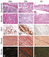
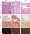
Although with intact mucosa at colonoscopy, chagasic megacolons have an overexpression of Gal-3
- PMID: 32159607
- PMCID: PMC7046340
- DOI: 10.31744/einstein_journal/2020AO5105
Although with intact mucosa at colonoscopy, chagasic megacolons have an overexpression of Gal-3
Abstract
Objective: To evaluate the density of anti-galectin-3-immunostained cells, collagen percentage, mast cell density and presence of pathological processes in intestinal muscle biopsies of patients.
Methods: Thirty-five patients who underwent intestinal biopsy were selected from 1997 to 2015. Patients were divided into three groups: chagasic patients with mucosal lesion (n=13), chagasic patients with intact mucosa (n=12) and non-chagasic patients with no mucosal lesion (n=10). Histological processing of the biopsied fragments and immunohistochemistry for galectin-3 were performed. Additional sections were stained with hematoxylin and eosin to evaluate the general pathological processes, picrosirius for evaluation of collagen and toluidine blue to evaluate the mast cell density.
Results: Patients of mucosal lesion group had a significantly higher frequency of ganglionitis and myositis when compared to the chagasic patients with intact mucosa and non-chagasic group. The density of anti-galectin-3-immunostained cells was significantly higher in the chagasic patients with intact mucosa group when compared to the non-chagasic group. The group of chagasic patients with intact mucosa presented a higher percentage of collagen in relation to the patients with mucosal lesion and to the non-chagasic group, with a significant difference. There was no significant difference in mast cell density among the three groups.
Conclusion: The higher density of anti-galectin-3-immunostained cells in patients in the chagasic patients with intact mucosa group suggested the need for greater attention in clinical evaluation of these patients, since this protein is associated with neoplastic transformation and progression.
Objetivo: Avaliar a densidade de células imunomarcadas por anti-galectina-3, a percentagem de colágeno, a densidade de mastócitos e a presença de processos patológicos na musculatura intestinal de pacientes biopsiados.
Métodos: Foram selecionados 35 pacientes submetidos à biópsia de intestino entre 1997 a 2015. Os pacientes foram divididos em três grupos: chagásicos com lesão de mucosa (n=13), chagásicos com mucosa íntegra (n=12) e não chagásicos sem lesão de mucosa (n=10). Foram realizados processamento histológico dos fragmentos biopsiados e imunohistoquímica para galectina-3. Cortes adicionais foram corados por hematoxilina e eosina, para avaliar os processos patológicos gerais, pelo picrosírius, para avaliação do colágeno, e pelo azul de toluidina, para avaliar a densidade de mastócitos.
Resultados: Os pacientes do grupo chagásicos com lesão de mucosa apresentaram frequência significativamente maior de ganglionite e miosite quando comparados aos dos grupos chagásico com mucosa íntegra e não chagásicos. A densidade das células imunomarcadas por anti-galectina-3 foi significativamente maior no grupo chagásicos com mucosa íntegra quando comparada ao grupo não chagásico. O grupo de chagásicos com mucosa íntegra apresentou maior percentagem de colágeno em relação aos grupos chagásicos com mucosa lesada e ao grupo de não chagásicos, com diferença significativa. Não houve diferença significativa com relação à densidade de mastócitos entre os três grupos.
Conclusão: A maior densidade de células imunomarcadas por anti-galectina-3 nos pacientes do grupo chagásico com mucosa íntegra sugere a necessidade de maior atenção na avaliação clínica desses pacientes, uma vez que essa proteína está associada com transformação e progressão neoplásica.
Conflict of interest statement
Figures


Similar articles
-
Evaluation of the immunohistochemical expression of Gal-1, Gal-3 and Gal-9 in the colon of chronic chagasic patients.Pathol Res Pract. 2017 Sep;213(9):1207-1214. doi: 10.1016/j.prp.2017.04.014. Epub 2017 Apr 20. Pathol Res Pract. 2017. PMID: 28554765
-
Morphometric study of eosinophils, mast cells, macrophages and fibrosis in the colon of chronic chagasic patients with and without megacolon.Parasitology. 2007 Jun;134(Pt 6):789-96. doi: 10.1017/S0031182007002296. Epub 2007 Feb 9. Parasitology. 2007. PMID: 17288632
-
Selective survival of calretinin- and vasoactive-intestinal-peptide-containing nerve elements in human chagasic submucosa and mucosa.Cell Tissue Res. 2012 Aug;349(2):473-81. doi: 10.1007/s00441-012-1406-8. Epub 2012 May 5. Cell Tissue Res. 2012. PMID: 22555304
-
Surgical treatment of chagasic megacolon with Duhamel-Habr-Gama technique modulated by frozen-section examination.Surg Infect (Larchmt). 2014 Aug;15(4):454-7. doi: 10.1089/sur.2012.181. Epub 2014 May 13. Surg Infect (Larchmt). 2014. PMID: 24824159 Review.
-
[Megas and cancer. Cancer of the large intestine in chagasic patients with megacolon].Arq Gastroenterol. 1989 Jan-Jun;26(1-2):13-6. Arq Gastroenterol. 1989. PMID: 2513795 Review. Portuguese.
Cited by
-
Galectins in Chagas Disease: A Missing Link Between Trypanosoma cruzi Infection, Inflammation, and Tissue Damage.Front Microbiol. 2022 Jan 3;12:794765. doi: 10.3389/fmicb.2021.794765. eCollection 2021. Front Microbiol. 2022. PMID: 35046919 Free PMC article. Review.
-
Biomarkers and Their Possible Functions in the Intestinal Microenvironment of Chagasic Megacolon: An Overview of the (Neuro)inflammatory Process.J Immunol Res. 2021 Apr 7;2021:6668739. doi: 10.1155/2021/6668739. eCollection 2021. J Immunol Res. 2021. PMID: 33928170 Free PMC article. Review.
References
-
- . Dubner S, Schapachnik E, Riera AR, Valero E. Chagas disease: state-of-the-art of diagnosis and management. Cardiol J. 2008;15(6):493-504. Review. - PubMed
- Dubner S, Schapachnik E, Riera AR, Valero E. Chagas disease: state-of-the-art of diagnosis and management. Cardiol J . 2008;15(6):493–504. Review. - PubMed
-
- . Pérez-Ayala A, Pérez-Molina JA, Norman F, Navarro M, Monge-Maillo B, Diaz-Menéndez M, et al. Chagas disease in Latin American migrants: a Spanish challenge. Clin Microbiol Infect. 2011;17(7):1108-13. - PubMed
- Pérez-Ayala A, Pérez-Molina JA, Norman F, Navarro M, Monge-Maillo B, Diaz-Menéndez M, et al. Chagas disease in Latin American migrants: a Spanish challenge. Clin Microbiol Infect . 2011;17(7):1108–1113. - PubMed
-
- . Dias JC, Ramos AN Jr, Gontijo ED, Luquetti A, Shikanai-Yasuda MA, Coura JR, Torres RM, Melo JR, Almeida EA, Oliveira W Jr, Silveira AC, Rezende JM, Pinto FS, Ferreira AW, Rassi A, Fragata AA Filho, Sousa AS, Correia D Filho, Jansen AM, Andrade GM, Britto CF, Pinto AY, Rassi A Jr, Campos DE, Abad-Franch F, Santos SE, Chiari E, Hasslocher-Moreno AM, Moreira EF, Marques DS, Silva EL, Marin-Neto JA, Galvão LM, Xavier SS, Valente SA, Carvalho NB, Cardoso AV, Silva RA, Costa VM, Vivaldini SM, Oliveira SM, Valente VD, Lima MM, Alves RV. II Consenso Brasileiro em Doença de Chagas, 2015. Epidemiol Serv Saude. 2016;25(spe):7-86. - PubMed
- Dias JC, Ramos AN, Jr, Gontijo ED, Luquetti A, Shikanai-Yasuda MA, Coura JR, Torres RM, Melo JR, Almeida EA, Oliveira W, Jr, Silveira AC, Rezende JM, Pinto FS, Ferreira AW, Rassi A, Fragata AA, Filho, Sousa AS, Correia D, Filho, Jansen AM, Andrade GM, Britto CF, Pinto AY, Rassi A, Jr, Campos DE, Abad-Franch F, Santos SE, Chiari E, Hasslocher-Moreno AM, Moreira EF, Marques DS, Silva EL, Marin JA, Neto, Galvão LM, Xavier SS, Valente SA, Carvalho NB, Cardoso AV, Silva RA, Costa VM, Vivaldini SM, Oliveira SM, Valente VD, Lima MM, Alves RV. II Consenso Brasileiro em Doença de Chagas, 2015. Epidemiol Serv Saude . 2016;25(spe):7–86. - PubMed
-
- . Crema E, Silva EC, Franciscon PM, Rodrigues Júnior V, Martins Júnior A, Teles CJ, et al. Prevalence of cholelithiasis in patients with chagasic megaesophagus. Rev Soc Bras Med Trop. 2011;44(3):324-6. - PubMed
- Crema E, Silva EC, Franciscon PM, Rodrigues V, Júnior, Martins A, Júnior, Teles CJ, et al. Prevalence of cholelithiasis in patients with chagasic megaesophagus. Rev Soc Bras Med Trop . 2011;44(3):324–326. - PubMed
MeSH terms
Substances
LinkOut - more resources
Full Text Sources
Medical

