. 2020 Apr 30;13(4):dmm040816.
doi: 10.1242/dmm.040816.

Zika virus non-structural protein NS4A restricts eye growth in Drosophila through regulation of JAK/STAT signaling

Affiliations

Zika virus non-structural protein NS4A restricts eye growth in Drosophila through regulation of JAK/STAT signaling

Sneh Harsh et al. Dis Model Mech. .

Abstract

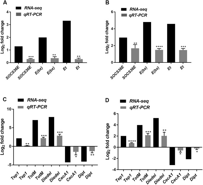
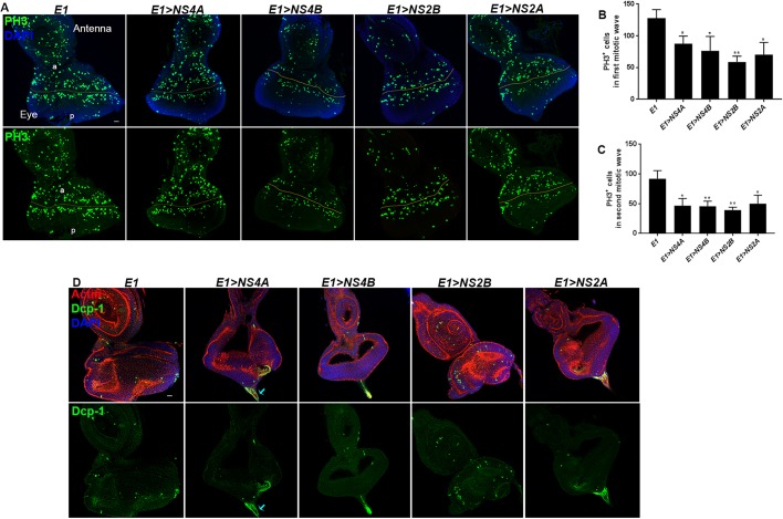
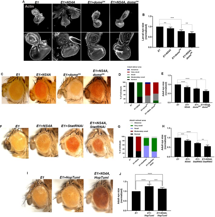
To gain a comprehensive view of the changes in host gene expression underlying Zika virus (ZIKV) pathogenesis, we performed whole-genome RNA sequencing (RNA-seq) of ZIKV-infected Drosophila adult flies. RNA-seq analysis revealed that ZIKV infection alters several and diverse biological processes, including stress, locomotion, lipid metabolism, imaginal disc morphogenesis and regulation of JAK/STAT signaling. To explore the interaction between ZIKV infection and JAK/STAT signaling regulation, we generated genetic constructs overexpressing ZIKV-specific non-structural proteins NS2A, NS2B, NS4A and NS4B. We found that ectopic expression of non-structural proteins in the developing Drosophila eye significantly restricts growth of the larval and adult eye and correlates with considerable repression of the in vivo JAK/STAT reporter, 10XStat92E-GFP At the cellular level, eye growth defects are associated with reduced rate of proliferation without affecting the overall rate of apoptosis. In addition, ZIKV NS4A genetically interacts with the JAK/STAT signaling components; co-expression of NS4A along with the dominant-negative form of domeless or StatRNAi results in aggravated reduction in eye size, while co-expression of NS4A in HopTuml (also known as hopTum ) mutant background partially rescues the hop-induced eye overgrowth phenotype. The function of ZIKV NS4A in regulating growth is maintained in the wing, where ZIKV NS4A overexpression in the pouch domain results in reduced growth linked with diminished expression of Notch targets, Wingless (Wg) and Cut, and the Notch reporter, NRE-GFP Thus, our study provides evidence that ZIKV infection in Drosophila results in restricted growth of the developing eye and wing, wherein eye phenotype is induced through regulation of JAK/STAT signaling, whereas restricted wing growth is induced through regulation of Notch signaling. The interaction of ZIKV non-structural proteins with the conserved host signaling pathways further advance our understanding of ZIKV-induced pathogenesis.This article has an associated First Person interview with the first author of the paper.

Keywords: Drosophila; Eye development; Host-pathogen interaction; JAK/STAT signaling; Zika virus.


Conflict of interest statement

Competing interestsThe authors declare no competing or financial interests.

Figures

Fig. 1.
Fig. 1.
Transcriptome analysis of ZIKV-infected flies through RNA-seq. (A) Overview of the experimental workflow. 5- to 6-day-old wild-type female flies (w1118 strain) were injected with the MR766 strain of ZIKV. PBS-injected flies served as negative controls. Total RNA was extracted at 4 and 8 days post-injection (dpi) for RNA-seq. (B) Transcriptome summary (number of reads and percentage mapped to D. melanogaster genome) from wild-type flies injected with PBS and ZIKV at 4 and 8 dpi. (C) Heat map showing the differentially expressed genes (DEGs) in PBS- and ZIKV-injected flies (in triplicate) at 4 and 8 dpi. (D) DEGs (downregulated/upregulated) in wild-type flies injected with ZIKV at 4 and 8 dpi when adjusted P-value is set at 0.05 and 0.1, respectively. (E) Venn diagrams showing the number of Drosophila genes that are differentially expressed (upregulated or downregulated) in wild-type flies injected with ZIKV at 4 and 8 dpi. Expression patterns are indicated (Up/Up, gene upregulation at both 4 and 8 dpi; Down/Down, gene downregulation at both timepoints; Up/Down, gene upregulation at 4 dpi and downregulation at 8 dpi; Down/Up, gene downregulation at 4 dpi and upregulation at 8 dpi). Significance was tested using a hypergeometric test: P-values for Up/Up, Down/Down, Up/Down and Down/Up trending genes were 2.065×10⁻³³, 1.361×10⁻¹¹, 3.050×10⁻⁹ and 5.485×10⁻¹¹, respectively.
Fig. 2.
Fig. 2.
Infection of adult flies with ZIKV induces diverse biological processes and molecular functions. (A,E) Representative enrichment of upregulated (log>1.2 fold) biological processes using Database for Annotation, Visualization and Integrated Discovery (DAVID) classification database at 4 and 8 dpi. (C,G) Representative enrichment of downregulated (log<−1.2 fold) biological processes using DAVID at 4 and 8 dpi. (B,D,F,H) GO-based molecular functions regulated by the DEGs at 4 dpi (B,D) and 8 dpi (F,H).
Fig. 3.
Fig. 3.
Validation of the enriched negative regulators of JAK/STAT pathway by qRT-PCR. (A,B) Log2 fold change (RNA-seq) and mRNA levels (qRT-PCR) of Socs36E, E(bx) and Et in wild-type adult flies infected with ZIKV at 4 and 8 dpi. (C,D) Log2 fold change (RNA-seq) and mRNA levels (qRT-PCR) of Tep1, TotM, Diedel, CecA1 and Dipt in wild-type adult flies infected with ZIKV at 4 and 8 dpi. All data were normalized to the housekeeping gene RpL32 and are shown relative to wild-type flies injected with PBS (sterile control). Three independent experiments were carried out with ten flies per sample in triplicate (‘*P<0.05, **P<0.01, ***P<0.001, ****P<0.0001). Bars represent the mean±s.d. Statistical analysis was performed using unpaired two-tailed Student's t-test.
Fig. 4.
Fig. 4.
ZIKV non-structural proteins induce restricted eye growth and are linked with downregulated JAK/STAT signaling. (A) Representative eye imaginal discs upon overexpression of ZIKV non-structural protein-coding genes driven under eye-specific eyeless-Gal4 (referred to as E1-Gal4) (E1>NS4A, E1>NS4B, E1>NS2B and E1>NS2A). Lower row shows the enlarged view of the eye imaginal discs. The cytoarchitecture was marked with Actin (gray). (B) Quantification of the size of eye imaginal discs upon E1-Gal4 driven overexpression of ZIKV non-structural protein coding genes, compared to E1-Gal4 alone. Bars show mean±s.d. (*P<0.05, **P<0.01; ns, not significant). (C) Representative images of the adult eye in the indicated genotypes. (D) Quantification of the size of adult eye upon overexpression of ZIKV non-structural protein-coding genes, compared to E1-Gal4. Bars show mean±s.d. (***P=0.0001, **P=0.0016, *P<0.05). (E) qRT-PCR analysis depicting the mRNA level of NS4A in the eye imaginal epithelia overexpressing ZIKV NS4A driven under E1-Gal4 (E1>NS4A), compared to E1-Gal4 alone. Bars show mean±s.d. (****P<0.0001). (F) qRT-PCR analysis depicting the mRNA level of chinmo, Mo25 and domeless in the eye imaginal disc where ZIKV NS4A was overexpressed. All data were normalized to the housekeeping gene RpL32 and are shown relative to E1-Gal4. Three independent experiments were carried out in triplicate and bars represent mean±s.d. (**P<0.05). (G) Expression of 10XStat92E-GFP (abbreviated to 10XStat-GFP, shown by red in merge and gray in separate channel) in eye imaginal epithelia carrying eyeless-specific overexpression of ZIKV NS4A compared to E1-Gal4 alone. The cytoarchitecture is marked with Actin (green). The 10XStat-GFP levels are marked by yellow arrows in both E1-Gal4- and E1>NS4A-carrying eye imaginal epithelia. (H) Quantification of 10XStat-GFP fluorescence intensity in the eye imaginal disc proper from E1-Gal4- and E1>NS4A-carrying larvae. Bars show mean±s.d. (**P=0.0015). Statistical analysis was performed using unpaired two-tailed Student's t-test. AU, arbitrary units. Scale bars: 100 μm.
Fig. 5.
Fig. 5.
ZIKV non-structural protein overexpression results in reduced rate of proliferation in eye imaginal epithelia. (A,D) Representative eye imaginal discs overexpressing ZIKV non-structural protein-coding genes driven under eye-specific E1-Gal4 (E1>NS4A, E1>NS4B, E1>NS2B and E1>NS2A). (A) The rate of proliferation was marked with anti-phosphohistone (PH3) staining (green). The yellow lines mark the morphogenetic furrow. The compartments are marked: a, anterior; p, posterior. In all images, nuclei were stained with DAPI (blue). (B) Quantification of the number of PH3-marked cells in the first mitotic wave or anterior region of eye discs upon ZIKV non-structural protein overexpression compared to E1-Gal4 alone. Bars show mean±s.d. (*P<0.05, **P<0.01). (C) Quantification of the number of PH3-marked cells in the second mitotic wave or posterior region of eye discs upon ZIKV non-structural protein overexpression compared to E1-Gal4 alone. Bars show mean±s.d. (*P<0.05, **P<0.01). (D) Cell death was indicated by anti-Dcp-1 staining (green); Actin was used to mark the cytoarchitecture (red). In all images, nuclei were stained with DAPI (blue). Statistical analysis for the graphs were performed using unpaired two-tailed Student's t-test. Scale bars: 100 μm.
Fig. 6.
Fig. 6.
ZIKV NS4A shows genetic interaction with different components of JAK/STAT signaling pathway. (A) Representative eye imaginal discs overexpressing ZIKV NS4A, a dominant negative form of domeless, and co-expression of the dominant-negative form of domeless and NS4A driven under the eye-specific driver, E1-Gal4 (E1>NS4A, E1>domeDN and E1>NS4A, domeDN, respectively), compared to E1-Gal4 alone. The lower row shows the enlarged view of the eye imaginal discs. Cytoarchitecture was marked with Actin (gray). (B) Quantification of the size of eye imaginal discs in the indicated genotypes. Bars show mean±s.d. (***P<0.0001, **P<0.05). (C) Representative images of the adult eye in E1>NS4A, E1>domeDN and E1>NS4A, domeDN, respectively. (D) Percentage of individuals displaying normal, moderate, small, very small or absence of adult retinal area in the indicated genotypes. (E) Quantification of the size of adult eye in E1>NS4A, E1>domeDN and E1>NS4A, domeDN, respectively, compared to E1-Gal4 alone. Bars show mean±s.d. (****P<0.0001, **P=0.0053). (F) Representative images of the adult eye overexpressing ZIKV NS4A, StatRNAi and co-expression of StatRNAi and NS4A driven under eye-specific E1-Gal4 (E1>NS4A, E1>StatRNAi and E1>NS4A, StatRNAi, respectively). (G) Penetrance of different eye phenotypes in the indicated genotypes. (H) Quantification of the size of adult eye in E1>NS4A, E1>StatRNAi and E1>NS4A, StatRNAi, respectively, compared to E1-Gal4 alone. Bars show mean±s.d. (****P<0.0001, ***P<0.001 and **P=0.0071). (I) Representative images of the adult eye upon E1-Gal4-driven overexpression of HopTuml and co-expression of NS4A and HopTuml (E1>HopTuml and E1>NS4A, HopTuml, respectively). (J) Quantification of the size of adult eye in E1>HopTuml and E1>NS4A, HopTuml, compared to E1-Gal4 alone. Bars show mean±s.d. (****P<0.0001 and ***P=0.0006). Statistical analysis was performed using unpaired two-tailed Student's t-test. Scale bars: 100 μm.
Fig. 7.
Fig. 7.
ZIKV NS4A overexpression results in thickening of veins in adult wing. (A) Representative wing imaginal discs upon overexpression of ZIKV non-structural protein-coding genes driven under wing-specific, engrailed-Gal4 (en>NS4A, en>NS2A, en>NS2B and en>NS4B). The engrailed marked compartment was tagged with RFP (enGal4>UAS-RFP). Wing disc-specific JAK/STAT target, CycA, is marked in green. (B) Anti-Dcp-1 was used to show cell death (green) in the indicated genotypes. (C) Representative images of the adult wing in en>NS4A, en>NS2A, en>NS2B and en>NS4B. The thickening and branching of veins are depicted with red arrows. Scale bars: 100 μm.
Fig. 8.
Fig. 8.
ZIKV NS4A overexpression results in notching of wing linked with downregulated Notch signaling. (A) Representative images of the adult wing upon overexpression of ZIKV NS4A driven under wing-specific, nubbin-Gal4 (nub>NS4A), compared to nub-Gal4 alone. (B-E) Representative wing imaginal discs displaying overexpression of ZIKV NS4A driven under nub-Gal4 (nub>NS4A), compared to nub-Gal4 alone. For clarity, the nubbin region is marked with a yellow outline. (B) Cell death was indicated with anti-Dcp-1 staining (blue in merge panel and shown in gray in separate channel). (C) Quantification of the size of the wing pouch upon overexpression of ZIKV non-structural protein NS4A under nub-Gal4 (nub>NS4A), compared to nub-Gal4 alone. The size of the wing pouch in each genotype was normalized with respect to the size of the whole wing disc. Bars show mean±s.d. (***P=0.0001). (D,E) Wingless and Cut, the targets of Notch signaling, are marked in red. Reduced expression of Wg and Cut upon NS4A overexpression (nub>NS4A) is indicated by yellow arrows. (F) Overexpression of NS4A in the wing pouch (nub>NS4A) significantly downregulated Notch signaling reporter NRE-GFP expression at the dorsal/ventral boundary, compared to nubbin-Gal4 alone. NRE-GFP is shown in red in the merge images and in gray in the separate channel. (G) The amount of Notch intracellular domain (NICD) was significantly reduced when ZIKV NS4A was overexpressed using nubbin-Gal4 (nub>NS4A), compared to nubbin-Gal4 alone. Notch protein level was marked with anti-NICD staining (red in merge channel and gray in the separate channel). In all images, cytoarchitecture was marked with Actin (green). Scale bars: 100 μm.

Similar articles

Cited by

References

    1. Agaisse H., Petersen U.-M., Boutros M., Mathey-Prevot B. and Perrimon N. (2003). Signaling role of hemocytes in Drosophila JAK/STAT-dependent response to septic injury. Dev. Cell 5, 441-450. 10.1016/S1534-5807(03)00244-2 - DOI - PubMed
    1. Apte-Sengupta S., Sirohi D. and Kuhn R. J. (2014). Coupling of replication and assembly in flaviviruses. Curr. Opin. Virol. 9, 134-142. 10.1016/j.coviro.2014.09.020 - DOI - PMC - PubMed
    1. Arbouzova N. I. and Zeidler M. P. (2006). JAK/STAT signalling in Drosophila: insights into conserved regulatory and cellular functions. Development 133, 2605-2616. 10.1242/dev.02411 - DOI - PubMed
    1. Bach E. A., Vincent S., Zeidler M. P. and Perrimon N. (2003). A sensitized genetic screen to identify novel regulators and components of the Drosophila janus kinase/signal transducer and activator of transcription pathway. Genetics 165, 1149-1166. - PMC - PubMed
    1. Bach E. A., Ekas L. A., Ayala-Camargo A., Flaherty M. S., Lee H., Perrimon N. and Baeg G.-H. (2007). GFP reporters detect the activation of the Drosophila JAK/STAT pathway in vivo. Gene Expr. Patterns 7, 323-331. 10.1016/j.modgep.2006.08.003 - DOI - PubMed

Publication types

MeSH terms