Resident plasmacytoid dendritic cells patrol vessels in the naïve limbus and conjunctiva
- PMID: 32109562
- PMCID: PMC7397780
- DOI: 10.1016/j.jtos.2020.02.005
Resident plasmacytoid dendritic cells patrol vessels in the naïve limbus and conjunctiva
Abstract
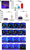
Plasmacytoid dendritic cells (pDCs) constitute a unique population of bone marrow-derived cells that play a pivotal role in linking innate and adaptive immune responses. While peripheral tissues are typically devoid of pDCs during steady state, few tissues do host resident pDCs. In the current study, we aim to assess presence and distribution of pDCs in naïve murine limbus and bulbar conjunctiva. Immunofluorescence staining followed by confocal microscopy revealed that the naïve bulbar conjunctiva of wild-type mice hosts CD45+ CD11clow PDCA-1+ pDCs. Flow cytometry confirmed the presence of resident pDCs in the bulbar conjunctiva through multiple additional markers, and showed that they express maturation markers, the T cell co-inhibitory molecules PD-L1 and B7-H3, and minor to negligible levels of T cell co-stimulatory molecules CD40, CD86, and ICAM-1. Epi-fluorescent microscopy of DPE-GFP×RAG1-/- transgenic mice with GFP-tagged pDCs indicated lower density of pDCs in the bulbar conjunctiva compared to the limbus. Further, intravital multiphoton microscopy revealed that resident pDCs accompany the limbal vessels and patrol the intravascular space. In vitro multiphoton microscopy showed that pDCs are attracted to human umbilical vein endothelial cells and interact with them during tube formation. In conclusion, our study shows that the limbus and bulbar conjunctiva are endowed with resident pDCs during steady state, which express maturation and classic T cell co-inhibitory molecules, engulf limbal vessels, and patrol intravascular spaces.
Keywords: Conjunctiva; Intravital microscopy; Limbus; Plasmacytoid dendritic cell; Vascular endothelial cell.
Copyright © 2020 Elsevier Inc. All rights reserved.
Figures


Similar articles
-
Intravital Multiphoton Microscopy of the Ocular Surface: Alterations in Conventional Dendritic Cell Morphology and Kinetics in Dry Eye Disease.Front Immunol. 2020 May 7;11:742. doi: 10.3389/fimmu.2020.00742. eCollection 2020. Front Immunol. 2020. PMID: 32457740 Free PMC article.
-
Plasmacytoid dendritic cells in the eye.Prog Retin Eye Res. 2021 Jan;80:100877. doi: 10.1016/j.preteyeres.2020.100877. Epub 2020 Jul 24. Prog Retin Eye Res. 2021. PMID: 32717378 Free PMC article. Review.
-
Murine plasmacytoid pre-dendritic cells generated from Flt3 ligand-supplemented bone marrow cultures are immature APCs.J Immunol. 2002 Dec 15;169(12):6711-9. doi: 10.4049/jimmunol.169.12.6711. J Immunol. 2002. PMID: 12471102
-
Enhanced activation of circulating plasmacytoid dendritic cells in patients with Chronic Obstructive Pulmonary Disease and experimental smoking-induced emphysema.Clin Immunol. 2018 Oct;195:107-118. doi: 10.1016/j.clim.2017.11.003. Epub 2017 Nov 8. Clin Immunol. 2018. PMID: 29127016
-
Imaging of plasmacytoid dendritic cell interactions with T cells.Blood. 2009 Jan 1;113(1):75-84. doi: 10.1182/blood-2008-02-139865. Epub 2008 Sep 25. Blood. 2009. PMID: 18818393
Cited by
-
New Therapeutic Approaches for Conjunctival Melanoma-What We Know So Far and Where Therapy Is Potentially Heading: Focus on Lymphatic Vessels and Dendritic Cells.Int J Mol Sci. 2022 Jan 27;23(3):1478. doi: 10.3390/ijms23031478. Int J Mol Sci. 2022. PMID: 35163401 Free PMC article. Review.
-
Intravital Multiphoton Microscopy of the Ocular Surface: Alterations in Conventional Dendritic Cell Morphology and Kinetics in Dry Eye Disease.Front Immunol. 2020 May 7;11:742. doi: 10.3389/fimmu.2020.00742. eCollection 2020. Front Immunol. 2020. PMID: 32457740 Free PMC article.
-
Dendritiform immune cells with reduced antigen-capture capacity persist in the cornea during the asymptomatic phase of allergic conjunctivitis.Eye (Lond). 2023 Sep;37(13):2768-2775. doi: 10.1038/s41433-023-02413-2. Epub 2023 Feb 6. Eye (Lond). 2023. PMID: 36747108 Free PMC article.
-
Increased dendritic cell density and altered morphology in allergic conjunctivitis.Eye (Lond). 2023 Oct;37(14):2896-2904. doi: 10.1038/s41433-023-02426-x. Epub 2023 Feb 6. Eye (Lond). 2023. PMID: 36747109 Free PMC article.
-
Epithelial Immune Cell Response to Initial Soft Contact Lens Wear in the Human Corneal and Conjunctival Epithelium.Invest Ophthalmol Vis Sci. 2023 Dec 1;64(15):18. doi: 10.1167/iovs.64.15.18. Invest Ophthalmol Vis Sci. 2023. PMID: 38099736 Free PMC article.
References
-
- Lennert K & Remmele W [Karyometric research on lymph node cells in man. I. Germinoblasts, lymphoblasts & lymphocytes]. Acta haematologica 19, 99–113 (1958). - PubMed
-
- Muller-Hermelink HK, Stein H, Steinmann G & Lennert K Malignant lymphoma of plasmacytoid T-cells. Morphologic and immunologic studies characterizing a special type of T-cell. The American journal of surgical pathology 7, 849–862 (1983). - PubMed
-
- Vollenweider R & Lennert K Plasmacytoid T-cell clusters in non-specific lymphadenitis. Virchows Archiv. B, Cell pathology including molecular pathology 44, 1–14 (1983). - PubMed
-
- Feller AC, Lennert K, Stein H, Bruhn HD & Wuthe HH Immunohistology and aetiology of histiocytic necrotizing lymphadenitis. Report of three instructive cases. Histopathology 7, 825–839 (1983). - PubMed
-
- Prasthofer EF, Prchal JT, Grizzle WE & Grossi CE Plasmacytoid T-cell lymphoma associated with chronic myeloproliferative disorder. The American journal of surgical pathology 9, 380–387 (1985). - PubMed
Publication types
MeSH terms
Grants and funding
LinkOut - more resources
Full Text Sources
Research Materials
Miscellaneous

