

Accelerated Partial Breast Irradiation: Macrophage Polarisation Shift Classification Identifies High-Risk Tumours in Early Hormone Receptor-Positive Breast Cancer
- PMID: 32075091
- PMCID: PMC7072550
- DOI: 10.3390/cancers12020446
Accelerated Partial Breast Irradiation: Macrophage Polarisation Shift Classification Identifies High-Risk Tumours in Early Hormone Receptor-Positive Breast Cancer
Abstract
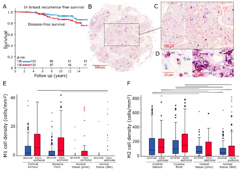
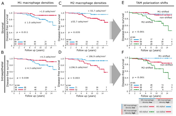
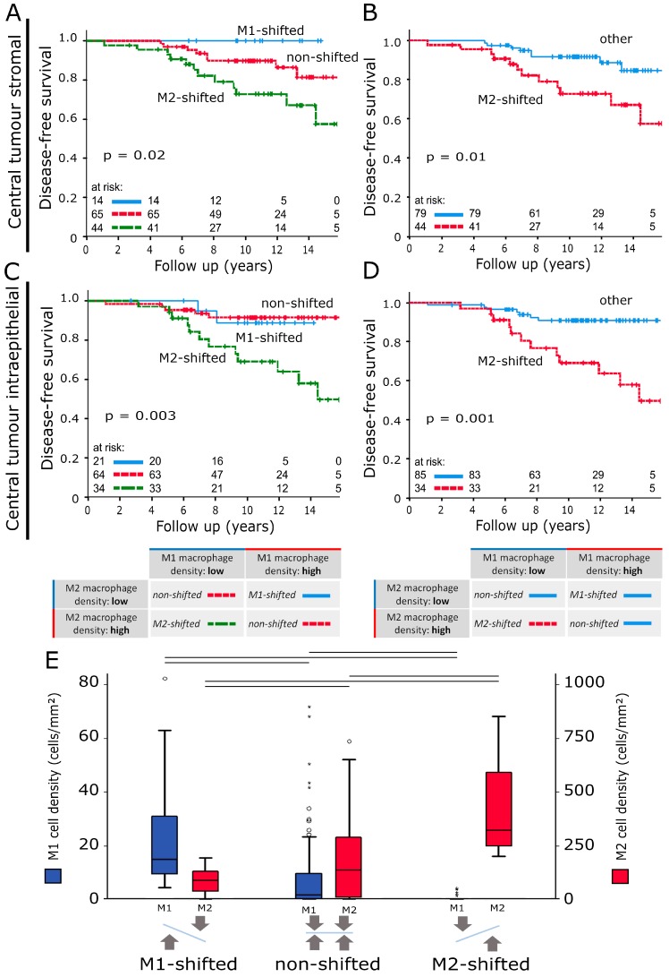
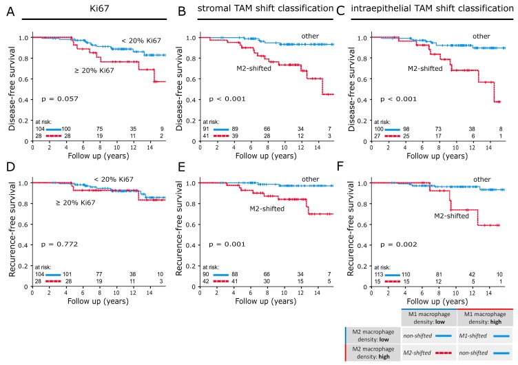
Studies have demonstrated correlations between accumulations of tumour-associated macrophages (TAMs), especially of M2-like phenotype, and increased mortality in advanced breast cancer. We investigated the prognostic potential of both main macrophage phenotypes in early hormone receptor-positive (HR+) breast cancer. The studied cohort of 136 patients participated in an institutional APBI phase II trial. Patient selection was characterized by HR+, small tumour size and no metastasis. Tissue microarrays from pre-RT resection samples were double stained for CD68/CD163 using immunohistochemistry. CD68+/CD163- cells were considered M1-like macrophages and CD68+/CD163+ was representative of M2-like macrophages. M1 and M2 macrophage densities were analysed semi-automatically in the stromal and intraepithelial tumour compartment. Low M1 and high M2 densities were strongly associated with decreased disease-free survival (DFS). Combined TAM phenotype densities were studied after defining a macrophage shift classification: M1-shifted (M1 high, M2 low) and non-shifted (M1 low, M2 low; M1 high, M2 high) tumours entailed a favourable outcome. In contrast, M2-shifted (M1 low, M2 high) TAM populations were associated with extremely reduced DFS. Thus, the full predictive potential of TAMs was revealed in a combined analysis of both phenotypes. The M2-shifted subgroup of tumours is classified as high-risk and probably not suitable for partial breast irradiation.
Keywords: CD163; CD68; accelerated partial breast irradiation; early breast cancer; hormone receptor-positive; prognosis; tumour associated macrophages.
Conflict of interest statement
The authors declare no conflict of interest.
Figures
Similar articles
-
Prognostic Value of Macrophage Phenotypes in Resectable Non-Small Cell Lung Cancer Assessed by Multiplex Immunohistochemistry.Neoplasia. 2019 Mar;21(3):282-293. doi: 10.1016/j.neo.2019.01.005. Epub 2019 Feb 10. Neoplasia. 2019. PMID: 30743162 Free PMC article.
-
Assessing the role of tumour-associated macrophage subsets in breast cancer subtypes using digital image analysis.Breast Cancer Res Treat. 2023 Feb;198(1):11-22. doi: 10.1007/s10549-022-06859-y. Epub 2023 Jan 9. Breast Cancer Res Treat. 2023. PMID: 36622544 Free PMC article.
-
Tumor-Associated Macrophages as Potential Prognostic Biomarkers of Invasive Breast Cancer.J Breast Cancer. 2019 Mar;22(1):38-51. doi: 10.4048/jbc.2019.22.e5. J Breast Cancer. 2019. PMID: 30941232 Free PMC article.
-
Breast Cancer Survival Outcomes and Tumor-Associated Macrophage Markers: A Systematic Review and Meta-Analysis.Oncol Ther. 2023 Mar;11(1):27-48. doi: 10.1007/s40487-022-00214-3. Epub 2022 Dec 9. Oncol Ther. 2023. PMID: 36484945 Free PMC article. Review.
-
Prognostic significance of CD68+ and CD163+ tumor associated macrophages in head and neck squamous cell carcinoma: A systematic review and meta-analysis.Oral Oncol. 2019 Jun;93:66-75. doi: 10.1016/j.oraloncology.2019.04.019. Epub 2019 Apr 28. Oral Oncol. 2019. PMID: 31109698 Review.
Cited by
-
Tumor-Associated Neutrophils Are a Negative Prognostic Factor in Early Luminal Breast Cancers Lacking Immunosuppressive Macrophage Recruitment.Cancers (Basel). 2024 Sep 15;16(18):3160. doi: 10.3390/cancers16183160. Cancers (Basel). 2024. PMID: 39335132 Free PMC article.
-
Estrogen receptor blockade and radiation therapy cooperate to enhance the response of immunologically cold ER+ breast cancer to immunotherapy.Breast Cancer Res. 2023 Jun 13;25(1):68. doi: 10.1186/s13058-023-01671-y. Breast Cancer Res. 2023. PMID: 37312163 Free PMC article.
-
Role of Tumor-Associated Myeloid Cells in Breast Cancer.Cells. 2020 Jul 27;9(8):1785. doi: 10.3390/cells9081785. Cells. 2020. PMID: 32726950 Free PMC article. Review.
-
M1 Macrophage and M1/M2 ratio defined by transcriptomic signatures resemble only part of their conventional clinical characteristics in breast cancer.Sci Rep. 2020 Oct 6;10(1):16554. doi: 10.1038/s41598-020-73624-w. Sci Rep. 2020. PMID: 33024179 Free PMC article.
-
Molecular Subtypes of Oral Squamous Cell Carcinoma Based on Immunosuppression Genes Using a Deep Learning Approach.Front Cell Dev Biol. 2021 Aug 5;9:687245. doi: 10.3389/fcell.2021.687245. eCollection 2021. Front Cell Dev Biol. 2021. PMID: 34422810 Free PMC article.
References
-
- International Agency for Research on Cancer, World Health Organization . Latest Global Cancer Data: Cancer Burden Rises to 18.1 Million New Cases and 9.6 Million Cancer Deaths in 2018. Volume 263 IARC; Lyon, France: 2018.
-
- Abo-Madyan Y., Welzel G., Sperk E., Neumaier C., Keller A., Clausen S., Schneider F., Ehmann M., Sutterlin M., Wenz F. Single-center long-term results from the randomized phase-3 targit-a trial comparing intraoperative and whole-breast radiation therapy for early breast cancer. Strahlenther. Onkol. 2019;195:640–647. doi: 10.1007/s00066-019-01438-5. - DOI - PubMed
LinkOut - more resources
Full Text Sources
Research Materials
