

Skeletal muscle in health and disease
- PMID: 32066552
- PMCID: PMC7044447
- DOI: 10.1242/dmm.042192
Skeletal muscle in health and disease
Abstract
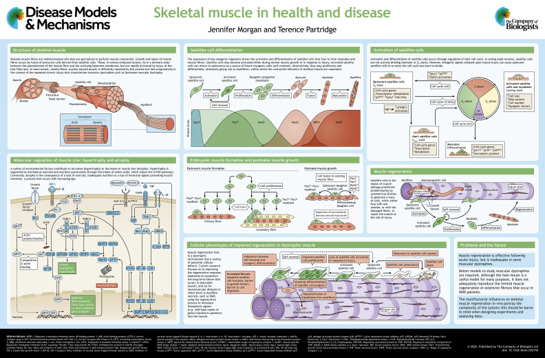
Skeletal muscle fibres are multinucleated cells that contain postmitotic nuclei (i.e. they are no longer able to divide) and perform muscle contraction. They are formed by fusion of muscle precursor cells, and grow into elongating myofibres by the addition of further precursor cells, called satellite cells, which are also responsible for regeneration following injury. Skeletal muscle regeneration occurs in most muscular dystrophies in response to necrosis of muscle fibres. However, the complex environment within dystrophic skeletal muscle, which includes inflammatory cells, fibroblasts and fibro-adipogenic cells, together with the genetic background of the in vivo model and the muscle being studied, complicates the interpretation of laboratory studies on muscular dystrophies. Many genes are expressed in satellite cells and in other tissues, which makes it difficult to determine the molecular cause of various types of muscular dystrophies. Here, and in the accompanying poster, we discuss our current knowledge of the cellular mechanisms that govern the growth and regeneration of skeletal muscle, and highlight the defects in satellite cell function that give rise to muscular dystrophies.
Keywords: Muscular dystrophy; Satellite cell; Skeletal muscle regeneration.
© 2020. Published by The Company of Biologists Ltd.
Conflict of interest statement
Competing interestsThe authors declare no competing or financial interests.
Similar articles
-
Skeletal Muscle Injury by Electroporation: A Model to Study Degeneration/Regeneration Pathways in Muscle.Methods Mol Biol. 2020;2063:157-169. doi: 10.1007/978-1-0716-0138-9_12. Methods Mol Biol. 2020. PMID: 31667769
-
Muscle Stem/Progenitor Cells and Mesenchymal Stem Cells of Bone Marrow Origin for Skeletal Muscle Regeneration in Muscular Dystrophies.Arch Immunol Ther Exp (Warsz). 2018 Oct;66(5):341-354. doi: 10.1007/s00005-018-0509-7. Epub 2018 Mar 13. Arch Immunol Ther Exp (Warsz). 2018. PMID: 29536116 Free PMC article. Review.
-
Muscular dystrophy: centronucleation may reflect a compensatory activation of defective myonuclei.J Biomed Sci. 1998;5(1):54-61. doi: 10.1007/BF02253356. J Biomed Sci. 1998. PMID: 9570514 Review.
-
Correlated NOS-Imu and myf5 expression by satellite cells in mdx mouse muscle regeneration during NOS manipulation and deflazacort treatment.Neuromuscul Disord. 2003 Jun;13(5):388-96. doi: 10.1016/s0960-8966(03)00029-4. Neuromuscul Disord. 2003. PMID: 12798794
-
Regulation of fibrosis in muscular dystrophy.Matrix Biol. 2018 Aug;68-69:602-615. doi: 10.1016/j.matbio.2018.01.014. Epub 2018 Feb 2. Matrix Biol. 2018. PMID: 29408413 Free PMC article. Review.
Cited by
-
Altered muscle niche contributes to myogenic deficit in the D2-mdx model of severe DMD.Cell Death Discov. 2023 Jul 4;9(1):224. doi: 10.1038/s41420-023-01503-0. Cell Death Discov. 2023. PMID: 37402716 Free PMC article.
-
Single-cell transcriptomic analysis of the identity and function of fibro/adipogenic progenitors in healthy and dystrophic muscle.iScience. 2023 Jul 23;26(8):107479. doi: 10.1016/j.isci.2023.107479. eCollection 2023 Aug 18. iScience. 2023. PMID: 37599828 Free PMC article.
-
Decoding the transcriptome of Duchenne muscular dystrophy to the single nuclei level reveals clinical-genetic correlations.Cell Death Dis. 2023 Sep 7;14(9):596. doi: 10.1038/s41419-023-06103-5. Cell Death Dis. 2023. PMID: 37673877 Free PMC article.
-
Study of the Expression and Function of Calcium-Sensing Receptor in Human Skeletal Muscle.Int J Mol Sci. 2021 Jul 6;22(14):7282. doi: 10.3390/ijms22147282. Int J Mol Sci. 2021. PMID: 34298895 Free PMC article.
-
Transcriptomic gene signatures measure satellite cell activity in muscular dystrophies.iScience. 2024 May 8;27(6):109947. doi: 10.1016/j.isci.2024.109947. eCollection 2024 Jun 21. iScience. 2024. PMID: 38840844 Free PMC article.
References
-
- Abu-Baker A., Kharma N., Perreault J., Grant A., Shekarabi M., Maios C., Dona M., Neri C., Dion P. A., Parker A. et al. (2019). RNA-based therapy utilizing oculopharyngeal muscular dystrophy transcript knockdown and replacement. Mol. Ther. Nucleic Acids 15, 12-25. 10.1016/j.omtn.2019.02.003 - DOI - PMC - PubMed
-
- Amthor H., Otto A., Vulin A., Rochat A., Dumonceaux J., Garcia L., Mouisel E., Hourde C., Macharia R., Friedrichs M. et al. (2009). Muscle hypertrophy driven by myostatin blockade does not require stem/precursor-cell activity. Proc. Natl. Acad. Sci. USA 106, 7479-7484. 10.1073/pnas.0811129106 - DOI - PMC - PubMed
Publication types
MeSH terms
LinkOut - more resources
Full Text Sources
