

Characterizing Membrane Association and Periplasmic Transfer of Bacterial Lipoproteins through Molecular Dynamics Simulations
- PMID: 32053772
- PMCID: PMC7139219
- DOI: 10.1016/j.str.2020.01.012
Characterizing Membrane Association and Periplasmic Transfer of Bacterial Lipoproteins through Molecular Dynamics Simulations
Abstract
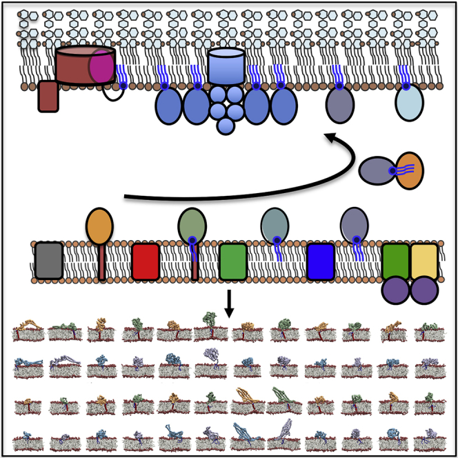
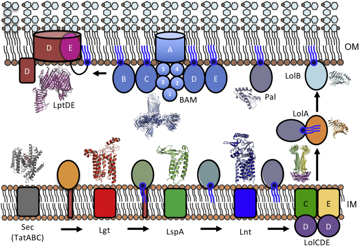
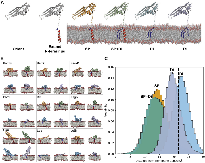
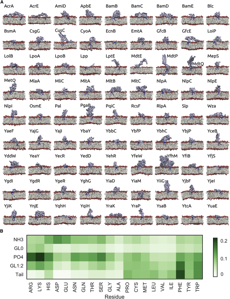
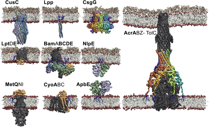
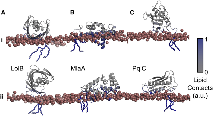
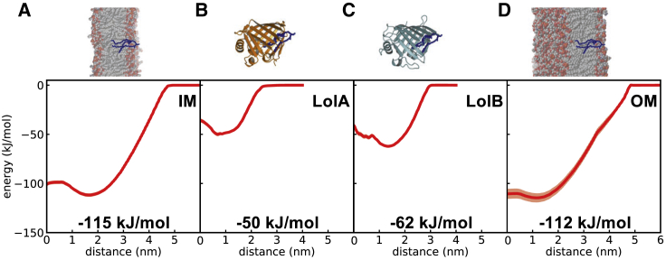
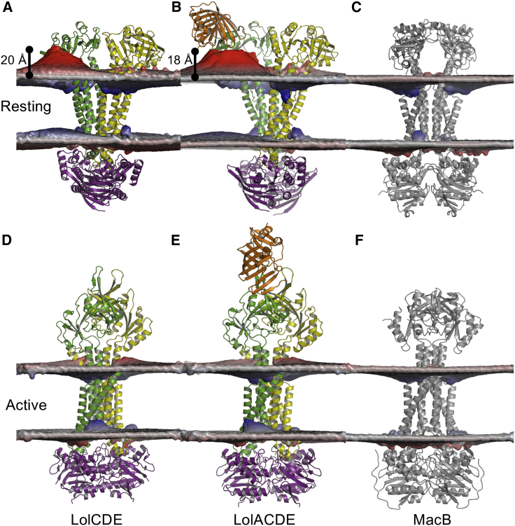
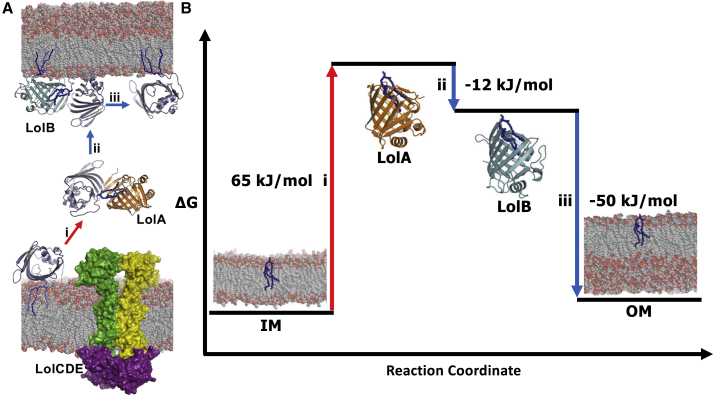
Escherichia coli lipoprotein precursors at the inner membrane undergo three maturation stages before transport by the Lol system to the outer membrane. Here, we develop a pipeline to simulate the membrane association of bacterial lipoproteins in their four maturation states. This has enabled us to model and simulate 81 of the predicted 114 E. coli lipoproteins and reveal their interactions with the host lipid membrane. As part of this set we characterize the membrane contacts of LolB, the lipoprotein involved in periplasmic translocation. We also consider the means and bioenergetics for lipoprotein localization. Our calculations uncover a preference for LolB over LolA and therefore indicate how a lipoprotein may be favorably transferred from the inner to outer membrane. Finally, we reveal that LolC has a role in membrane destabilization, thereby promoting lipoprotein transfer to LolA.
Keywords: antibiotics; biogenesis; lipoprotein; membrane proteins; molecular simulation.
Copyright © 2020 The Authors. Published by Elsevier Ltd.. All rights reserved.
Conflict of interest statement
Declaration of Interests The authors declare no competing interests.
Figures

Similar articles
-
Structural investigation of the interaction between LolA and LolB using NMR.J Biol Chem. 2009 Sep 4;284(36):24634-43. doi: 10.1074/jbc.M109.001149. Epub 2009 Jun 22. J Biol Chem. 2009. PMID: 19546215 Free PMC article.
-
Model of mouth-to-mouth transfer of bacterial lipoproteins through inner membrane LolC, periplasmic LolA, and outer membrane LolB.Proc Natl Acad Sci U S A. 2009 Apr 7;106(14):5877-82. doi: 10.1073/pnas.0900896106. Epub 2009 Mar 23. Proc Natl Acad Sci U S A. 2009. PMID: 19307584 Free PMC article.
-
Structural basis of lipoprotein recognition by the bacterial Lol trafficking chaperone LolA.Proc Natl Acad Sci U S A. 2022 Sep 6;119(36):e2208662119. doi: 10.1073/pnas.2208662119. Epub 2022 Aug 29. Proc Natl Acad Sci U S A. 2022. PMID: 36037338 Free PMC article.
-
ABC transporters involved in the biogenesis of the outer membrane in gram-negative bacteria.Biosci Biotechnol Biochem. 2011;75(6):1044-54. doi: 10.1271/bbb.110115. Epub 2011 Jun 13. Biosci Biotechnol Biochem. 2011. PMID: 21670534 Review.
-
Sorting of lipoproteins to the outer membrane in E. coli.Biochim Biophys Acta. 2004 Nov 11;1694(1-3):IN1-9. Biochim Biophys Acta. 2004. PMID: 15672528 Review.
Cited by
-
An octameric PqiC toroid stabilises the outer-membrane interaction of the PqiABC transport system.EMBO Rep. 2024 Jan;25(1):82-101. doi: 10.1038/s44319-023-00014-4. Epub 2024 Jan 16. EMBO Rep. 2024. PMID: 38228789 Free PMC article.
-
Effect of magnesium sulfate in oxidized lipid bilayers properties by using molecular dynamics.Biochem Biophys Rep. 2021 Apr 28;26:100998. doi: 10.1016/j.bbrep.2021.100998. eCollection 2021 Jul. Biochem Biophys Rep. 2021. PMID: 33997315 Free PMC article.
-
What have molecular simulations contributed to understanding of Gram-negative bacterial cell envelopes?Microbiology (Reading). 2022 Mar;168(3):001165. doi: 10.1099/mic.0.001165. Microbiology (Reading). 2022. PMID: 35294337 Free PMC article. Review.
-
Periplasmic Targets for the Development of Effective Antimicrobials against Gram-Negative Bacteria.ACS Infect Dis. 2020 Sep 11;6(9):2337-2354. doi: 10.1021/acsinfecdis.0c00384. Epub 2020 Aug 24. ACS Infect Dis. 2020. PMID: 32786281 Free PMC article. Review.
-
Structural basis for bacterial lipoprotein relocation by the transporter LolCDE.Nat Struct Mol Biol. 2021 Apr;28(4):347-355. doi: 10.1038/s41594-021-00573-x. Epub 2021 Mar 29. Nat Struct Mol Biol. 2021. PMID: 33782615
References
-
- Abellon-Ruiz J., Kaptan S.S., Basle A., Claudi B., Bumann D., Kleinekathofer U., van den Berg B. Structural basis for maintenance of bacterial outer membrane lipid asymmetry. Nat. Microbiol. 2017;2:1616–1623. - PubMed
-
- Abraham M.J., Murtola T., Schulz R., Páll S., Smith J.C., Hess B., Lindahl E. GROMACS: high performance molecular simulations through multi-level parallelism from laptops to supercomputers. SoftwareX. 2015;1-2:19–25.
-
- Abramson J., Riistama S., Larsson G., Jasaitis A., Svensson-Ek M., Laakkonen L., Puustinen A., Iwata S., Wikstrom M. The structure of the ubiquinol oxidase from Escherichia coli and its ubiquinone binding site. Nat. Struct. Biol. 2000;7:910–917. - PubMed
-
- Arnarez C., Mazat J.P., Elezgaray J., Marrink S.J., Periole X. Evidence for cardiolipin binding sites on the membrane-exposed surface of the cytochrome bc1. J. Am. Chem. Soc. 2013;135:3112–3120. - PubMed
Publication types
MeSH terms
Substances
Grants and funding
- BB/S003339/1/BB_/Biotechnology and Biological Sciences Research Council/United Kingdom
- BB/L01386X/1/BB_/Biotechnology and Biological Sciences Research Council/United Kingdom
- MR/S009213/1/MRC_/Medical Research Council/United Kingdom
- WT_/Wellcome Trust/United Kingdom
- BB/R002517/1/BB_/Biotechnology and Biological Sciences Research Council/United Kingdom
LinkOut - more resources
Full Text Sources
Molecular Biology Databases
Research Materials
