. 2020 Feb 12;5(1):e00869-19.
doi: 10.1128/mSphere.00869-19.

Toxoplasma Cathepsin Protease B and Aspartyl Protease 1 Are Dispensable for Endolysosomal Protein Digestion

Affiliations

Toxoplasma Cathepsin Protease B and Aspartyl Protease 1 Are Dispensable for Endolysosomal Protein Digestion

Christian McDonald et al. mSphere. .

Abstract

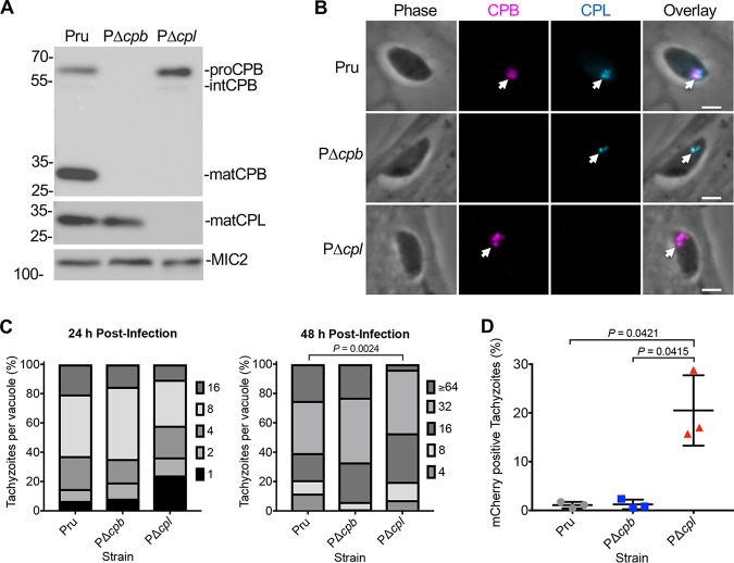
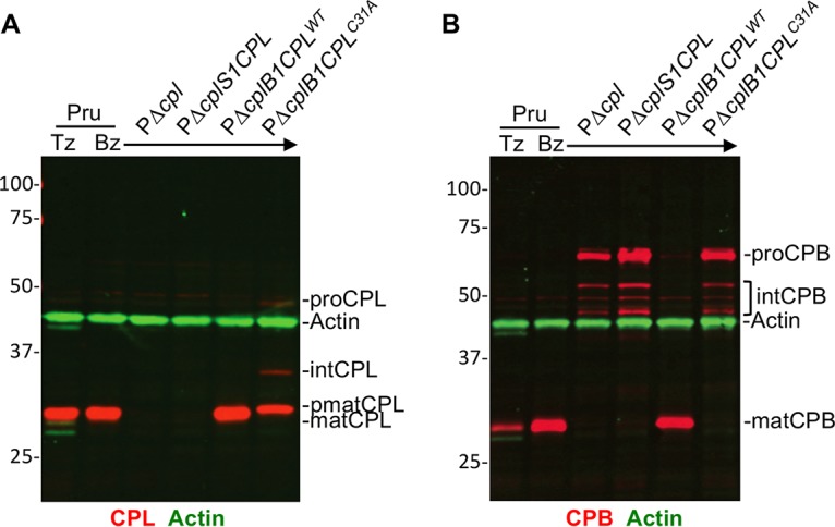
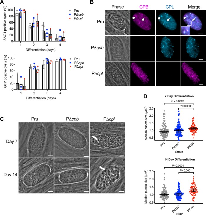
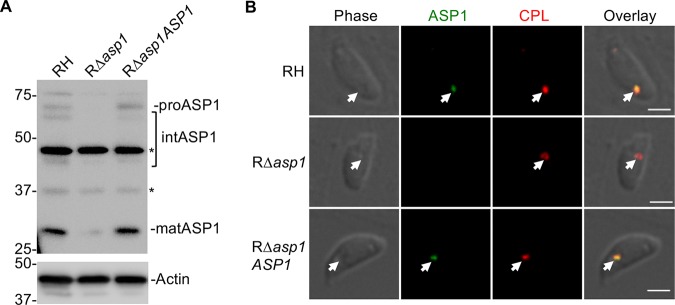
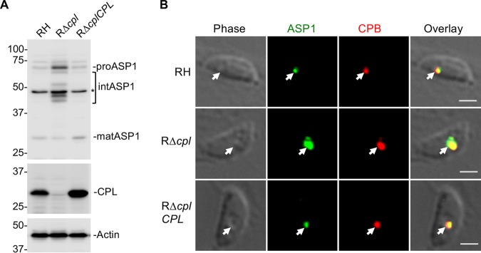
The lysosome-like vacuolar compartment (VAC) is a major site of proteolysis in the intracellular parasite Toxoplasma gondii Previous studies have shown that genetic ablation of a VAC-residing cysteine protease, cathepsin protease L (CPL), resulted in the accumulation of undigested protein in the VAC and loss of parasite viability during the chronic stage of infection. However, since the maturation of another VAC localizing protease, cathepsin protease B (CPB), is dependent on CPL, it remained unknown whether these defects result directly from ablation of CPL or indirectly from a lack of CPB maturation. Likewise, although a previously described cathepsin D-like aspartyl protease 1 (ASP1) could also play a role in proteolysis, its definitive residence and function in the Toxoplasma endolysosomal system were not well defined. Here, we demonstrate that CPB is not necessary for protein turnover in the VAC and that CPB-deficient parasites have normal growth and viability in both the acute and chronic stages of infection. We also show that ASP1 depends on CPL for correct maturation, and it resides in the T. gondii VAC, where, similar to CPB, it plays a dispensable role in protein digestion. Taken together with previous work, our findings suggest that CPL is the dominant protease in a hierarchy of proteolytic enzymes within the VAC. This unusual lack of redundancy for CPL in T. gondii makes it a single exploitable target for disrupting chronic toxoplasmosis.IMPORTANCE Roughly one-third of the human population is chronically infected with the intracellular single-celled parasite Toxoplasma gondii, but little is known about how this organism persists inside people. Previous research suggested that a parasite proteolytic enzyme, termed cathepsin protease L, is important for Toxoplasma persistence; however, it remained possible that other associated proteolytic enzymes could also be involved in the long-term survival of the parasite during infection. Here, we show that two proteolytic enzymes associated with cathepsin protease L play dispensable roles and are dependent on cathepsin L to reach maturity, which differs from the corresponding enzymes in humans. These findings establish a divergent hierarchy of proteases and help focus attention principally on cathepsin protease L as a potential target for interrupting Toxoplasma chronic infection.

Keywords: Toxoplasma gondii; autophagy; cathepsin; lysosome; proteases.


Figures

FIG 1
FIG 1
CPB does not play a major role in tachyzoite growth and protein turnover. (A) Immunoblot of tachyzoite lysates probed for T. gondii CPL and CPB along with MIC2 as a loading control. Protein bands are labeled as proform (pro), intermediate form (int), or mature form (mat) based on their molecular weights. (B) Recently invaded intracellular tachyzoites were stained with anti-CPB (violet) and anti-CPL (blue). Arrowheads indicate labeling of the VAC. Scale bars, 2 μm. (C) T. gondii tachyzoites were cultured in HFF monolayers. Samples were collected at 24 and 48 h postinfection, fixed, and stained with anti-SAG1 antibody, and the number of parasites per vacuole was quantified. Percentages of various replication stages for each strain are quantified. Results represent means from 2 or 3 biological replicates. The total numbers of parasitophorous vacuoles counted across 3 biological replicates were the following: Pru, 147 and 152; PΔcpb, 144 and 145; PΔcpl, 153 and 136 (values are for 24 and 48 h postinfection, respectively). Only statistically significant differences are represented. Statistical significance was determined by unpaired two-tailed Student’s t test performed on the mean number of parasites per vacuole for each strain across 3 biological replicates. (D) Assay quantification of tachyzoite ingestion of mCherry showing means ± standard deviations from 3 biological replicates. The data generated were analyzed using the following number of tachyzoites per replicate: Pru, n = 469, 464, and 231; PΔcpb, n = 374, 613, and 213; and PΔcpl, n = 427, 299, and 216. All strains were compared, and only significant differences are shown. Unpaired two-tailed t test with Welch’s corrections was performed on the means from 3 biological replicates.
FIG 2
FIG 2
T. gondii CPB maturation is dependent on the expression of active CPL. Li-COR Western blots of tachyzoite and bradyzoite cell lysates probed for T. gondii CPL (red in panel A), CPB (red in panel B), and actin (green in panels A and B) as the loading control. Protein bands are labeled as proform (pro), intermediate form (int), pseudomature form (pmat), and mature form (mat) based on their molecular weights.
FIG 3
FIG 3
CPL, not CPB, is the major protease necessary for the turnover of autophagosomes in the VAC in bradyzoites. (A) The rate of parasite differentiation from the tachyzoite stage to the bradyzoite stage was determined in vitro by assessment of the tachyzoite-specific antigen SAG1 and GFP under the control of a bradyzoite LDH2 promoter. Infected monolayers were cultured for the indicated days, fixed, probed for SAG1, and quantified. Bars indicate means ± standard deviations from 3 biological replicates. Experiments analyzed 105, 106, 153, and 151 cysts for Pru, 117, 123, 149, and 110 cysts for PΔcpb, and 75, 108, 155, and 130 cysts for PΔcpl on days 1, 2, 3, and 4, respectively. All strains were compared, and only statistical significance is shown. Unpaired two-tailed t test with Welch’s corrections was performed on the means from 3 biological replicates. (B) Immunofluorescence localization of CPB and CPL in bradyzoites. Parasite strains were differentiated for 7 days, fixed, and stained with anti-CPB (violet) and anti-CPL (blue). The inset in the merged image of Pru shows enlargement of the boxed region. Scale bars, 5 μm. (C) Phase-contrast microscopy was used to image bradyzoites in vitro following 7- and 14-day cultures. Enlarged dark puncta seen in PΔcpl bradyzoites are indicative of defective protein degradation in the VAC (arrows). Scale bars, 5 μm. (D) The size of the puncta in bradyzoites was measured. The following numbers of cysts across 3 biological replicates were used to analyze each strain: Pru (day 7, 109 cysts; day 14, 98 cysts), PΔcpb (day 7, 106 cysts; day 14, 102 cysts), and PΔcpl (day 7, 105 cysts; day 14, 97 cysts). Lines represent medians with 95% confidence intervals. For this data set, ROUT with a Q value of 0.1% was used to identify and remove 1 outlier each for Pru and PΔcpl for 14 days postdifferentiation. Since the data did not fit a normal distribution, a nonparametric Kruskal-Wallis test with Dunn’s multiple comparisons was used to compare the medians of data combined from 3 biological replicates. All strains were compared, and only significant differences are shown.
FIG 4
FIG 4
T. gondii ASP1, a cathepsin D ortholog that localizes to the VAC. (A) Immunoblot detection of T. gondii ASP1 in tachyzoites. Protein bands are labeled as proform (pro), intermediate form (int), and mature form (mat) based on their molecular weights. Polypeptides marked with asterisks are nonspecific bands. (B) Immunofluorescence assay of tachyzoites after fixation, staining, and probing for T. gondii ASP1 (green) and CPL (red). Arrowheads indicate colocalization of ASP1 with CPL in the VAC. Scale bars, 2 μm.
FIG 5
FIG 5
T. gondii immature ASP1 accumulates in tachyzoites lacking active CPL in the VAC. (A) Immunoblot of tachyzoite lysates probed for ASP1 and CPL along with actin as a loading control. For ASP1, based on molecular weights, protein bands are labeled as proform (pro), intermediate form (int), and mature form (mat). (B) Immunofluorescence assay to detect T. gondii ASP1 (green) and CPB (red) in RH, RΔcpl, and RΔcplCPL tachyzoites. Arrowheads show localization of ASP1 with CPB in the VAC. Scale bars, 2 μm.
FIG 6
FIG 6
ASP1 is dispensable for tachyzoite replication or ingestion. (A) Parasite lysates of tachyzoites and bradyzoites were immunoblotted with antibodies against T. gondii ASP1 and CPL along with actin as a loading control. For ASP1, based on molecular weights, protein bands are labeled as proform (pro), intermediate form (int), and mature form (mat). (B) T. gondii tachyzoites were cultured in HFF monolayers. Samples were collected at 24 and 48 h postinfection, fixed, and stained with anti-SAG1 antibody, and the numbers of parasites per vacuole were quantified. Percentages of various replication stages for each strain are quantified. The total number of parasitophorous vacuoles counted across 2 biological replicates was the following: Pru, 147 and 152; PΔcpl, 153 and 136; PΔasp1, 159 and 128; and PΔasp1Δcpl, 163 and 142 (all values are for 24 and 48 h postinfection, respectively). Only statistically significant differences are represented. Statistical significance was determined by unpaired two-tailed Student’s t test performed on the mean number of parasites per vacuole for each strain across 3 biological replicates. (C) Assay quantitation of tachyzoite ingestion of mCherry showing means ± standard deviations from 4 biological replicates. The data generated were analyzed using the following number of tachyzoites per replicate: Pru (n = 205, 219, 223, and 207), PΔcpl (n = 224, 242, 224, and 223), PΔasp1 (n = 218, 220, 220, and 210), and PΔasp1Δcpl (n = 203, 235, 228, and 246). All strains were compared, and only significant differences are shown. Unpaired two-tailed t test with Welch’s corrections was performed to determine statistical significance.
FIG 7
FIG 7
T. gondii ASP1 has a limited role in protein turnover of autophagosomes. (A) The rate of parasite differentiation from the tachyzoite stage to the bradyzoite stage was determined in vitro by assessment of the tachyzoite-specific antigen SAG1 and GFP under the control of a bradyzoite LDH2 promoter. Infected monolayers were cultured for the indicated days, fixed, probed for SAG1, and quantified. Bars indicate means ± standard deviations from 3 biological replicates. Experiments analyzed 105, 106, 153, and 151 cysts for Pru, 75, 108, 155, and 130 cysts for PΔcpl, 119, 124, 189, and 146 cysts for PΔasp1 and 78, 110, 112, and 161 cysts for PΔasp1Δcpl on days 1, 2, 3, and 4, respectively. All strains were compared, and only statistical significance is shown. Unpaired two-tailed t test with Welch’s corrections was performed on the means across 3 biological replicates. (B) Phase-contrast microscopy was used to image bradyzoites in vitro following 7- and 14-day culture under differentiation conditions. Enlarged dark puncta indicative of defective protein degradation in VACs are visible in PΔcpl bradyzoites and PΔasp1Δcpl cysts (arrows). Scale bars, 5 μm. (C) The size of the punctae in bradyzoites was measured. The following numbers of cysts across 3 biological replicates were used to analyze each strain: Pru (day 7, 109 cysts; day 14, 98 cysts), PΔcpl (day 7, 105 cysts; day 14, 97 cysts), PΔasp1 (day 7, 85 cysts; day 14, 97 cysts), PΔasp1Δcpl (day 7, 90 cysts; day 14, 97 cysts). Lines represent medians with 95% confidence intervals. For this data set, ROUT with a Q value of 0.1% was used to identify and remove 1 outlier from PΔasp1 for 7 days postdifferentiation and 1 outlier each for Pru and PΔcpl for 14 days postdifferentiation. Since the data did not fit a normal distribution, we used a nonparametric Kruskal-Wallis test with Dunn’s multiple comparisons to compare the medians of data combined from 3 biological replicates. All strains were compared, and only significant differences are shown.
FIG 8
FIG 8
CPL is a central protease in bradyzoite viability. (A) Schematic illustration of the workflow used to examine bradyzoite viability. Experiments conducted were used to generate the data shown in panels B and C. Details are provided in Materials and Methods. (B) Plaque assays were performed on bradyzoites to determine the viability of each strain, based on their ability to undergo the lytic cycle following forced encystation. Plaque zones were visualized following crystal violet staining of the HFF monolayer. (C) Quantitation of the plaque assay described for panel B. Quantitative PCR analysis was used to determine the number of parasites applied to fresh HFF monolayers for the bradyzoite plaque assay, allowing for the number of plaques per genome to be calculated. Data were normalized to those for the parental strain (Pru) and are shown as a percentage. Bars indicate means ± standard deviations from 3 or 4 biological replicates. Only significant differences between Pru and subsequent strains are indicated. Mann-Whitney test was used to assess statistical significance.

Similar articles

Cited by

References

    1. Montoya J, Liesenfeld O. 2004. Toxoplasmosis. Lancet 363:1965–1976. doi:10.1016/S0140-6736(04)16412-X. - DOI - PubMed
    1. Siqueira-Neto JL, Debnath A, McCall L-I, Bernatchez JA, Ndao M, Reed SL, Rosenthal PJ. 2018. Cysteine proteases in protozoan parasites. PLoS Negl Trop Dis 12:e0006512. doi:10.1371/journal.pntd.0006512. - DOI - PMC - PubMed
    1. Robbins JR, Zeldovich VB, Poukchanski A, Boothroyd JC, Bakardjiev AI. 2012. Tissue barriers of the human placenta to infection with Toxoplasma gondii. Infect Immun 80:418–428. doi:10.1128/IAI.05899-11. - DOI - PMC - PubMed
    1. Stoka V, Turk V, Turk B. 2016. Lysosomal cathepsins and their regulation in aging and neurodegeneration. Ageing Res Rev 32:22–37. doi:10.1016/j.arr.2016.04.010. - DOI - PubMed
    1. Neurath H, Walsh KA. 1976. Role of proteolytic enzymes in biological regulation (a review). Proc Natl Acad Sci U S A 73:3825–3832. doi:10.1073/pnas.73.11.3825. - DOI - PMC - PubMed

Publication types

LinkOut - more resources