Physical and Antibacterial Properties of Peppermint Essential Oil Loaded Poly (ε-caprolactone) (PCL) Electrospun Fiber Mats for Wound Healing
- PMID: 32039166
- PMCID: PMC6988806
- DOI: 10.3389/fbioe.2019.00346
Physical and Antibacterial Properties of Peppermint Essential Oil Loaded Poly (ε-caprolactone) (PCL) Electrospun Fiber Mats for Wound Healing
Abstract
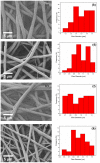
The aim of this study was to fabricate and characterize various concentrations of peppermint essential oil (PEP) loaded on poly(ε-caprolactone) (PCL) electrospun fiber mats for healing applications, where PEP was intended to impart antibacterial activity to the fibers. SEM images illustrated that the morphology of all electrospun fiber mats was smooth, uniform, and bead-free. The average fiber diameter was reduced by the addition of PEP from 1.6 ± 0.1 to 1.0 ± 0.2 μm. Functional groups of the fibers were determined by Raman spectroscopy. Gas chromatography-mass spectroscopy (GC-MS) analysis demonstrated the actual PEP content in the samples. In vitro degradation was determined by measuring weight loss and their morphology change, showing that the electrospun fibers slightly degraded by the addition of PEP. The wettability of PCL and PEP loaded electrospun fiber mats was measured by determining contact angle and it was shown that wettability increased with the incorporation of PEP. The antimicrobial activity results revealed that PEP loaded PCL electrospun fiber mats exhibited inhibition against Staphylococcus aureus (gram-positive) and Escherichia coli (gram-negative) bacteria. In addition, an in-vitro cell viability assay using normal human dermal fibroblast (NHDF) cells revealed improved cell viability on PCL, PCLPEP1.5, PCLPEP3, and PCLGEL6 electrospun fiber mats compared to the control (CNT) after 48 h cell culture. Our findings showed for the first time PEP loaded PCL electrospun fiber mats with antibiotic-free antibacterial activity as promising candidates for wound healing applications.
Keywords: antibacterial activity; electrospinning; peppermint essential oil; poly (ε-caprolactone); wound healing.
Copyright © 2019 Unalan, Slavik, Buettner, Goldmann, Frank and Boccaccini.
Figures


Similar articles
-
The Drug-Loaded Electrospun Poly(ε-Caprolactone) Mats for Therapeutic Application.Nanomaterials (Basel). 2021 Apr 4;11(4):922. doi: 10.3390/nano11040922. Nanomaterials (Basel). 2021. PMID: 33916638 Free PMC article.
-
Evaluation of Electrospun Poly(ε-Caprolactone)/Gelatin Nanofiber Mats Containing Clove Essential Oil for Antibacterial Wound Dressing.Pharmaceutics. 2019 Nov 1;11(11):570. doi: 10.3390/pharmaceutics11110570. Pharmaceutics. 2019. PMID: 31683863 Free PMC article.
-
Fabrication of Biocompatible Electrospun Poly(ε-caprolactone)/Gelatin Nanofibers Loaded with Pinus radiata Bark Extracts for Wound Healing Applications.Polymers (Basel). 2022 Jun 9;14(12):2331. doi: 10.3390/polym14122331. Polymers (Basel). 2022. PMID: 35745907 Free PMC article.
-
Electrospun essential oil-doped chitosan/poly(ε-caprolactone) hybrid nanofibrous mats for antimicrobial food biopackaging exploits.Carbohydr Polym. 2019 Nov 1;223:115108. doi: 10.1016/j.carbpol.2019.115108. Epub 2019 Jul 19. Carbohydr Polym. 2019. PMID: 31426968
-
Fabrication of a Polycaprolactone/Chitosan Nanofibrous Scaffold Loaded with Nigella sativa Extract for Biomedical Applications.BioTech (Basel). 2023 Feb 12;12(1):19. doi: 10.3390/biotech12010019. BioTech (Basel). 2023. PMID: 36810446 Free PMC article.
Cited by
-
The Drug-Loaded Electrospun Poly(ε-Caprolactone) Mats for Therapeutic Application.Nanomaterials (Basel). 2021 Apr 4;11(4):922. doi: 10.3390/nano11040922. Nanomaterials (Basel). 2021. PMID: 33916638 Free PMC article.
-
Insights into Theranostic Properties of Titanium Dioxide for Nanomedicine.Nanomicro Lett. 2020 Jan 14;12(1):22. doi: 10.1007/s40820-019-0362-1. Nanomicro Lett. 2020. PMID: 34138062 Free PMC article. Review.
-
Electrospun Polymer Nanofibers with Antimicrobial Activity.Polymers (Basel). 2022 Apr 20;14(9):1661. doi: 10.3390/polym14091661. Polymers (Basel). 2022. PMID: 35566830 Free PMC article. Review.
-
Biological properties of polycaprolactone and barium titanate composite in biomedical applications.Sci Prog. 2023 Oct-Dec;106(4):368504231215942. doi: 10.1177/00368504231215942. Sci Prog. 2023. PMID: 38031343 Free PMC article. Review.
-
Antibacterial and Antibiofilm Properties of Native Australian Plant Endophytes against Wound-Infecting Bacteria.Microorganisms. 2024 Aug 19;12(8):1710. doi: 10.3390/microorganisms12081710. Microorganisms. 2024. PMID: 39203552 Free PMC article.
References
-
- Abedalwafa M., Wang F., Wang L., Li C. (2013). Biodegradable poly-epsilon-caprolactone (PCL) for tissue engineering applications: a review. Rev. Adv. Mater. Sci. 34, 123–140.
-
- Agnes Mary S., Giri Dev V. R. (2015). Electrospun herbal nanofibrous wound dressings for skin tissue engineering. J. Text. Inst. 106, 886–895. 10.1080/00405000.2014.951247 - DOI
-
- Amiri S., Rahimi A. (2019). Poly (ε-caprolactone) electrospun nanofibers containing cinnamon essential oil nanocapsules: a promising technique for controlled release and high solubility. J. Indus. Text. 48, 1527–1544. 10.1177/1528083718764911 - DOI
-
- Balasubramanian K., Kodam K. M. (2014). Encapsulation of therapeutic lavender oil in an electrolyte assisted polyacrylonitrile nanofibres for antibacterial applications. RSC Adv. 4, 54892–54901. 10.1039/C4RA09425E - DOI
LinkOut - more resources
Full Text Sources
Miscellaneous

