. 2020 Mar 12;5(5):e134172.
doi: 10.1172/jci.insight.134172.

Extracellular CIRP as an endogenous TREM-1 ligand to fuel inflammation in sepsis

Affiliations

Extracellular CIRP as an endogenous TREM-1 ligand to fuel inflammation in sepsis

Naomi-Liza Denning et al. JCI Insight. .

Abstract

Extracellular cold-inducible RNA-binding protein (eCIRP) is a recently discovered damage-associated molecular pattern. Understanding the precise mechanism by which it exacerbates inflammation is essential. Here we identified that eCIRP is a new biologically active endogenous ligand of triggering receptor expressed on myeloid cells-1 (TREM-1), fueling inflammation in sepsis. Surface plasmon resonance revealed a strong binding affinity between eCIRP and TREM-1, and fluorescence resonance energy transfer assay confirmed eCIRP's interaction with TREM-1 in macrophages. Targeting TREM-1 by its siRNA or a decoy peptide, LP17, or by using TREM-1-/- mice dramatically reduced eCIRP-induced inflammation. We developed a potentially novel 7-aa peptide derived from human eCIRP, M3, which blocked the interaction of TREM-1 and eCIRP. M3 suppressed inflammation induced by eCIRP or agonist TREM-1 antibody cross-linking in murine macrophages or human peripheral blood monocytes. M3 also inhibited eCIRP-induced systemic inflammation and tissue injury. Treatment with M3 further protected mice from sepsis, improved acute lung injury, and increased survival. Thus, we have discovered a potentially novel TREM-1 ligand and developed a new peptide, M3, to block eCIRP-TREM-1 interaction and improve outcomes in sepsis.

Keywords: Bacterial infections; Immunology; Inflammation; Innate immunity; Macrophages.


Conflict of interest statement

Conflict of interest: PW is an inventor of patent applications (WO/2010/120726 and 61/881.798) covering the fundamental concept of targeting cold-inducible RNA-binding protein for the treatment of inflammatory diseases, licensed by TheraSource LLC. PW is a cofounder of TheraSource LLC.

Figures

Figure 1
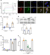
Figure 1. eCIRP binds TREM-1 to promote inflammation.
(A) SPR between rmCIRP and rmTREM-1. Anti-his antibody was used to capture rmCIRP-his. rmTREM-1 was injected as an analyte in concentrations of 0 to 500 nM. (B) RAW264.7 cells were treated with rmCIRP (5 μg/mL) at 4°C for 10 minutes, fixed in a nonpermeabilized fashion, and stained with primary antibodies against CIRP, TREM-1, and CD-11b as well as fluorescently labeled secondary antibodies. Confocal microscopy images were obtained with a 63× objective. Colocalization is indicated by the yellow color. (C) After the staining protocol described in B, cell-associated fluorescence was measured. The transfer of fluorescence was calculated as FRET units. Data are expressed as mean ± SEM obtained from 3 independent experiments; n = 8–9/group. Groups compared by unpaired t test (*P < 0.01 vs. CD11b). (D) RAW264.7 cells were stimulated with rmCIRP (1 μg/mL) for 10 minutes. Extracted proteins were immunoprecipitated by using anti-DAP12 antibody, followed by Western blotting using phospho-Tyr (p-Tyr; 4G10) and DAP12 antibody. Extracted total proteins obtained from RAW264.7 cells stimulated with rmCIRP (1 μg/mL) for 10 minutes were subjected to Western blotting using p-Syk, Syk, and β-actin antibodies. Representative Western blots for phosphotyrosine (4G10), DAP12, p-Syk, Syk, and β-actin are shown. Phosphotyrosine (p-DAP12) and p-Syk expression in each sample was normalized to DAP12 or Syk or β-actin expression and the mean values of 0 minutes of rmCIRP-treated groups were standardized as one for comparison. Data are expressed as mean ± SEM (n = 5 samples/group). The groups were compared by 1-way ANOVA and Tukey’s method (*P < 0.05 vs. PBS). (E) RAW264.7 cells were transfected as shown. Cells were stimulated with PBS control or 1 μg/mL rmCIRP. After 6 hours, TNF-α in the supernatant was analyzed by ELISA. Data are expressed as mean ± SEM (n = 3 samples/group). Multiple groups were compared by 1-way ANOVA and Tukey’s method (*P < 0.05 vs. respective PBS group; #P < 0.01 vs. rmCIRP-treated nontransfected cells). (F) RAW264.7 cells were stimulated with PBS or rmCIRP (1 μg/mL). Simultaneously cells were treated with various doses of LP17 or LP17-Sc1. After 24 hours, TNF-α in culture supernatants was measured by ELISA. Data are expressed as mean ± SEM obtained from 5 independent experiments (n = 3–10 wells/group). The groups were compared by Kruskal-Wallis test with Dunn’s method (*P < 0.05 vs. PBS; #P < 0.05 vs. rmCIRP). FRET, fluorescence resonance energy transfer; PerC, peritoneal cavity; ELISA, enzyme-linked immunosorbent assay; PBS, phosphate-buffered saline.
Figure 2
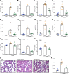
Figure 2. LP17 inhibits eCIRP-induced inflammation in vivo.
Adult C57BL/6 mice were randomly assigned to sham, vehicle (PBS), or treatment group. rmCIRP at a dose of 5 mg/kg BW or equivalent volume of normal saline was administered i.v. via retro-orbital injection. LP17 at a dose of 5 mg/kg BW or vehicle was given i.p. at the time of rmCIRP injection. At 5 hours after rmCIRP injection, mice were euthanized, and blood and tissue were collected for analysis. (A) AST, (B) ALT, and (C) LDH were determined using specific colorimetric enzymatic assays. Serum (D) IL-6 and (E) IL-1β were measured by ELISA. Lung mRNA levels of (F) TNF-α, (G) IL-1β, and (H) IL-6 were measured by real-time PCR (RT-PCR). Equal amounts of total lung protein (250–350 μg) were loaded into respective ELISA wells for assessment of lung protein levels of (I) TNF-α, (J) IL-1β, and (K) IL-6. (L) Representative images of H&E-stained lung tissue at original magnification ×200. (M) Lung injury score calculated at original magnification ×400. n = 5 high-powered fields/group. Data are expressed as mean ± SEM. n = 5–7 mice/group. The groups were compared by 1-way ANOVA and Tukey’s method (*P < 0.05 vs. sham, and #P < 0.05 vs. vehicle mice). ALT, alanine aminotransferase; AST, aspartate aminotransferase; LDH, lactate dehydrogenase.
Figure 3
Figure 3. TREM-1 deficiency ameliorates eCIRP-mediated inflammation.
Primary peritoneal macrophages were isolated from WT and TREM-1–/– mice and were stimulated with PBS or rmCIRP (1 μg/mL). After 24 hours, (A) TNF-α and (B) IL-6 in culture supernatants were measured by ELISA. Data are expressed as mean ± SEM; n = 12 wells/group. Multiple groups were compared by 1-way ANOVA and Tukey’s method (*P < 0.05 vs. respective PBS group; #P < 0.001 vs. rmCIRP-treated WT cells). Adult C57BL/6 WT and TREM-1–/– mice were given rmCIRP at a dose of 5 mg/kg BW or equivalent volume PBS. After 5 hours, serum (C) IL-6 and (D) IL-1β were measured by ELISA. Data are expressed as mean ± SEM. n = 3 mice/group. Multiple groups were compared by 1-way ANOVA and Tukey’s method (*P < 0.05 vs. respective PBS group; #P < 0.05 vs. rmCIRP-treated WT mice).
Figure 4
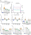
Figure 4. M3, a small CIRP-derived peptide, inhibits eCIRP and TREM-1 interaction.
(A) Partial aa sequence of CIRP highlighting an area of similarity with PGLYRP1. RAW264.7 cells were treated with 10 μg/mL of peptides M1, M2, or M3 for 30 minutes, then stimulated with rmCIRP (1 μg/mL). After 24 hours, TNF-α in culture supernatants was measured. Data are expressed as mean ± SEM obtained from 2 independent experiments (n = 5–7 wells/peptide group) and were compared by 1-way ANOVA and Tukey’s method (*P < 0.05 vs. unstimulated cells, and #P < 0.05 vs. rmCIRP-treated cells). (B) SPR between rmTREM-1 and M3. (C) RAW264.7 cells and (D) primary peritoneal macrophages were treated with M3 or M3-Sc1 (both 10 μg/mL) for 30 minutes. Cells were then stimulated with PBS or rmCIRP (5 μg/mL), and FRET analysis was performed as described in Figure 1C. Data are expressed as mean ± SEM obtained from 3 independent experiments; n = 7–9/group, compared by 1-way ANOVA and Tukey’s method (*P < 0.05 vs. CD11b + rmCIRP, and #P < 0.05 vs. TREM-1 + rmCIRP). (E) To activate RAW264.7 cells through TREM-1, 96-well plates were precoated with 20 μg/mL of an agonist anti–TREM-1 mAb. Then, 5 × 10⁴ cells/well were premixed with PBS control or M3 (10 μg/mL) or scramble peptide (10 μg/mL) for 30 minutes, then plated. TNF-α production was measured in the culture supernatants after an additional 24 hours of incubation. The experiment was performed 2 independent times with n = 5–6 wells per group. Scramble peptide groups, n = 5/group. Data are expressed as mean ± SEM and were compared by 1-way ANOVA and Tukey’s method (*P < 0.05 vs. uncoated; #P < 0.05 vs. TREM-1 Ab + PBS). (F and G) RAW264.7 cells were treated with M3 or scramble peptide for 30 minutes, then stimulated with PBS or rmCIRP (1 μg/mL). After 24 hours, (F) TNF-α and (G) IL-6 in culture supernatants were assessed. Data are expressed as mean ± SEM obtained from 3 independent experiments and compared by 1-way ANOVA and Tukey’s method (*P < 0.05 vs. PBS-treated cells; #P < 0.05 vs. rmCIRP + PBS). (H) Human macrophages were treated with M3 for 30 minutes, then stimulated with PBS or rmCIRP (1 μg/mL). After 24 hours, TNF-α in culture supernatants was measured. Data are expressed as mean ± SEM (n = 5 wells/group) and were compared by Kruskal-Wallis test with Dunn’s method (*P < 0.05 vs. PBS-treated cells; #P < 0.05 vs. rmCIRP + PBS). Ab, antibody.
Figure 5
Figure 5. M3 inhibits eCIRP- and LPS-mediated inflammation.
Adult C57BL/6 mice were randomly assigned to sham, vehicle (PBS), or treatment group. rmCIRP at a dose of 5 mg/kg BW or equivalent volume normal saline was administered. M3 at a dose of 10 mg/kg BW or vehicle was given i.p. at the time of rmCIRP injection. At 5 hours after rmCIRP injection, mice were euthanized, and blood and tissue were collected for analysis. Lung mRNA and protein levels of (A and B) TNF-α, (E and F) IL-1β, and (I and J) IL-6 were measured by RT-PCR and ELISA. Serum levels of (C) IL-6 and (D) IL-1β were measured by ELISA. Data are expressed as mean ± SEM. The groups were compared by 1-way ANOVA and Tukey’s method (*P < 0.05 vs. sham, and #P < 0.05 vs. vehicle mice). Adult C57BL/6 mice were i.p. injected with LPS at a dose of 15 mg/kg BW or equivalent volume normal saline (sham). M3 at a dose of 10 mg/kg BW or vehicle (PBS) was given simultaneously. After 90 minutes, serum was collected, and ELISA was used to measure (G) IL-6 and (H) TNF-α. Data are expressed as mean ± SEM. n = 4–5 mice/group. The groups were compared by 1-way ANOVA and Tukey’s method (*P < 0.05 vs. sham, and #P < 0.05 vs. vehicle-treated mice). C57BL/6 mice were injected i.p. with 15 mg/kg BW LPS and simultaneously given M3 i.p. at a dose of 10 mg/kg BW or equivalent volume vehicle (PBS). (K) Mice were monitored for survival for 7 days. n = 20 mice/group; *P < 0.05 vs. LPS + vehicle (PBS), log-rank (Mantel-Cox) test.
Figure 6
Figure 6. M3 protects mice from polymicrobial sepsis.
Adult C57BL/6 mice were randomly assigned to sham laparotomy, CLP plus vehicle (PBS), or CLP plus treatment group. At the time of CLP treatment, mice received an i.p. instillation of 10 mg/kg BW M3 before abdominal closure. Vehicle groups received an equivalent volume of normal saline. Analysis of serum and tissue in graphs (A–G and I–J) were obtained 20 hours after double-puncture CLP. (A) AST and (B) LDH were determined using specific colorimetric enzymatic assays. Serum (C) IL-6 and (D) TNF-α were measured by ELISA. Lung mRNA levels of (E) IL-6, (F) TNF-α, and (G) KC were measured by RT-PCR. Data are expressed as mean ± SEM (n = 5–8 mice/group) and were compared by 1-way ANOVA and Tukey’s method (A, B, D, and F) or Kruskal-Wallis test with Dunn’s method (C, E, and G) (*P < 0.05 vs. sham, and #P < 0.05 vs. vehicle-treated mice). (H) Kaplan-Meier survival curve generated from treatment (M3) and vehicle CLP mice during the 10-day monitoring period after reduced-severity CLP with simultaneous M3 treatment is shown. n = 20 mice in each group; *P < 0.05 vs. vehicle, determined by log-rank test. (I) Representative images of H&E-stained lung tissue at original magnification ×200. (J) Lung injury score calculated at original magnification ×400. n = 4 high-powered fields/group. Data are expressed as mean ± SEM and compared by 1-way ANOVA and Tukey’s method (*P < 0.05 vs. sham, and #P < 0.05 vs. vehicle mice).
Figure 7
Figure 7. Summary of findings.
Sepsis causes an increased release of eCIRP. The endogenous ligand eCIRP recognizes TREM-1 and activates intracellular signaling molecules DAP12 and Syk, leading to increased expression of proinflammatory mediators that cause excessive inflammation and remote tissue injury. eCIRP increases TREM-1 expression, possibly via positive feedback induction. A small peptide, M3, derived from human eCIRP abrogates eCIRP–TREM-1 interaction, thereby leading to decreased inflammation and attenuated ALI.

Similar articles

Cited by

References

    1. Rittirsch D, Flierl MA, Ward PA. Harmful molecular mechanisms in sepsis. Nat Rev Immunol. 2008;8(10):776–787. doi: 10.1038/nri2402. - DOI - PMC - PubMed
    1. Aziz M, Jacob A, Yang WL, Matsuda A, Wang P. Current trends in inflammatory and immunomodulatory mediators in sepsis. J Leukoc Biol. 2013;93(3):329–342. doi: 10.1189/jlb.0912437. - DOI - PMC - PubMed
    1. Denning NL, Aziz M, Gurien SD, Wang P. DAMPs and NETs in sepsis. Front Immunol. 2019;10:2536. - PMC - PubMed
    1. Nishiyama H, Itoh K, Kaneko Y, Kishishita M, Yoshida O, Fujita J. A glycine-rich RNA-binding protein mediating cold-inducible suppression of mammalian cell growth. J Cell Biol. 1997;137(4):899–908. doi: 10.1083/jcb.137.4.899. - DOI - PMC - PubMed
    1. Qiang X, et al. Cold-inducible RNA-binding protein (CIRP) triggers inflammatory responses in hemorrhagic shock and sepsis. Nat Med. 2013;19(11):1489–1495. doi: 10.1038/nm.3368. - DOI - PMC - PubMed

Publication types