. 2020 Mar 1;318(3):G490-G503.
doi: 10.1152/ajpgi.00229.2019. Epub 2020 Jan 27.

Group II p21-activated kinase, PAK4, is needed for activation of focal adhesion kinases, MAPK, GSK3, and β-catenin in rat pancreatic acinar cells

Affiliations

Group II p21-activated kinase, PAK4, is needed for activation of focal adhesion kinases, MAPK, GSK3, and β-catenin in rat pancreatic acinar cells

Irene Ramos-Álvarez et al. Am J Physiol Gastrointest Liver Physiol. .

Abstract

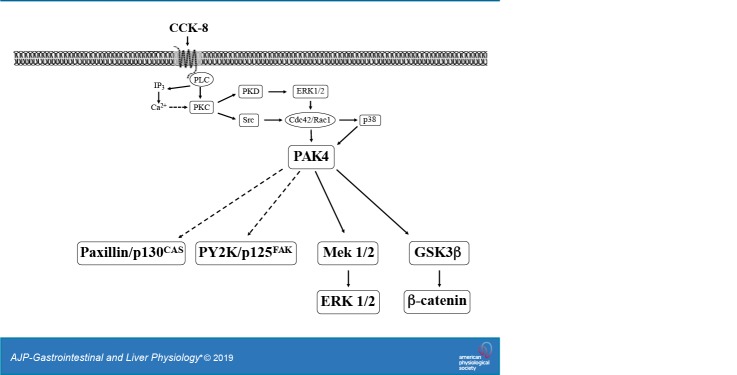
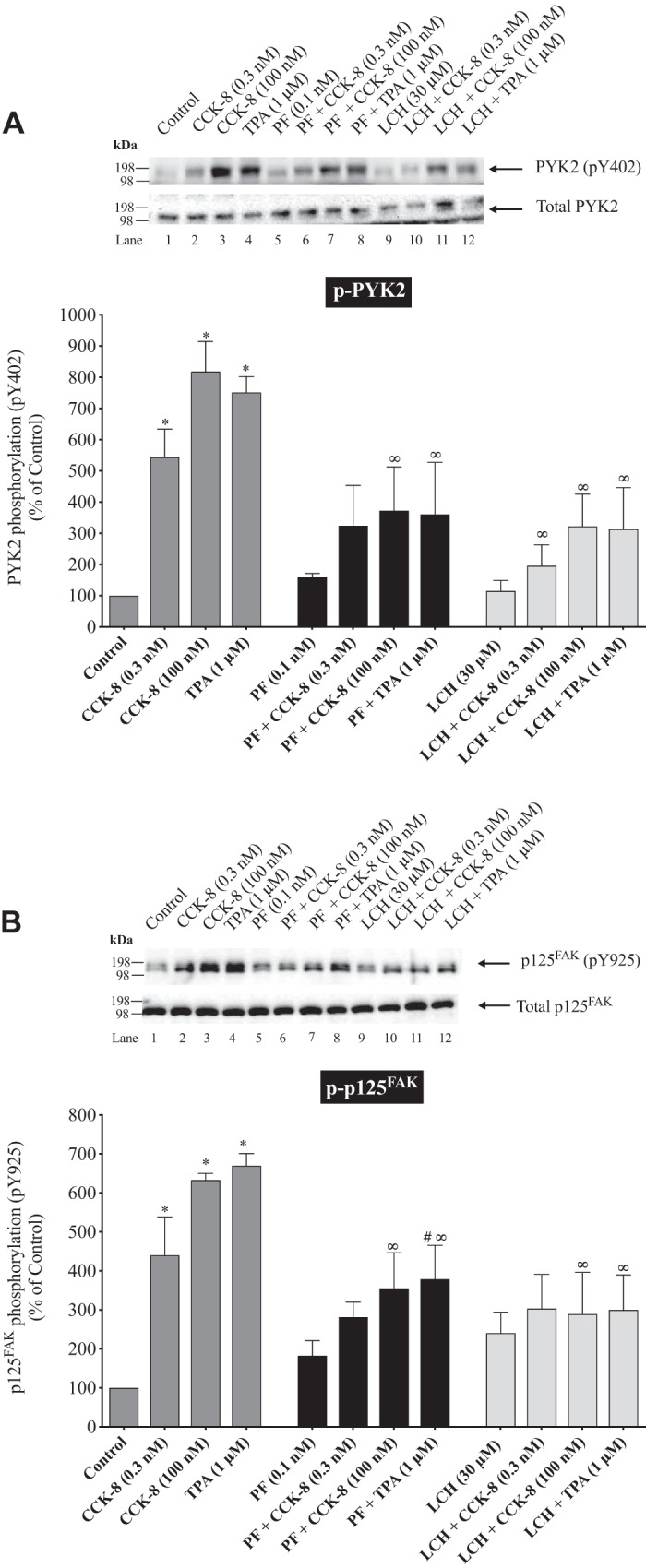
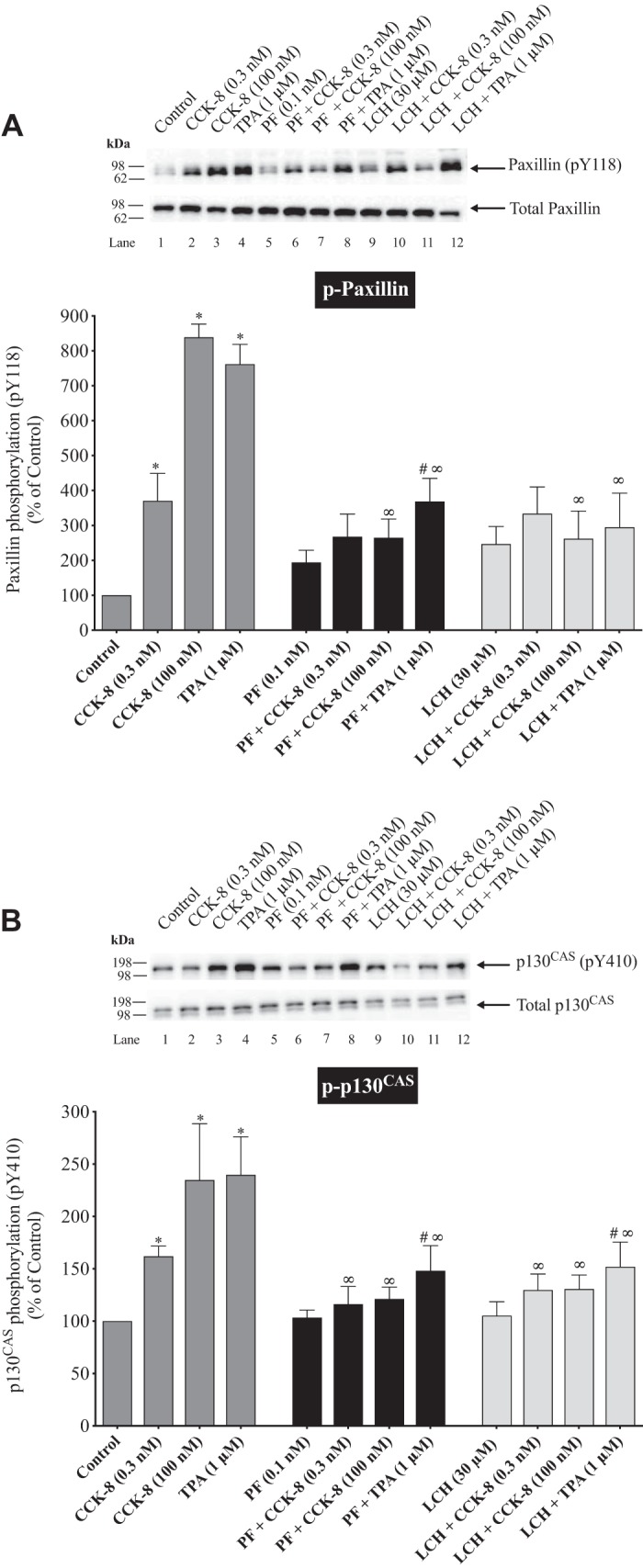
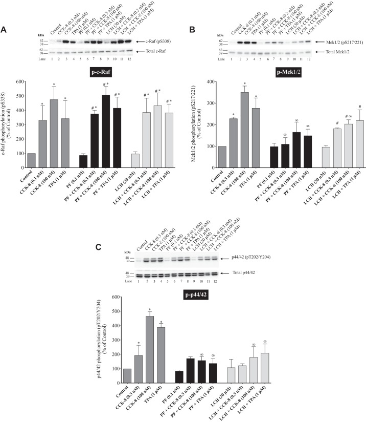
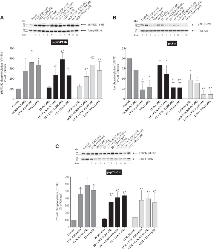
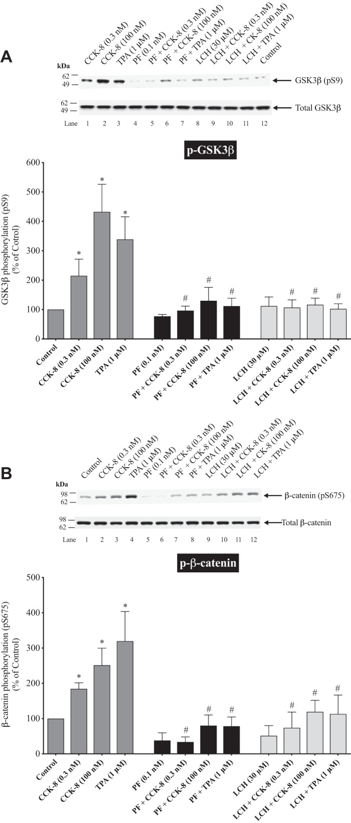
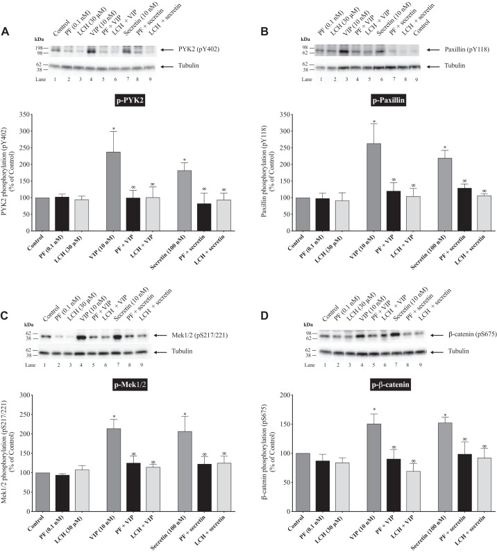
PAK4 is the only member of the Group II p21-activated kinases (PAKs) present in rat pancreatic acinar cells and is activated by gastrointestinal hormones/neurotransmitters stimulating PLC/cAMP and by various pancreatic growth factors. However, little is known of the role of PAK4 activation in cellular signaling cascades in pancreatic acinar cells. In the present study, we examined the role of PAK4's participation in five different cholecystokinin-8 (CCK-8)-stimulated signaling pathways (PI3K/Akt, MAPK, focal adhesion kinase, GSK3, and β-catenin), which mediate many of its physiological acinar-cell effects, as well as effects in pathophysiological conditions. To define PAK4's role, the effect of two different PAK4 inhibitors, PF-3758309 and LCH-7749944, was examined under experimental conditions that only inhibited PAK4 activation and not activation of the other pancreatic PAK, Group I PAK2. The inhibitors' effects on activation of these five signaling cascades by both physiological and pathophysiological concentrations of CCK, as well as by 12-O-tetradecanoylphobol-13-acetate (TPA), a PKC-activator, were examined. CCK/TPA activation of focal adhesion kinases(PYK2/p125FAK) and the accompanying adapter proteins (paxillin/p130CAS), Mek1/2, and p44/42, but not c-Raf or other MAPKs (JNK/p38), were mediated by PAK4. Activation of PI3K/Akt/p70s6K was independent of PAK4, whereas GSK3 and β-catenin stimulation was PAK4-dependent. These results, coupled with recent studies showing PAK4 is important in pancreatic fluid/electrolyte/enzyme secretion and acinar cell growth, show that PAK4 plays an important role in different cellular signaling cascades, which have been shown to mediate numerous physiological and pathophysiological processes in pancreatic acinar cells.NEW & NOTEWORTHY In pancreatic acinar cells, cholecystokinin (CCK) or 12-O-tetradecanoylphobol-13-acetate (TPA) activation of focal adhesion kinases (p125FAK,PYK2) and its accompanying adapter proteins, p130CAS/paxillin; Mek1/2, p44/42, GSK3, and β-catenin are mediated by PAK4. PI3K/Akt/p70s6K, c-Raf, JNK, or p38 pathways are independent of PAK4 activation.

Keywords: CCK; MAPK; PAK4; cell signaling; focal adhesion kinase; pancreatic acini.


Conflict of interest statement

No conflicts of interest, financial or otherwise, are declared by the authors.

Figures

None
Graphical abstract
Fig. 1.
Fig. 1.
Effect of PF-3758309 and LCH-7749944, PAK (p21-activated kinases) 4 inhibitors, on the ability of cholecystokinin (CCK-8) or 12-O-tetradecanoylphobol-13-acetate (TPA) to stimulate focal adhesion kinases: PYK2 (A) and p125FAK (B). Isolated pancreatic acini were incubated in the absence or presence of PF-3758309 (0.1 nM) or LCH-7749944 (30 µM) for 3 h and then incubated with no addition (control), CCK-8 (0.3 and 100 nM) for 3 min or TPA (1 µM) for 5 min and then lysed. Western blots were analyzed using anti-pY402 PYK2, anti-pY925 p125FAK, and, as a loading control, anti-total PYK2 or p125FAK. Bands were visualized using chemiluminescence and quantified by densitometry. Top: results of a representative blot of four independent experiments are shown. Bottom: quantitative results show means ± SE of at least four independent experiments. Results are expressed as a percentage of basal stimulation of the control Group. *P < 0.05 compared with the Control Group. #P < 0.05 compared with inhibitors alone (PF-3758309 or LCH-7749944). ∞P < 0.05 compared with stimulants without inhibitors.
Fig. 2.
Fig. 2.
Effect of PF-3758309 and LCH-7749944, PAK4 inhibitors, on the ability of cholecystokinin (CCK-8) or 12-O-tetradecanoylphobol-13-acetate (TPA) to stimulate the adapter proteins; paxillin (A) and p130CAS (B). Experimental conditions were as described in Fig. 1. Western blots were analyzed using anti-pY118 paxillin and anti-Y410 p130CAS. Bands were visualized using chemiluminescence and quantified by densitometry. Top: results of a representative blot of four independent experiments are shown. Bottom: quantitative results show means ± SE of at least four independent experiments. Results are expressed as a percentage of basal stimulation of the Control Group. *P < 0.05 compared with the Control Group. #P < 0.05 compared with inhibitors alone (PF-3758309 or LCH-7749944). ∞P < 0.05 compared with stimulants without inhibitors.
Fig. 3.
Fig. 3.
Effect of PF-3758309 and LCH-7749944, PAK4 inhibitors, on the ability of cholecystokinin (CCK-8) or 12-O-tetradecanoylphobol-13-acetate (TPA) to stimulate various kinases in the mitogen-activated kinase pathway: c-raf (A), Mek1/2 (B) and p44/42 C: experimental conditions were as described in Fig. 1. Western blots were analyzed using anti-pS338 c-Raf, anti-pS217/221 Mek1/2, and anti-pT202/Y204 p44/42. Bands were visualized using chemiluminescence and quantified by densitometry. Top: results of a representative blot of four independent experiments are shown. Bottom: quantitative results show means ± SE of at least four independent experiments. Results are expressed as % of basal stimulation of the Control Group. *P < 0.05 compared with the Control Group. #P < 0.05 compared with inhibitors alone (PF-3758309 or LCH-7749944). ∞P < 0.05 compared with stimulants without inhibitors.
Fig. 4.
Fig. 4.
Effect of PF-3758309 and LCH-7749944, PAK4 inhibitors, on the ability of CCK-8 or 12-O-tetradecanoylphobol-13-acetate (TPA) to stimulate various kinases in the mitogen-activated pathway: p38 (A) and JNK (B). Experimental conditions were as described in Fig. 1. Western blots were analyzed using anti-pT180/Y182 p38 and anti-pT183/Y185 JNK. Bands were visualized using chemiluminescence and quantified by densitometry. Top: results of a representative blot of four independent experiments are shown. Bottom: quantitative results show means ± SE of at least four independent experiments. Results are expressed as a percentage of basal stimulation of the Control Group. *P < 0.05 compared with the Control Group. #P < 0.05 compared with inhibitors alone (PF-3758309 or LCH-7749944).
Fig. 5.
Fig. 5.
Effect of PF-3758309 and LCH-7749944, PAK4 inhibitors, on the ability of cholecystokinin (CCK-8) or 12-O-tetradecanoylphobol-13-acetate (TPA) to stimulate various kinases in the PI3K pathway: p85 (A), Akt (B), and p70s6k (C). Experimental conditions were as described in Fig. 1, except for Akt studies where incubation times were 15 min with CCK-8/TPA. Western blots were analyzed using anti-pY458 p85PI3K, anti-pS473 Akt, and anti-pT389 p70s6k. Bands were visualized using chemiluminescence and quantified by densitometry. Top: results of a representative blot of four independent experiments are shown. Bottom: quantitative results show means ± SE of at least four independent experiments. Results are expressed as a percentage of basal stimulation of the Control Group. *P < 0.05 compared with the Control Group. #P < 0.05 compared with inhibitors alone (PF-3758309 or LCH-7749944).
Fig. 6.
Fig. 6.
Effect of PF-3758309 and LCH-7749944, PAK4 inhibitors, on the ability of cholecystokinin (CCK-8) or 12-O-tetradecanoylphobol-13-acetate (TPA) to stimulate GSK3 and β-catenin. Experimental conditions were as described in Fig. 1, except incubation times for CCK-8 and TPA were 2 h. Western blots were analyzed using anti-pS9 GSK3 and anti-pS675 β-catenin. Bands were visualized using chemiluminescence and quantified by densitometry. Top: results of a representative blot of four independent experiments are shown. Bottom: quantitative results show means ± SE of at least four independent experiments. Results are expressed as a percentage of basal stimulation of the Control Group. *P < 0.05 compared with the Control Group. #P < 0.05 compared with inhibitors alone (PF-3758309 or LCH-7749944).
Fig. 7.
Fig. 7.
Effect of PF-3758309 and LCH-7749944, PAK4 inhibitors, on the ability of vasoactive intestinal peptide (VIP) or secretin to stimulate the focal adhesion kinase PYK2 (A), the adapter protein paxillin (B), Mek1/2 (C), and β-catenin (D). Experimental conditions were as described in Figs. 1, 2, 3, and 6, except incubation times for VIP (10 nM) and secretin (10 nM) were 15 min. Western blots were analyzed using anti-pY402 PYK2, anti-pY118 Paxillin, anti-pS217/221 Mek1/2, and anti-pS675 β-catenin, and anti-tubulin was used as a loading control. Bands were visualized using chemiluminescence and quantified by densitometry. Top: results of a representative blot of four independent experiments are shown. Bottom: quantitative results show means ± SE of at least four independent experiments. Results are expressed as a percentage of basal stimulation of the control Group. *P < 0.05 compared with the control Group; ∞P < 0.05 compared with stimulants without inhibitors.

Similar articles

Cited by

References

    1. Ahmed T, Shea K, Masters JR, Jones GE, Wells CM. A PAK4-LIMK1 pathway drives prostate cancer cell migration downstream of HGF. Cell Signal 20: 1320–1328, 2008. doi:10.1016/j.cellsig.2008.02.021. - DOI - PubMed
    1. Aparicio IM, Garcia-Marin LJ, Andreolotti AG, Bodega G, Jensen RT, Bragado MJ. Hepatocyte growth factor activates several transduction pathways in rat pancreatic acini. Biochim Biophys Acta 1643: 37–46, 2003. doi:10.1016/j.bbamcr.2003.08.007. - DOI - PubMed
    1. Bachmann VA, Riml A, Huber RG, Baillie GS, Liedl KR, Valovka T, Stefan E. Reciprocal regulation of PKA and Rac signaling. Proc Natl Acad Sci USA 110: 8531–8536, 2013. doi:10.1073/pnas.1215902110. - DOI - PMC - PubMed
    1. Barac A, Basile J, Vázquez-Prado J, Gao Y, Zheng Y, Gutkind JS. Direct interaction of p21-activated kinase 4 with PDZ-RhoGEF, a G protein-linked Rho guanine exchange factor. J Biol Chem 279: 6182–6189, 2004. doi:10.1074/jbc.M309579200. - DOI - PubMed
    1. Barrett A, Pellet-Many C, Zachary IC, Evans IM, Frankel P. p130Cas: a key signalling node in health and disease. Cell Signal 25: 766–777, 2013. doi:10.1016/j.cellsig.2012.12.019. - DOI - PubMed

Publication types

MeSH terms