Traction force microscopy for understanding cellular mechanotransduction
- PMID: 31964473
- PMCID: PMC7061206
- DOI: 10.5483/BMBRep.2020.53.2.308
Traction force microscopy for understanding cellular mechanotransduction
Abstract
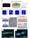
Under physiological and pathological conditions, mechanical forces generated from cells themselves or transmitted from extracellular matrix (ECM) through focal adhesions (FAs) and adherens junctions (AJs) are known to play a significant role in regulating various cell behaviors. Substantial progresses have been made in the field of mechanobiology towards novel methods to understand how cells are able to sense and adapt to these mechanical forces over the years. To address these issues, this review will discuss recent advancements of traction force microscopy (TFM), intracellular force microscopy (IFM), and monolayer stress microscopy (MSM) to measure multiple aspects of cellular forces exerted by cells at cell-ECM and cell-cell junctional intracellular interfaces. We will also highlight how these methods can elucidate the roles of mechanical forces at interfaces of cell-cell/cell-ECM in regulating various cellular functions. [BMB Reports 2020; 53(2): 74-81].
Conflict of interest statement
The authors have no conflicting interests.
Figures


Similar articles
-
Traction force microscopy - Measuring the forces exerted by cells.Micron. 2021 Nov;150:103138. doi: 10.1016/j.micron.2021.103138. Epub 2021 Aug 12. Micron. 2021. PMID: 34416532 Review.
-
Finite element analysis of traction force microscopy: influence of cell mechanics, adhesion, and morphology.J Biomech Eng. 2013 Jul 1;135(7):71009. doi: 10.1115/1.4024467. J Biomech Eng. 2013. PMID: 23720059 Free PMC article.
-
A Biologist's Guide to Traction Force Microscopy Using Polydimethylsiloxane Substrate for Two-Dimensional Cell Cultures.STAR Protoc. 2020 Aug 28;1(2):100098. doi: 10.1016/j.xpro.2020.100098. eCollection 2020 Sep 18. STAR Protoc. 2020. PMID: 33111126 Free PMC article.
-
2.5D Traction Force Microscopy: Imaging three-dimensional cell forces at interfaces and biological applications.Int J Biochem Cell Biol. 2023 Aug;161:106432. doi: 10.1016/j.biocel.2023.106432. Epub 2023 Jun 7. Int J Biochem Cell Biol. 2023. PMID: 37290687 Review.
-
In silico CDM model sheds light on force transmission in cell from focal adhesions to nucleus.J Biomech. 2016 Sep 6;49(13):2625-2634. doi: 10.1016/j.jbiomech.2016.05.031. Epub 2016 Jun 4. J Biomech. 2016. PMID: 27298154
Cited by
-
The extracellular matrix mechanics in the vasculature.Nat Cardiovasc Res. 2023 Aug;2(8):718-732. doi: 10.1038/s44161-023-00311-0. Epub 2023 Aug 10. Nat Cardiovasc Res. 2023. PMID: 39195965 Review.
-
Combined Traction Force-Atomic Force Microscopy Measurements of Neuronal Cells.Biomimetics (Basel). 2022 Oct 8;7(4):157. doi: 10.3390/biomimetics7040157. Biomimetics (Basel). 2022. PMID: 36278714 Free PMC article.
-
Mechanical properties of human tumour tissues and their implications for cancer development.Nat Rev Phys. 2024 Apr;6(4):269-282. doi: 10.1038/s42254-024-00707-2. Epub 2024 Mar 19. Nat Rev Phys. 2024. PMID: 38706694 Free PMC article.
-
Focusing on Mechanoregulation Axis in Fibrosis: Sensing, Transduction and Effecting.Front Mol Biosci. 2022 Mar 11;9:804680. doi: 10.3389/fmolb.2022.804680. eCollection 2022. Front Mol Biosci. 2022. PMID: 35359592 Free PMC article. Review.
-
Cell Response in Free-Packed Granular Systems.ACS Appl Mater Interfaces. 2022 Sep 14;14(36):40469-40480. doi: 10.1021/acsami.1c24095. Epub 2022 Aug 31. ACS Appl Mater Interfaces. 2022. PMID: 36044384 Free PMC article.
References
Publication types
MeSH terms
Substances
LinkOut - more resources
Full Text Sources
Research Materials
Miscellaneous

