. 2020 Feb 26;40(9):1849-1861.
doi: 10.1523/JNEUROSCI.1838-19.2020. Epub 2020 Jan 16.

Repeated Exposure to Multiple Concurrent Stresses Induce Circuit Specific Loss of Inputs to the Posterior Parietal Cortex

Affiliations

Repeated Exposure to Multiple Concurrent Stresses Induce Circuit Specific Loss of Inputs to the Posterior Parietal Cortex

Yaaqov Libovner et al. J Neurosci. .

Abstract

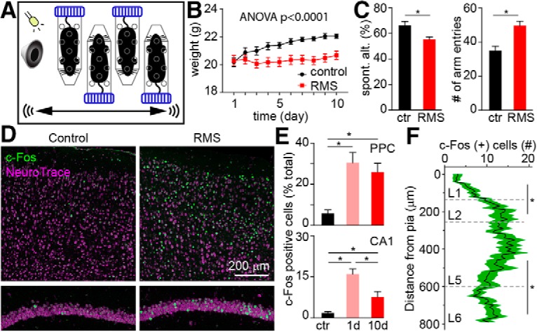
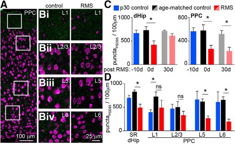
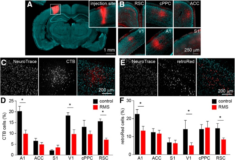
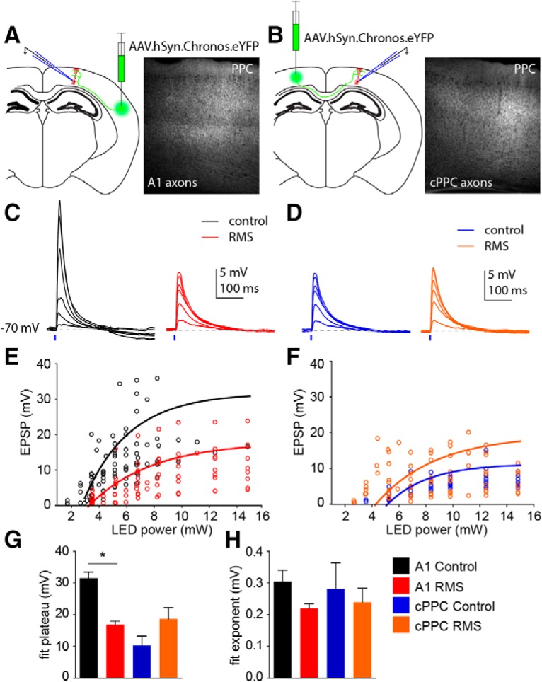
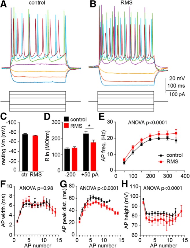
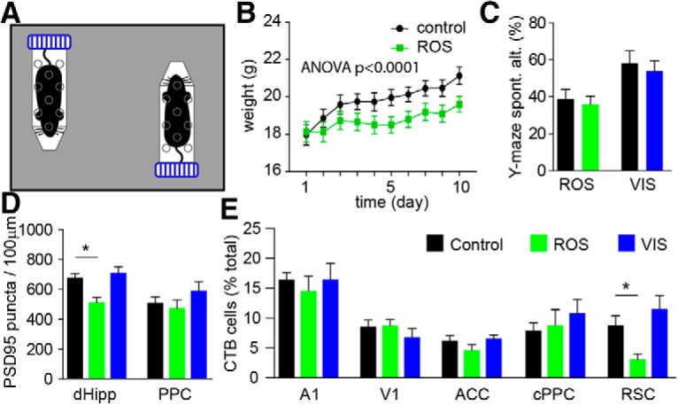
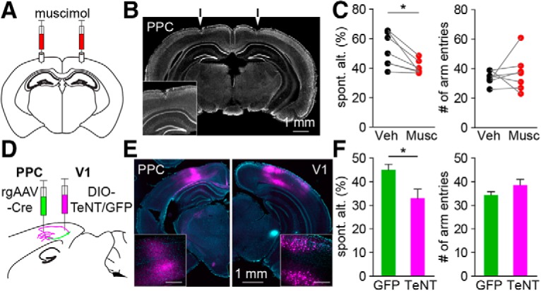
Severe loss of excitatory synapses in key brain regions is thought to be one of the major mechanisms underlying stress-induced cognitive impairment. To date, however, the identity of the affected circuits remains elusive. Here we examined the effect of exposure to repeated multiple concurrent stressors (RMS) on the connectivity of the posterior parietal cortex (PPC) in adolescent male mice. We found that RMS led to layer-specific elimination of excitatory synapses with the most pronounced loss observed in deeper cortical layers. Quantitative analysis of cortical projections to the PPC revealed a significant loss of sensory and retrosplenial inputs to the PPC while contralateral and frontal projections were preserved. These results were confirmed by decreased synaptic strength from sensory, but not from contralateral, projections in stress-exposed animals. Functionally, RMS disrupted visuospatial working memory performance, implicating disrupted higher-order visual processing. These effects were not observed in mice subjected to restraint-only stress for an identical period of time. The PPC is considered to be a cortical hub for multisensory integration, working memory, and perceptual decision-making. Our data suggest that sensory information streams targeting the PPC may be impacted by recurring stress, likely contributing to stress-induced cognitive impairment.SIGNIFICANCE STATEMENT Repeated exposure to stress profoundly impairs cognitive functions like memory, attention, or decision-making. There is emerging evidence that stress not only impacts high-order regions of the brain, but may affect earlier stages of cognitive processing. Our work focuses on the posterior parietal cortex, a brain region supporting short-term memory, multisensory integration, and decision-making. We show evidence that repeated stress specifically damages sensory inputs to this region. This disruption of synaptic connectivity is linked to working memory impairment and is specific to repeated exposure to multiple stressors. Altogether, our data provide a potential alternative explanation to ailments previously attributed to downstream, cognitive brain structures.

Keywords: chronic stress; multimodal stress; posterior parietal cortex; retrograde tracing; synapse loss; visuospatial working memory.


Figures

Figure 1.
Figure 1.
Repeated exposure to multiple concurrent stressors impairs spatial working memory and activates the PPC. A, Schematic of the RMS paradigm. B, Weight change of control (black) and stressed mice (red) during the 10 d exposure to RMS. Data points indicate the mean weight (±SEM) of mice on each day. C, Bar graphs indicate the mean (±SEM) spontaneous alternation rate (left) and the number of arm entries (right) in control (black) and RMS (red) mice. D, Example confocal images of c-Fos labeling (green) in PPC (top row) and CA1 (bottom row) from control animals and mice exposed to RMS. E, Bar graphs represent the mean (±SEM) proportion of c-Fos-labeled neurons in the PPC (top) and the hippocampus (bottom) in control mice (black) and in mice exposed to RMS for 1 d (salmon) or for 10 d (red). F, Histogram representing the distribution of c-Fos+ neurons in the PPC. Mean number of c-Fos+ cells (solid black line) ± SEM (green shading), gray dashed lines mark approximate layers in the PPC. *p < 0.05, one-way ANOVA with Bonferroni's multiple-comparison test.
Figure 2.
Figure 2.
Repeated, multimodal stress causes excitatory synapse loss in the PPC. A, Example confocal image of PSD-95 (green) and NeuroTrace (magenta)-stained PPC section. Bi–Biv, Representative crops for layer 1 (Bi), layer 2/3 (Bii), layer 5 (Biii), and layer 6 (Biv) of PPC. C, Bar graphs represent the mean (±SEM) PSD-95 puncta densities in the dorsal hippocampus (dHipp, left), or in the PPC (right). Puncta were quantified at the starting time point in P30 control mice (blue), immediately after the 10 d RMS in P40 control (black) and stressed (red) animals or following a 30 d rest period in P70 control (gray) and stressed (salmon) mice. D, Bar graphs represent the mean (±SEM) PSD-95 puncta densities in SR in the hippocampus and layers 1, 2/3, 5, and 6 in the PPC in unstressed P30 (blue), age-matched control (black), and stressed (red) mice. *p < 0.05, one-way ANOVA with Bonferroni's multiple-comparison test; ns, not significant.
Figure 3.
Figure 3.
Circuit-specific loss of neuronal projections to the PPC following RMS. A, representative image of the CTB-555 injection site. Inset, High-magnification image of the injection location. B, Representative images of CTB-555-labeled projection cells in the RSC, the cPPC, the ACC, the V1, the A1, and the S1. C, Representative confocal image of NeuTrace (cyan) CTB-555 (red)-labeled cells in the auditory cortex. D, Bar graphs represent the mean (±SEM) proportion of CTB-555-labeled projection cells in cortical regions of control (black) and stressed (red) mice. E, Representative confocal image of NeuTrace (cyan) red retrobead (red)-labeled cells in the auditory cortex. F, Bar graphs represent the mean (±SEM) proportion of retrobead-labeled projection cells in different cortical regions of control (black) and stressed (red) mice. RSC, retrosplenial cortices. *p < 0.05, one-way ANOVA with Bonferroni's multiple-comparison test.
Figure 4.
Figure 4.
Circuit-specific reduction of synaptic strength by repeated, multimodal stress. A, B, Schematic illustrating the experiment (left) and wide-field image showing Chronos-expressing auditory (A) and cPPC (B) fibers in the recorded PPC hemisphere (right). C, Example EPSP recordings evoked by stimulating auditory fibers in control (black) and stressed (red) animals. D, Example EPSP recordings evoked by stimulating cPPC fibers in control (blue) and stressed (orange) animals. E, Scatter plot showing the auditory EPSP magnitude–LED power relationship in control (black) and stressed (red) animals. Solid lines represent the reconstructed exponential fit from the mean of the calculated coefficients. F, Scatter plot showing the cPPC EPSP magnitude–LED power relationship in control (blue) and stressed (orange) animals. Solid lines represent the reconstructed exponential fit from the mean of the calculated coefficients. G, Bars represent the mean (±SEM) plateau from the exponential fit. H, Bars represent the mean (±SEM) exponent from the exponential fit. *p < 0.05, Student's t test.
Figure 5.
Figure 5.
Circuit-specific effect of RMS on excitatory and inhibitory currents in the PPC. A, Left, Average IPSC (dashed lines) and EPSC (solid lines) responses to optogenetic activation of A1 fibers in control (black) and RMS (red) mice. Right, Average IPSC (dashed lines) and EPSC (solid lines) responses to optogenetic activation of cPPC afferents in control (blue) and RMS (orange) mice. B, C, Population data showing increasing current amplitude in response to increasing LED power in control (black and blue) and stressed (red and orange) animals. Solid lines represent the mean exponential fit to the data. D, Bars show the mean (±SEM) plateau of exponentials fitted to EPSCs. E, Bars show mean (±SEM) plateau of exponential fitted to IPSCs. *p < 0.05, Student's t test.
Figure 6.
Figure 6.
RMS increases the excitability of layer 5 pyramidal neurons in the PPC. A, Example membrane potential responses to current pulses in a control mouse. B, Example membrane potential responses to current pulses in a stressed mouse. C, Comparison of resting membrane potential in control (black) and stressed (red) mice. D, Comparison of input resistance in control (black) and stressed (red) mice. E–H, Comparison of action potential firing frequency (E), action potential width (F), distance between action potential peaks (G), and action potential heights (H) in control and stressed mice. All bars represent the mean (±SEM); statistical comparisons: two-way ANOVA, p values are displayed above each graph. *p < 0.05, Student's t-test.
Figure 7.
Figure 7.
Single-modality stress paradigms have minimal effect on PPC. A, Schematic of ROS. B, Weight change of control (black) and stressed mice (green) during the 10 d exposure to ROS. Data points indicate the mean weight (±SEM) of mice on each day; figure shows result of two-way ANOVA test. C, Bars represent spontaneous alternation percentage (±SEM) in control (black), ROS (green) and VIS (blue) exposed mice. D, Bar graphs represent the mean (±SEM) PSD-95 puncta densities in the dorsal hippocampus (dHipp) and in the PPC in control (black), ROS (green), and VIS (blue) mice immediately after stress exposure. E, Bar graphs represent the mean (±SEM) proportion of CTB-555-labeled projection cells in different cortical regions of control (black), ROS (green), and VIS (blue) mice. *p < 0.05, one-way ANOVA with Bonferroni's multiple-comparison test.
Figure 8.
Figure 8.
PPC activity and V1 long-range connectivity play a key role in navigating the Y-maze. A, Schematic of cortical inhibition via muscimol. B, Representative coronal section; arrows show the location of cannula implant. Inset, Higher-magnification image of the PPC. C, Paired comparison of spontaneous alternation (left) and total number of arm entries (right) after saline (black) or muscimol (red) injection into the PPC. *p < 0.05, paired t test. D, Schematic of the TeNT experiment. E, Example images showing expression in V1 cell bodies (right) and afferent fibers in the PPC (left). Insets show magnified images of V1 and PPC. Scale bar, inset, 300 μm. F, Bars represent mean (±SEM) spontaneous alternation percentage (left) and total number of arm entries (right) in GFP control (green) and TeNT-expressing (magenta) animals. *p < 0.05, paired t test.

Similar articles

Cited by

References

    1. Akrami A, Kopec CD, Diamond ME, Brody CD (2018) Posterior parietal cortex represents sensory history and mediates its effects on behaviour. Nature 554:368–372. 10.1038/nature25510 - DOI - PubMed
    1. Andersen RA, Cui H (2009) Intention, action planning, and decision making in parietal-frontal circuits. Neuron 63:568–583. 10.1016/j.neuron.2009.08.028 - DOI - PubMed
    1. Andersen RA, Snyder LH, Bradley DC, Xing J (1997) Multimodal representation of space in the posterior parietal cortex and its use in planning movements. Annu Rev Neurosci 20:303–330. 10.1146/annurev.neuro.20.1.303 - DOI - PubMed
    1. Anderson EM, Gomez D, Caccamise A, McPhail D, Hearing M (2019) Chronic unpredictable stress promotes cell-specific plasticity in prefrontal cortex D1 and D2 pyramidal neurons. Neurobiol Stress 10:100152. 10.1016/j.ynstr.2019.100152 - DOI - PMC - PubMed
    1. Andres AL, Regev L, Phi L, Seese RR, Chen Y, Gall CM, Baram TZ (2013) NMDA receptor activation and calpain contribute to disruption of dendritic spines by the stress neuropeptide CRH. J Neurosci 33:16945–16960. 10.1523/JNEUROSCI.1445-13.2013 - DOI - PMC - PubMed

Publication types

MeSH terms