. 2020 Jan 14;21(2):539.
doi: 10.3390/ijms21020539.

Hemmule: A Novel Structure with the Properties of the Stem Cell Niche

Affiliations

Hemmule: A Novel Structure with the Properties of the Stem Cell Niche

Vitaly Vodyanoy et al. Int J Mol Sci. .

Abstract

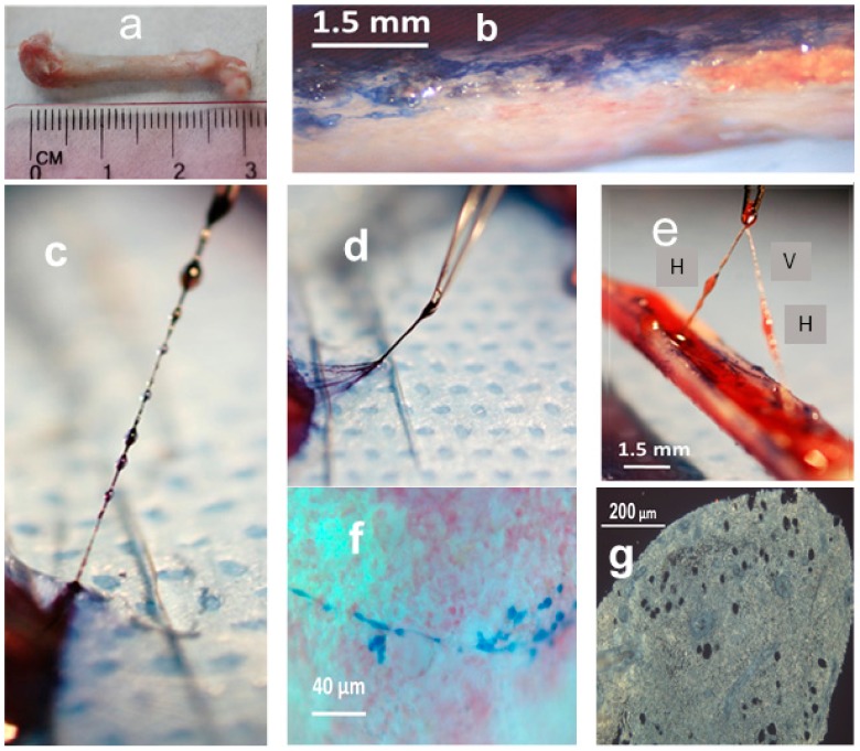
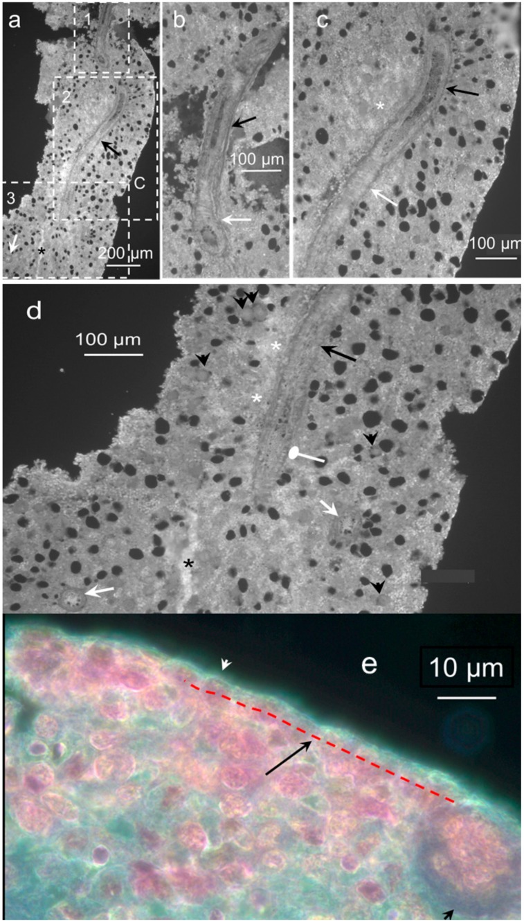
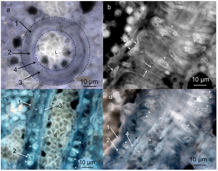
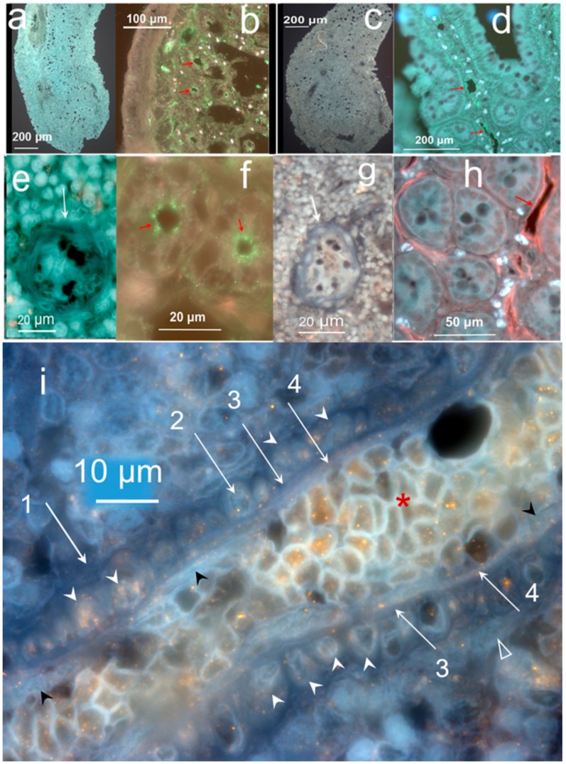
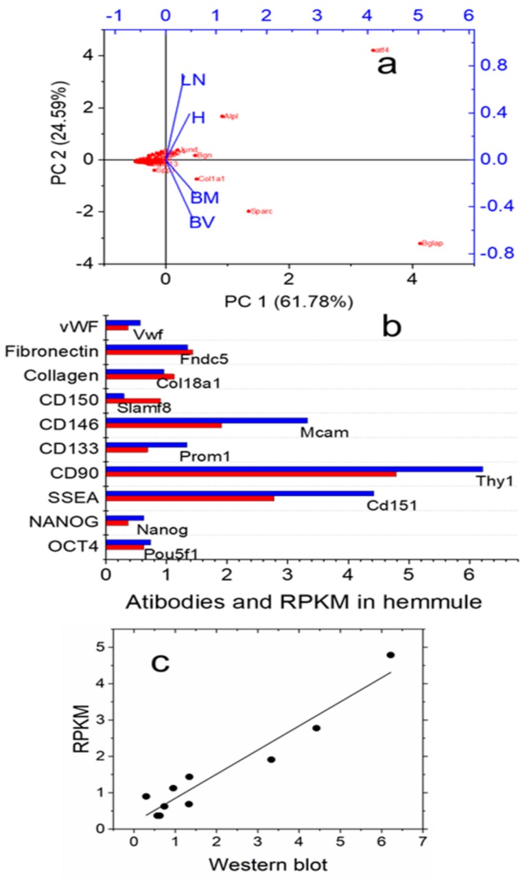
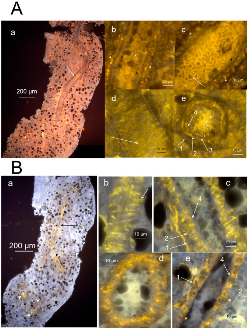
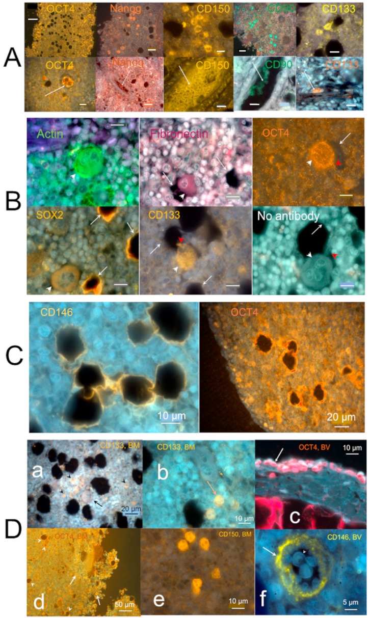
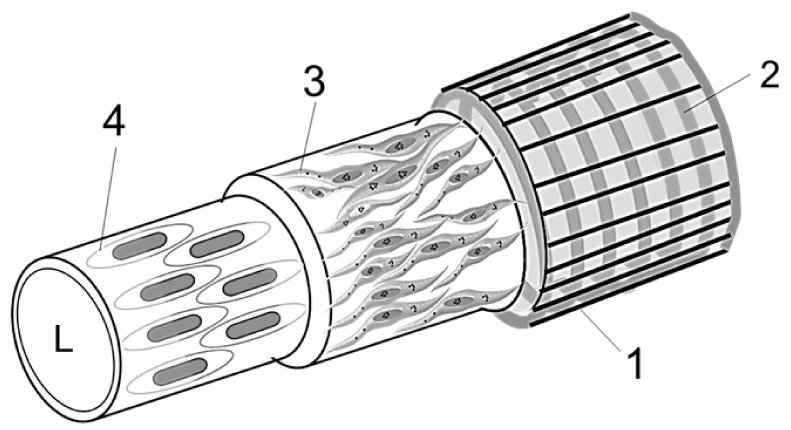
Stem cells are nurtured and regulated by a specialized microenvironment known as stem cell niche. While the functions of the niches are well defined, their structure and location remain unclear. We have identified, in rat bone marrow, the seat of hematopoietic stem cells-extensively vascularized node-like compartments that fit the requirements for stem cell niche and that we called hemmules. Hemmules are round or oval structures of about one millimeter in diameter that are surrounded by a fine capsule, have afferent and efferent vessels, are filled with the extracellular matrix and mesenchymal, hematopoietic, endothelial stem cells, and contain cells of the megakaryocyte family, which are known for homeostatic quiescence and contribution to the bone marrow environment. We propose that hemmules are the long sought hematopoietic stem cell niches and that they are prototypical of stem cell niches in other organs.

Keywords: bone marrow; endothelial; hematopoietic; megakaryocyte; mesenchymal; node; quiescence; vascular.


Conflict of interest statement

The authors declare no conflict of interest.

Figures

Figure 1
Figure 1
The hemmule and vessels taken from the freshly split rat femur bone. (a) Femur bone. (b) A split bone exposes a bone marrow stained by the trypan blue. (c) A series of hemmules are connected to the vessel. The lower hemmule is attached to the end hemmule inside the bone marrow. The hemmules and vessels are stained with trypan blue. The distance between the centers of the background dots is 1.5 mm. (d) The efferent vessel of the lower end hemmule is composed of multiple subvessels. (e) These hemmules are extracted from the bone marrow, while both ends of the vessel are attached to the end hemmules in the bone marrow. H—hemmule, V—vessel. (f) A thin branched vessel in the non-fixed bone marrow is stained by trypan blue. (g) A slice of the fixed hemmule is extracted from the bone marrow. Openings are voids of large size cells.
Figure 2
Figure 2
Longitudinal section of hemmule. A section of 6 μm from the rat femur bone marrow. (a) The section shows a portion of the central vessel passing in the longitudinal direction (black arrow). A smaller longitudinal vessel is labeled using an asterisk. The lumen of one transversal vessel is identified by a white arrow. Letter C depicts a fragment of the capsule. The section is divided by three areas of interest, marked as 1, 2, and 3 and shown in panels (bd), respectively. (b) The magnified area 1 from panel (a) depicts a top part of the central vessel. The lower portion of the vessel (white arrow) illustrates the lumen with thick walls. A higher part of the vessel (black arrow) cuts open to expose a central duct with thick walls. (c) The magnified area 2 from panel (a). A white arrow reveals part of the vessel with a longitudinal ribbed wall surface. A black arrow points to the cut open vessel with thick walls. A white asterisk denotes an area with a high concentration of small vessels. (d) The magnified area 3 from panel (a). The central longitudinal vessel (long black arrow) is shown to be cut open (round head white arrow) before going down transversely into the slide. Two more large transverse vessels lumens are visible in this section (white arrows). The slice is highly vascular, depicting the density of small vessels (or channels) in several parts of the slide and exemplified by the areas marked by white asterisks. A relatively large vessel or a channel is labeled using a black asterisk. The entire area of the slice is covered with round holes, which shows the footprints of missing large size cells. Some of these cells are in close proximity to the holes (short black arrows). (e) The magnified portion of hemmule. White arrowhead shows the capsule. Black arrow illustrates the layer of cell around the hemmule border (red dotted line). Black arrowhead shows a cross-section of the transversal vessel. Sections a-d are not stained; section e is stained by Hematoxylin and eosin stain (H&E).
Figure 3
Figure 3
High magnification darkfield images of the vessel inside the hemmule. (a) Transversal section of the hemmule vessel. The wall of the vessels is composed of several optically distinguished Layers 1–4; L—lumen. (b) The longitudinal section of the vessel: arrowheads—vascular smooth muscle cells comprised the thick fibers-like loops (Layer 2), solid arrows—longitudinal thin fiber treads, and transversal fiber, respectively, dotted arrows—longitudinal fibroblastic cells and their processes belonging to the vessel external Layer 1. (c) The longitudinal section cuts the middle portion of the vessel. White arrows-layers 1–4, arrowheads—cross-sections of vascular smooth muscle cells of Layer 2, white asterisk—represent a fibroblastic cell of the Layer 1, and black asterisks denote endothelial cells of Layer 4. (d) The longitudinal section cuts the vessel wall in the vicinity of Layer 3, the subendothelial layer, primarily comprising of finely dispersed smooth muscle cells and bundles of fibrils. White asterisks—dispersed smooth muscle cells of Layer 3, arrows—Layers 1, 2, and 4, respectively, arrowheads—cross-sections of vascular smooth muscle cells of Layer 2, black asterisk—endothelial cell in Layer 4. No sections are stained. Pseudo color is caused by auto fluorescence.
Figure 4
Figure 4
Hemmule does not interact with RECA-1 antibody (blood vessel endothelial cell marker) and with LYVE-1 (lymphatic vessel endothelial cell and hyaluronan receptor marker). Immunofluorescence staining of hemmule and small intestine. (a,e) Fluorescence images of a rat hemmule cross section treated with RECA-1 polyclonal antibodies and Goat Anti-Rabbit IgG as secondary antibodies visualized by Alexa 488. The white arrow in panel e shows lumens of the hemmule vessel. (b,f). Fluorescence image of a cross-section of small intestine is labeled with RECA-1 antibodies (positive control). The vascular profiles are located on the luminal aspect of the inner circular. Red arrows denote endothelial lining of blood vessels (green). (c,g). Fluorescence images of a rat hemmule cross-section treated with LYVE-1 polyclonal antibodies and Goat Anti-Rabbit IgG as secondary antibodies and visualized by Alexa 488. The white arrow in g indicates lumen of the hemmule vessels. (d,h) Fluorescence image of a cross-section of small intestine labeled with LYVE 1 antibodies (positive control), visualized with Alexa 488 (d) and Alexa 555 (h). Red srrows show endothelial lining of lymphatic vessels. (i) Fluorescence images of cells within the longitudinal cross-sections of hemmule are immunostained with the anti-LYVE-1 antibody. The longitudinal section cuts the middle portion of the vessel. Red star—the intra-vessel cells stained by the anti-LYVE-1 antibody; white arrows—Layers 1–4; white arrowheads—cross-sections of vascular smooth muscle cells of Layer 2; black arrowhead—endothelial cells of the Layer 4; open triangle—fibroblastic cell of Layer 1. Layer 4 of endothelial cells is not stained by anti-LYVE-1 antibody.
Figure 5
Figure 5
Principal component analyses (PCA), Western blot, and RNA-Seq. Protein expression obtained by Western blot and reads assigned per kilobase per million mapped reads (RPKM) correlation. (a) Biplot graph of the principal component analysis using gene expression (RPKM values) for the selected niche genes, hematopoietic progenitor, mesenchymal, and osteogenic genes. H, BV, BM, and LN are hemmule, blood vessel, bone narrow, and lymph node, respectively. (b) The lineup of relative levels of protein expressions (blue) and normalized RNA reads, RPKM (red). (c) The linear correlation between RPKM and relative levels of proteins. Pearson’s r = 0.96; Adj. R-square = 0.91.
Figure 6
Figure 6
Layered structure of hemmule. (A) The sections of hemmule immunostained with the anti-CD150 antibody (a). The longitudinal cross-section. The inside portion of the large longitudinal vessel cells’ (arrow) lumen indicates positive staining involving anti-CD150 antibody. A small longitudinal channel is labeled by a black asterisk. Many stained cells are scattered in the entire area of the section. The lumens of transversal vessels are illustrated by white arrowheads. (b) The magnified fragment of the longitudinal vessel section. White arrowheads labels the cross-section of transversally positioned vascular smooth muscle of Layer 2. Black arrowheads marks the innermost Layer 4 of endothelial cells. The white arrow is shown to labels one of the cells inside the vessel of about 3 μm in diameter. (c) Another magnified fragment of the longitudinal vessel section. A white arrowhead is shown to label the cross-section of transversally positioned vascular smooth muscle cells of Layer 2. (d) Another fragment of the longitudinal vessel section. The area of the vessel between two dotted lines reveals transversal vascular smooth muscle cells (white arrow). (e) The transversal section of the vessel. Arrow 1 and 4 label the outmost and innermost layers of the vessel, respectively. The unstained Layer 3 is positioned between Layers 2 and 4. The cells inside the vessel are labeled by a white asterisk. L—Lumen. (B) The cross-section is immunostained with the anti-CD146 antibody. (a) The cells of longitudinal and transversal vessel walls (arrows and arrowheads, respectively) show binding of the antibody. (b). The magnified fragment of the longitudinal cross-section of the vessel is illustrated in the previou panel. The transverse thick vascular smooth muscle cells “ribs” is exemplified by one section and signified by the arrow. Many longitudinal fibers on the vessel’s long axis are visible in this fragment. (c). Another magnified latitudinal fragment of the section. Arrows 1 depict the layer of fibroblastic cells (Layer 1), whereas the layer of vascular smooth muscle cells (Layer 2) is illustrated by arrow 2. The layer of endothelial cells (Layer 4) is not stained (arrow 4). (d) The transversal section of the vessel. The vascular smooth muscle cells of Layer 2 are heavily stained. (e) Another magnified latitudinal fragment of the section. Arrows label Layers 1 and 2, respectively. Layer 4 is not stained (arrow 4).
Figure 7
Figure 7
Individual hemmule cells stained by stem cell antibodies. (A) Fluorescent images of individual stem cell progenitors in the hemmule, which express embryonic cell markers. The top sections show individual stem cell progenitors being scattered in the hemmule cross-sections. The lower sections denote the progenitor cells inside the hemmule vessels L—lumen. Bars in the top panels, Oct4, 50 μm; Nanog, 20 μm; CD150, 10 μm; CD90, 20 μm; CD133, 10 μm. Bars in lower panels: Oct4, 20 μm; Nanog, 20 μm; CD150, 10 μm; CD90, 10 μm; CD133, 10 μm. (B) Large size cells in the hemmule belong to the megakaryocyte family. Some of these large sized cells illustrate small cytoplasmic extension (depicted by red arrowhead) and are in close proximity to the pores—the optically empty rounded regions (arrows). The formation of the most of these pores occurs when the large sized cells are displaced during the thin histological sample slicing. The rims of these pores may help retain molecules from these cells, which is evidenced by the staining by the antibodies. Bars: 10 μm. (C) Borders of the pores are stained by antibodies. (D) Both bone marrow and blood vessel cells stained by stem cell antibodies (positive controls). (a) Anti-CD133 positive cells in the bone marrow: the large size cell (arrow) and scattered cells (arrowheads). (b) Anti-CD133 positive scattered cells in bone marrow. (c) Anti-OCT4 antibody positive cells (arrow) on the surface of the blood vessel. (d) Anti-OCT4 antibody positive cells (arrows) and stained pores in bone marrow. (e) Anti-CD150 antibody cells in the bone marrow. (f) Anti-CD146 antibody stains the arteriole in the bone marrow (arrow).
Figure 8
Figure 8
Diagram idealization of a vessel inside a hemmule. It comprises of four Layers: 1—the outermost layer of thin longitudinal fibers; 2—the adjacent layer is a continuous fibrous helix, or circumferentially oriented spiral that is composed of transversally positioned cells; 3—this layer comprises of thinly dispersed longitudinally oriented muscle-like cells; and 4—the innermost layer consisting of endothelial cells. L—Lumen.

Similar articles

Cited by

References

    1. Davis H., Guo X., Lambert S., Stancescu M., Hickman J.J. Small Molecule Induction of Human Umbilical Stem Cells into Myelin Basic Protein Positive Oligodendrocytes in a Defined Three-Dimensional Environment. ACS Chem. Neurosci. 2011;3:31–39. doi: 10.1021/cn200082q. - DOI - PMC - PubMed
    1. Scadden D.T. The stem-cell niche as an entity of action. Nature. 2006;441:1075–1079. doi: 10.1038/nature04957. - DOI - PubMed
    1. Li L., Xie T. Stem cell niche: Structure and function. Annu. Rev. Cell Dev. Biol. 2005;21:605–631. doi: 10.1146/annurev.cellbio.21.012704.131525. - DOI - PubMed
    1. Adams G.B., Scadden D.T. The hematopoietic stem cell in its place. Nat. Immunol. 2006;7:333–337. doi: 10.1038/ni1331. - DOI - PubMed
    1. Hooper A.T., Butler J.M., Nolan D.J., Kranz A., Iida K., Kobayashi M., Kopp H.G., Shido K., Petit I., Yanger K., et al. Engraftment and reconstitution of hematopoiesis is dependent on VEGFR2-mediated regeneration of sinusoidal endothelial cells. Cell Stem Cell. 2009;4:263–274. doi: 10.1016/j.stem.2009.01.006. - DOI - PMC - PubMed