. 2020 Apr 1;130(4):2129-2145.
doi: 10.1172/JCI132691.

Epithelial splicing regulatory protein 2-mediated alternative splicing reprograms hepatocytes in severe alcoholic hepatitis

Affiliations

Epithelial splicing regulatory protein 2-mediated alternative splicing reprograms hepatocytes in severe alcoholic hepatitis

Jeongeun Hyun et al. J Clin Invest. .

Abstract

Severe alcoholic hepatitis (SAH) is a deadly liver disease without an effective medical therapy. Although SAH mortality is known to correlate with hepatic accumulation of immature liver cells, why this occurs and how it causes death are unclear. Here, we demonstrate that expression of epithelial splicing regulatory protein 2 (ESRP2), an RNA-splicing factor that maintains the nonproliferative, mature phenotype of adult hepatocytes, was suppressed in both human SAH and various mouse models of SAH in parallel with the severity of alcohol consumption and liver damage. Inflammatory cytokines released by excessive alcohol ingestion reprogrammed adult hepatocytes into proliferative, fetal-like cells by suppressing ESRP2. Sustained loss of ESRP2 permitted reemergence of a fetal RNA-splicing program that attenuates the Hippo signaling pathway and thus allows fetal transcriptional regulators to accumulate in adult liver. We further showed that depleting ESRP2 in mice exacerbated alcohol-induced steatohepatitis, enabling surviving hepatocytes to shed adult hepatocyte functions and become more regenerative, but threatening overall survival by populating the liver with functionally immature hepatocytes. Our findings revealed a mechanism that explains why liver failure develops in patients with the clinical syndrome of SAH, suggesting that recovery from SAH might be improved by limiting adult-to-fetal reprogramming in hepatocytes.

Keywords: Cell Biology; Cytokines; Hepatology; Molecular pathology; RNA processing.


Conflict of interest statement

Conflict of interest: The authors have declared that no conflict of interest exists.

Figures

Figure 1
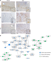
Figure 1. Hepatocytes are fetal like in patients with SAH.
(A) IHC for C/EBPα, YAP, and TAZ in explanted liver tissues of patients with SAH and healthy human liver. Representative images are shown. Scale bars: 100 μm. Original magnification, ×100; ×400 (higher magnification images). (B) An enrichment map consisted of KEGG pathways and annotations of tissue expression that are involved by significantly upregulated (green circles) or downregulated (blue circles) genes in liver tissues of SAH patients compared with healthy controls (n = 5 each) by RNA-Seq analysis. The node size is proportionate to the number of genes for each category, and thickness of edges is proportionate to the number of shared genes between categories.
Figure 2
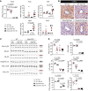
Figure 2. ESRP2 expression and ESRP2-mediated adult RNA-splicing program are downregulated in livers of patients with SAH.
(A) The log2 fold changes of ESRP2 and direct YAP/TAZ target genes (CYR61, CTGF, PTGS2) assessed by RNA-Seq in liver explants from SAH patients relative to healthy controls (n = 5 each). (B) Quantitative reverse transcription PCR (qRT-PCR) analysis for ESRP2 and YAP/TAZ target genes (AREG, CYR61, CTGF, PTGS2) in human SAH livers and healthy controls. (C) Immunoblots for ESRP2 normalized to GAPDH as a loading control in lysates of liver tissues from SAH patients and healthy controls. (D) IHC for ESRP2 in explanted liver tissues of SAH patients and healthy human liver. (E) Alternative splicing of ESRP2 target mRNAs including CSNK1D, NF2, SLK, and FLNB between liver tissues of SAH patients and healthy controls. Upper bands include the exon (red part), while the lower bands skip the exon. Exon lengths are shown beside the gene name, and the differences in PSI values are shown as mean ± SEM below each image. (FH) qRT-PCR analysis (F), immunoblot (G), and IHC (H) for IGF2BP3 in the livers of SAH patients and healthy controls. qRT-PCR results are graphed as dot plots (SAH, black squares; healthy controls, white circles) showing mean ± SEM (red bars) (n = 5 individuals/group). See full, unedited blots for C and G and full, unedited gels for E in the supplemental material. Statistical analysis was performed by using 2-tailed Student’s t test between 2 groups (n = 5 individuals/group, P values are specified otherwise, **P < 0.001). Representative images are shown for IHC. Scale bars: 100 μm (original magnification, ×100), 20 μm (original magnification, ×400).
Figure 3
Figure 3. Fetal RNA splicing occurs as increase of alcohol exposure and liver damage in mouse model of AH.
The Pearson’s r correlation analysis between the PSI of Csnk1d, Nf2, Slk, or Kras in liver tissues and the levels of blood alcohol, total histologic score, or inflammation of control (Ctrl) or AH mice of the Tsukamoto-French model (r, correlation coefficient). Results are graphed as dot plots (AH, black squares; controls, white circles). MNC, mononuclear cell; PMN, polymorphonuclear neutrophil.
Figure 4
Figure 4. Adult hepatocytes are reprogrammed to fetal-like cells in proinflammatory cytokine-dependent spheroid culture systems.
(A) qRT-PCR for Esrp2 and YAP/TAZ target genes (Areg and Ptgs2) in freshly isolated primary mouse hepatocytes (pHEP) and primary hepatocytes cultured under spheroid-forming system for 18 days continuously treated with TNF-α, IL-1β, or TNF-α + IL-1β. Results are graphed as box and whiskers plots (min to max), and means are indicated as plus signs. (B and C) Alternative splicing of Nf2, Slk, Flnb, and Kras in primary hepatocytes as specified. The representative gel images are shown from at least 3 replicates (B). See full, unedited gels in the supplemental material. Results are graphed as box and whiskers plots (min to max), and means are indicated as plus signs (C). (D) Images of hepatocyte spheroids stained for YAP (left, red) and TAZ (right, red) visualized by whole-mount immunofluorescence. Nuclear counterstaining was done by DAPI (blue). Scale bars: 50 μm. (E) scRNA-Seq analysis for Esrp2, Areg, and Ptgs2 in primary hepatocytes (PRIMARY) and hepatocyte spheroids under TNF-α–mediated expansion medium (EXP) or TNF-α–withdrawn induction medium (IND) as described in a previous publication (38).
Figure 5
Figure 5. Fetal reprogramming of hepatocytes is mediated by ESRP2-regulated RNA-splicing program.
(A) qRT-PCR for Esrp2, Areg, and Ptgs2 in pHEP and TNF-α–treated hepatocyte spheroids either transduced with adenoviruses expressing GFP (TNF-α + AdGFP) or mouse ESRP2 (TNF-α + AdESRP2). Results are graphed as box and whiskers plots (min to max), and means are indicated as plus signs. (B and C) Alternative splicing of Nf2, Slk, Flnb, and Kras in primary hepatocytes as specified; pHEP samples and the band images of pHEP are the same as those used for Figure 4B (B). See full, unedited gels in the supplemental material. Results are graphed as box and whiskers plots (min to max), and means are indicated as plus signs (C). All statistical analyses were performed by 1-way ANOVA with Tukey’s corrections (n = 3 biological replicates/group).
Figure 6
Figure 6. Fetal reprogrammed hepatocytes lose adult liver-specific functions.
(A) A schematic of ammonia (NH4+) detoxification pathway by hepatocytes in the liver. Glu, glutamate; Gln, glutamine; CP, carbamoyl phosphate. (B) qRT-PCR analysis for Cps1 and Glul in freshly isolated primary mouse hepatocytes and primary hepatocytes cultured under spheroid-forming system for 18 days continuously treated with TNF-α, IL-1β, or TNF-α + IL-1β. (C) The levels of urea, glutamine, and ammonia in conditioned medium of primary hepatocytes as specified. Results are graphed as box and whiskers plots (min to max), and means are indicated as plus signs. Statistical analysis was performed by 1-way ANOVA with Tukey’s corrections (n = 3 biological replicates/group). (D) scRNA-seq analysis for Cps1 and Glul in primary hepatocytes and hepatocyte spheroids under TNF-α–mediated expansion medium or TNF-α–withdrawn induction medium, as described in a previous publication (38). (E) qRT-PCR analysis for CPS1 and GLUL in human livers with SAH and healthy controls. Results are graphed as dot plots (SAH, black squares; healthy controls, white circles) showing mean ± SEM (red bars) (n = 5 individuals/group). Statistical analysis was performed by using 2-tailed Student’s t test between 2 groups. (F) qRT-PCR analysis for Cps1 and Glul in mouse livers from WT and Esrp2-KO mice at baseline. Results are graphed as dot plots (Esrp2-KO, black circles; WT, white circles) showing mean ± SEM (red bars) (n = 3–5 individuals/group). Statistical analysis was performed by using 2-tailed Student’s t test between 2 groups. (G) The serum levels of ammonia, urea, and albumin (ALB) in healthy WT and Esrp2-KO mice. Mean ± SEM results are graphed, and statistical analysis was performed by using 2-tailed Student’s t test between 2 groups (n = 3–5 mice/group).
Figure 7
Figure 7. Loss of ESRP2 promotes adult-to-fetal switch in Gao binge model.
(A) qRT-PCR analysis for Esrp2 and YAP/TAZ target genes (Areg, Fgf1, Itgb2, and Birc5) in WT and Esrp2-KO mice exposed to 10 days feeding and 1 binge drinking of ethanol (10d + 1B) and pair-fed mice. (B) IHC for ESRP2 in liver sections from these mice. Scale bars: 100 μm. (CD) Alternative splicing for HK mRNAs and EMT-related genes (Slk, Arhgef10l, Flnb, and Kras) in WT and Esrp2-KO Gao binge mouse models (C). See full, unedited gels in the supplemental material. All results are graphed as dot plots (WT pair, white circles; WT 10d + 1B, white squares; Esrp2-KO pair, black circles; Esrp2-KO 10d + 1B, black squares) showing mean ± SEM (red bars) (n = 3–7 individuals/group) (D). All statistical analyses were performed by 2-way ANOVA with Tukey’s corrections.
Figure 8
Figure 8. ESRP2-depleted hepatocytes become proliferative ductal-like cells after alcohol exposure.
IHC for CCND1 and SOX9 in liver sections of WT and Esrp2-KO mice with or without alcohol exposure. The numbers of CCND1+ hepatocytes (Hep) and SOX9+ liver cells (hepatocytes [Hep]; ductal cells [Duct]) were counted in more than 10 randomly selected ×100 or ×400 fields per section, respectively. Scale bars: 100 μm. Results are graphed as dot plots (WT pair, white circles; WT 10d + 1B, white squares; Esrp2-KO pair, black circles; Esrp2-KO 10d + 1B, black squares) showing mean ± SEM (red bars) (n = 3–7 individuals/group). Statistical analyses were performed by 2-way ANOVA with Tukey’s corrections.
Figure 9
Figure 9. Depleting ESRP2 exacerbates alcohol-induced steatohepatitis in mice.
(A) The serum levels of ALT in WT and Esrp2-KO mice treated with alcohol as per the Gao binge model. Results are graphed as dot plots (WT pair: white circles with white bar; WT 10d + 1B, white squares with white bar; Esrp2-KO pair, black circles with gray bar; Esrp2-KO 10d + 1B, black squares with gray bar) showing mean ± SEM (red bars) (n = 3–7 individuals/group). (B) Hepatic TG content in WT and Esrp2-KO mice with or without alcohol exposure. (C) H&E and ORO staining in liver sections of these mice. Yellow bracket indicates ductular reaction. Scale bars: 20 μm. (DF) qRT-PCR analysis for Pparγ (D), inflammatory cytokines (Tnfα and Il1β) and immune cell markers (Cd68 and Ly6G) (E), and fibrogenic markers (Col1α1 and Snail) (F) in WT and Esrp2-KO mice exposed to alcohol or pair-fed control diet. All results are graphed as dot plots (WT pair, white circles; WT 10d + 1B, white squares; Esrp2-KO pair, black circles; Esrp2-KO 10d + 1B, black squares) showing mean ± SEM (red bars) (n = 3–7 individuals/group). All statistical analyses were performed by 2-way ANOVA with Tukey’s corrections.
Figure 10
Figure 10. Model for liver failure in severe acute alcoholic hepatitis (SAH).
Inflammatory cytokines, such as TNF-α and IL-1β, suppress ESRP2, an adult RNA-splicing factor. Loss of ESRP2 permits fetal RNA-splicing variants of HKs to accumulate. Because fetal HKs are relatively inactive, phosphorylation and clearance of their targets, YAP/TAZ, are impaired, allowing these fetal transcription factors to accumulate and reactivate fetal gene expression programs. Thus, the surviving adult hepatocytes are reprogrammed to become more fetal like and regenerative, but less mature. This provokes loss of critical adult hepatocyte functions, including ammonia detoxification and clotting factor biosynthesis, and causes the clinical features of liver failure.

Similar articles

Cited by

References

    1. Louvet A, Mathurin P. Alcoholic liver disease: mechanisms of injury and targeted treatment. Nat Rev Gastroenterol Hepatol. 2015;12(4):231–242. doi: 10.1038/nrgastro.2015.35. - DOI - PubMed
    1. Yu CH, Xu CF, Ye H, Li L, Li YM. Early mortality of alcoholic hepatitis: a review of data from placebo-controlled clinical trials. World J Gastroenterol. 2010;16(19):2435–2439. doi: 10.3748/wjg.v16.i19.2435. - DOI - PMC - PubMed
    1. Orntoft NW, Sandahl TD, Jepsen P, Vilstrup H. Short-term and long-term causes of death in patients with alcoholic hepatitis in Denmark. Clin Gastroenterol Hepatol. 2014;12(10):1739–44.e1. doi: 10.1016/j.cgh.2014.04.020. - DOI - PubMed
    1. Altamirano J, et al. Acute kidney injury is an early predictor of mortality for patients with alcoholic hepatitis. Clin Gastroenterol Hepatol. 2012;10(1):65–71.e3. doi: 10.1016/j.cgh.2011.09.011. - DOI - PubMed
    1. Jung Y, et al. Accumulation of hedgehog-responsive progenitors parallels alcoholic liver disease severity in mice and humans. Gastroenterology. 2008;134(5):1532–1543. doi: 10.1053/j.gastro.2008.02.022. - DOI - PMC - PubMed

Publication types