

Exosomes secreted by prostate cancer cells under hypoxia promote matrix metalloproteinases activity at pre-metastatic niches
- PMID: 31943365
- PMCID: PMC7189745
- DOI: 10.1002/mc.23157
Exosomes secreted by prostate cancer cells under hypoxia promote matrix metalloproteinases activity at pre-metastatic niches
Abstract
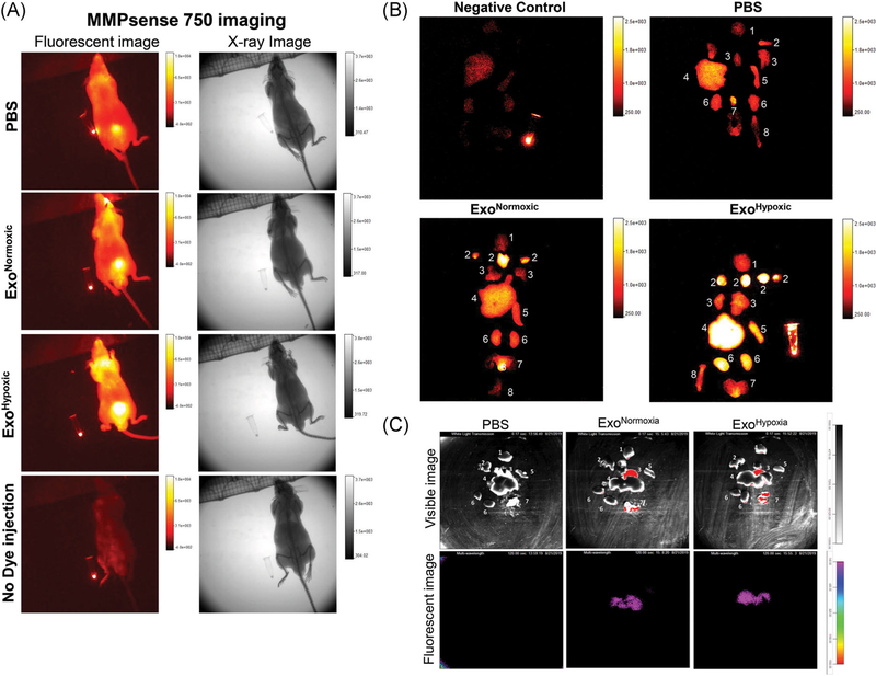
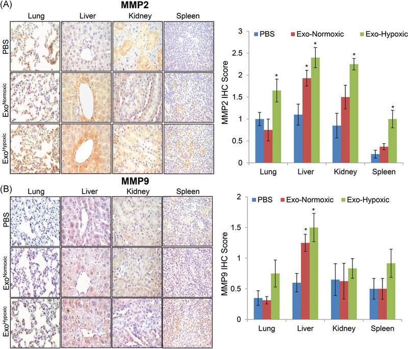
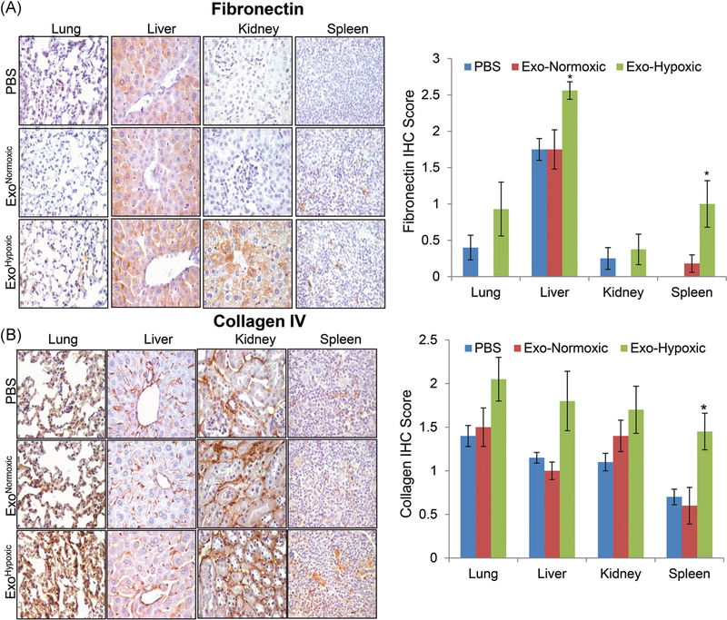
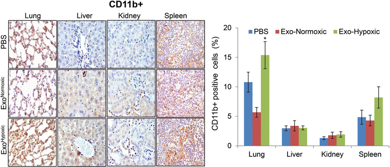
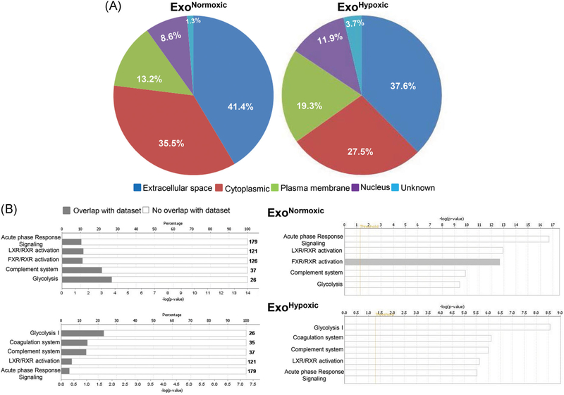
Approximately, 30 000 men die from prostate cancer (PCa) every year in the United States, mainly due to the metastasis. Thus, the key events associated with PCa metastasis are under rigorous investigation, with recent studies showing that preparation of pre-metastatic niches (PMN) in distant organs is an important step. However, the molecular basis for PMN preparation is still unclear. Hypoxia in primary tumors promotes aggressiveness; however, its precise role in metastasis is not clear. We recently reported that exosomes secreted by PCa cells under hypoxia promote stemness and invasiveness in naïve PCa cells; however, whether these extracellular vesicles also influence PMN remains unknown. In the present study, we isolated exosomes from human PCa PC3 cells under normoxic (21% O2 , exosomes secreted under normoxic condition [ExoNormoxic ]) and hypoxic (1% O2 , exosomes secreted under hypoxic condition [ExoHypoxic ]) conditions, and characterized their effect (10 µg exosomes, intraperitoneal (IP) treatment every 48 hours for 4 weeks) on key biomarkers associated with PMN in nude mice. Whole animal fluorescence imaging showed that ExoHypoxic treatment promotes matrix metalloproteinases (MMPs) activity in several putative metastatic sites. Histological studies confirmed that ExoHypoxic treatment enhanced the level of MMP2, MMP9, and extracellular matrix proteins (fibronectin and collagen) as well as increased the number of CD11b+ cells at selective PMN sites. Furthermore, proteomic profiling of exosomes by liquid chromatography/mass spectrometry identified cargo proteins in ExoNormoxic and ExoHypoxic as well as distinct canonical pathways targeted by them. These results suggest that exosomes secreted by PCa cells under hypoxia plausibly remodel distant PMN, and thus, could be a potential target to control metastatic PCa.
Keywords: exosomes; hypoxia; matrix metalloproteinases; pre-metastatic niches; proteomics.
© 2020 Wiley Periodicals, Inc.
Conflict of interest statement
CONFLICT OF INTERESTS
The authors declare that there are no conflict of interests.
Figures
Similar articles
-
Exosomes secreted under hypoxia enhance invasiveness and stemness of prostate cancer cells by targeting adherens junction molecules.Mol Carcinog. 2015 Jul;54(7):554-65. doi: 10.1002/mc.22124. Epub 2013 Dec 17. Mol Carcinog. 2015. PMID: 24347249 Free PMC article.
-
Exosomal microRNA profiling to identify hypoxia-related biomarkers in prostate cancer.Oncotarget. 2018 Feb 17;9(17):13894-13910. doi: 10.18632/oncotarget.24532. eCollection 2018 Mar 2. Oncotarget. 2018. PMID: 29568403 Free PMC article.
-
Role of Exosomes in Prostate Cancer Metastasis.Int J Mol Sci. 2021 Mar 29;22(7):3528. doi: 10.3390/ijms22073528. Int J Mol Sci. 2021. PMID: 33805398 Free PMC article. Review.
-
Exosomes in hypoxia-induced remodeling of the tumor microenvironment.Cancer Lett. 2020 Sep 28;488:1-8. doi: 10.1016/j.canlet.2020.05.018. Epub 2020 May 27. Cancer Lett. 2020. PMID: 32473240 Review.
-
Hypoxia-induced exosome secretion promotes survival of African-American and Caucasian prostate cancer cells.Sci Rep. 2018 Mar 1;8(1):3853. doi: 10.1038/s41598-018-22068-4. Sci Rep. 2018. PMID: 29497081 Free PMC article.
Cited by
-
Stress-induced extracellular vesicles: insight into their altered proteomic composition and probable physiological role in cancer.Mol Cell Biochem. 2024 Sep 20. doi: 10.1007/s11010-024-05121-x. Online ahead of print. Mol Cell Biochem. 2024. PMID: 39302488 Review.
-
Extracellular vesicle and lipoprotein diagnostics (ExoLP-Dx) with membrane sensor: A robust microfluidic platform to overcome heterogeneity.Biomicrofluidics. 2024 Jul 24;18(4):041301. doi: 10.1063/5.0218986. eCollection 2024 Jul. Biomicrofluidics. 2024. PMID: 39056024 Free PMC article.
-
Extracellular vesicles and particles impact the systemic landscape of cancer.EMBO J. 2022 Sep 15;41(18):e109288. doi: 10.15252/embj.2021109288. Epub 2022 Sep 2. EMBO J. 2022. PMID: 36052513 Free PMC article. Review.
-
Role of the Hypoxic-Secretome in Seed and Soil Metastatic Preparation.Cancers (Basel). 2022 Nov 30;14(23):5930. doi: 10.3390/cancers14235930. Cancers (Basel). 2022. PMID: 36497411 Free PMC article. Review.
-
Immune determinants of the pre-metastatic niche.Cancer Cell. 2023 Mar 13;41(3):546-572. doi: 10.1016/j.ccell.2023.02.018. Cancer Cell. 2023. PMID: 36917952 Free PMC article. Review.
References
-
- Kingsley LA, Fournier PG, Chirgwin JM, Guise TA. Molecular biologyof bone metastasis. Mol Cancer Ther. 2007;6:2609–2617. - PubMed
-
- Siegel RL, Miller KD, Jemal A. Cancer statistics, 2019. CA Cancer J Clin. 2019;69:7–34. - PubMed
-
- Sceneay J, Smyth MJ, Moller A. The pre-metastatic niche: finding common ground. Cancer Metastasis Rev. 2013;32:449–464. - PubMed
-
- Hood JL, San RS, Wickline SA. Exosomes released by melanoma cells prepare sentinel lymph nodes for tumor metastasis. Cancer Res. 2011; 71:3792–3801. - PubMed
Publication types
MeSH terms
Substances
Grants and funding
LinkOut - more resources
Full Text Sources
Medical
Research Materials
Miscellaneous
