. 2020 Jun;380(3):435-448.
doi: 10.1007/s00441-019-03163-y. Epub 2020 Jan 13.

Interaction with ectopic cochlear crista sensory epithelium disrupts basal cochlear sensory epithelium development in Lmx1a mutant mice

Affiliations

Interaction with ectopic cochlear crista sensory epithelium disrupts basal cochlear sensory epithelium development in Lmx1a mutant mice

David H Nichols et al. Cell Tissue Res. 2020 Jun.

Abstract

The LIM homeodomain transcription factor Lmx1a shows a dynamic expression in the developing mouse ear that stabilizes in the non-sensory epithelium. Previous work showed that Lmx1a functional null mutants have an additional sensory hair cell patch in the posterior wall of a cochlear duct and have a mix of vestibular and cochlear hair cells in the basal cochlear sensory epithelium. In E13.5 mutants, Sox2-expressing posterior canal crista is continuous with an ectopic "crista sensory epithelium" located in the outer spiral sulcus of the basal cochlear duct. The medial margin of cochlear crista is in contact with the adjacent Sox2-expressing basal cochlear sensory epithelium. By E17.5, this contact has been interrupted by the formation of an intervening non-sensory epithelium, and Atoh1 is expressed in the hair cells of both the cochlear crista and the basal cochlear sensory epithelium. Where cochlear crista was formerly associated with the basal cochlear sensory epithelium, the basal cochlear sensory epithelium lacks an outer hair cell band, and gaps are present in its associated Bmp4 expression. Further apically, where cochlear crista was never present, the cochlear sensory epithelium forms a poorly ordered but complete organ of Corti. We propose that the core prosensory posterior crista is enlarged in the mutant when the absence of Lmx1a expression allows JAG1-NOTCH signaling to propagate into the adjacent epithelium and down the posterior wall of the cochlear duct. We suggest that the cochlear crista propagates in the mutant outer spiral sulcus because it expresses Lmo4 in the absence of Lmx1a.

Keywords: Cochlea; Crista; Ear; Lmx1a; Mouse.


Conflict of interest statement

Conflict of interest The authors declare that they have no conflict(s) of interest.

Figures

Fig. 1
Fig. 1
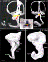
Bmp4 and Sox2 are expressed in the cochlear crista. (a, a’) Whole mounts of E11.0 wild type (a) and mutant Lmx1a null mice (a’) ears stained for Bmp4 expression. Blue asterisks mark posterior crista and red asterisks cochlear Bmp4 expression. Bmp4 expression is absent from the utriculo-saccular constriction of the wild-type ear (black arrow in a), but present in the mutant (blue arrow in a’). (b, b’) Whole mounts of E13.5 wild-type (b) and mutant (b’) ears stained for Bmp4 expression. Black arrow marks the utriculo-saccular constriction in b. Blue arrow mark the cochlear crista, and the black arrow the basal cochlear sensory epithelium in b’. Black (b) and red (b’) asterisks marks the apical tip of the cochlea. (c, c’) Horizontal sections across the cochlear ducts of wild-type (c) and Lmx1a null mutant (c’) ears stained for Bmp4 expression. To the right of the blue arrow crista sensory epithelium (cochlear crista) is stained in the outer spiral sulcus; red arrows mark cochlear sensory epithelium. (d, d’) Whole mounts of E13.5 wild-type (d) and mutant (d’) ears stained for Sox2 expression. Blue asterisks mark the posterior crista, and red asterisks mark the apical tip of the cochlear sensory epithelium. Blue arrow in d’ lies over the cochlear crista. (e, e’) Horizontal sections across the cochlear ducts of E13.5 wild-type (e) and mutant (e’) ears stained for Sox2 expression. Blue arrow marks crista sensory epithelium (cochlear crista) in the outer spiral sulcus, red arrows mark cochlear sensory epithelium. All bars are 100 μm
Fig. 2
Fig. 2
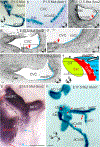
The cochlear crista differentiates Atoh1-expressing hair cells. (a, a’) Whole mounts of E17.5 ears stained for Atoh1 expression in Lmx1a null mice. Note that the enlarged medial arm of the posterior crista (medPC) includes an Atoh1-expressing extension of the cochlear crista (red curved arrow) along its ventral margin. This extension is not present on the ventral margin of the smaller lateral arm (latPC). Boxed area in a’ marks a region in which the cochlear crista ends in scattered cells and the previously associated basal CoSE retains patches of outer hair cells (OHC). (b, b’) Adjacent phase contrast (b) and toluidine blue stained (b’) sections in the plane marked B in a’. Red arrows mark basal cochlear sensory epithelium and blue arrows, cochlear crista. Note cupula-like matrix beneath the blue arrows.(c, c’) Selected phase contrast serial sections across the length of the posterior crista (c) and the width of the cochlear crista (c’). Black arrows mark cupular matrix, blue arrows, hair cells, and yellow arrows mark a supporting cell layer. Abbreviations: bCoSE, basal cochlear sensory epithelium; coCr, cochlear crista; latPC, lateral arm of the posterior crista; medPC, medial arm of the posterior crista; OHC, outer hair cell patch. Bars are 100 μm (a, a’), and 10 μm (b, b’, c, c’)
Fig. 3
Fig. 3
Utriculo-saccular and cochleo-saccular constrictions fail to form in the mutant. (a) Lateral view of the lumen of an E12.5 wild-type ear. Yellow and red bands mark the sites of the utriculo-saccular and cochleo-saccular constrictions respectively. (b) Lateral view of an E14.5 wild-type ear. Note that the constrictions now isolate the base of the cochlear duct from the utricle and saccule (Utr, Sac a/b inset). Black ovals mark the approximate site of a papilla neglecta. Note the segregation of the saccule is fully distinct. (a/b inset) Flat mount of an E14.5 wild-type ear stained for Lmx1a expression. Yellow and red lines mark strong expression in the planes of the utriculo-saccular and cochleo-saccular constrictions, respectively. (c) Lateral view of the lumen of an E14.5 mutant. An arrow marks the cochlear crista, and the site of the transition from basal to apical cochlear sensory epithelia. (d) Lateral view of an E16.5 mutant ear showing an enlarged but unchanged shape. Abbreviations: AC/HC, anterior + horizontal crista; coCr, cochlear crista; CVC, common vestibular cavity; Sac, sacccule; PC, posterior crista; Utr, utricle. All bars are 100 μm
Fig. 4
Fig. 4
Mutant cochlear and crista sensory epithelia make contact. (a, b) P1.5 Whole mount (a) and section (b) stained for Atoh1 expression in hair cells. Note the crista is parted from the basal cochlear sensory epithelium (bCoSE) at this age and is not included. Black line in (a) marks the plane of section in b where the basal CoSE approaches the saccular macula (Sac) and joins it in an adjacent section (not shown). (c, d, d’,e) Selected horizontal serial sections cut from an E13.5 ear stained for Sox2 expression in planes indicated in f. Blue lines approximate crista sensory epithelium. Red asterisks mark the abneural edge of the basal cochlear sensory epithelium. Red arrows mark the region of contact between crista and cochlear sensory epithelia. The cochlear sensory epithelium is artifactually broken in (d). (d’) shows the boxed area in (d). In (e), the plane of section is dorsal to the cochlear sensory epithelium. (f) Diagram illustrating the association of the crista sensory epithelium with the basal cochlear sensory epithelium along the dashed red line. Here the cochlear sensory epithelium will lack an outer hair cell band and cochlear Bmp4 expression will be patchy. (g) Whole mount of an E16.5 posterior and cochlear crista stained for Sox2 expression. White arrows mark the proposed directions of crista propagation by lateral induction. Blue arrows mark distal patches of Sox2 expression. Note the broad and elongated medial arm of the posterior crista (medPC). Dotted black line marks the now separating zone of contact with the basal cochlear epithelium. (h) By E17.5, the cochlear crista and medial posterior cristae are separated from the basal cochlear sensory epithelium, but the former contact zone can still be recognized (double headed red arrows). Abbreviations: aCoSE, apical cochlear sensory epithelium; AC/HC, anterior and horizontal cristae; bCoSE, basal cochlear sensory epithelium; coCr, cochlear crista; CVC, common vestibular cavity; latPC, lateral arm of the posterior crista; medPC, medial arm of the posterior crista; Sac, saccular macula; Utr, utricle. Bars in a, g, and h are 100 μm. All others are 10 μm
Fig. 5
Fig. 5
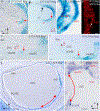
A Nog-expressing epithelium separates basal cochlear sensory epithelium from crista sensory epithelium. (a) Section across the cochlear duct of an E13.5 wild-type ear stained for Nog expression (red arrow). Red asterisk marks the site of the cochlear sensory epithelium. (b) Similar section across an E17.5 wild-type duct stained for Nog expression in the outer spiral sulcus. Red asterisk marks the site of the cochlear sensory epithelium. A black arrow marks the edge of the stria vascularis identified by black melanocytes in its forming intermediate layer. A red arrow marks the outer spiral sulcus. (c) Atoh1 stained cochlear duct from the E17.5 mutant ear in Fig. 2a’. Note several hair cells (e.g., red arrow) are located in the epithelium separating the cochlear sensory epithelium from the crista sensory epithelium. (d) Flat mount of the basal CoSE of a P1.5 mutant immunostained for MYOSIN VIIa to label maturing hair cells. White arrow marks the abneural margin of the basal cochlear sensory epithelium. Note scattered hair cells to the right of the margin. (e) Horizontal section across the posterior crista and basal cochlear sensory epithelium of an E12.5 mutant ear. The medial arm of the crista (blue asterisk) and cochlear sensory epithelium (red asterisk) are in direct planar contact (red arrow). Nog expression stains the interface between the two. (f) similar section from an E13.5 ear. The contacting edge of the crista is thinning. (g) Horizontal section across the common vestibular cavity (CVC) of an E17.5 mutant ear stained for Nog expression. A red asterisk marks the abneural (lateral) margin of the basal cochlear sensory epithelium, and a blue asterisk marks the facing margin of the cochlear crista. The double headed arrow parallels the Nog stained epithelium separating the cochlear sensory epithelium from the crista sensory epithelium. (h) Vertical section across the common vestibular cavity of an E17.5 mutant ear stained for Sox2 expression. The double-headed red arrow parallels the unstained non-sensory epithelium separating the cochlear sensory epithelium from the crista. Abbreviations: bCoSE, basal cochlear sensory epithelium; coCr, cochlear crista; CVC, common vestibular cavity; latPC, lateral arm of the posterior crista; medPC, medial arm of the posterior crista; Sac, saccular macula. Bars in c, d are 100 μm. All others are 10 μm
Fig. 6
Fig. 6
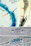
The mutant basal cochlear sensory epithelium is altered. (a, b) Whole mounts of E17.5 (a) and E18.5 (b) basal-apical cochlear sensory epithelial junctions. Red arrows mark the inner hair cell bands, and black arrows the blunt end terminations of the outer hair cell bands at the junctions. The black line in b suggests unbroken cochlear Bmp4 expression in the apical cochlear sensory epithelium (solid line) and patchy expression in the basal cochlear sensory epithelium (dashed line). (c, d) Sections across the basal cochlear sensory epithelium of an E16.5 mutant ear stained for Sox2 expression (c, bright field) and an E18.5 mutant ear stained for Atoh1 (d, phase contrast) expression. Black arrows bracket hair cell layers and white arrows, supporting cell layers. In d note that the inner hair cell row of the basal CoSE is eight hair cells wide and the tunnel of Corti is missing. Abbreviations: aCoSE, apical cochlear sensory epithelium; bCoSE, basal cochlear sensory epithelium; IHC, inner hair cells; OHC, outer hair cells. All bars are 25 μm
Fig. 7
Fig. 7
Basal cochlear Bmp4 expression is patchy in the mutant. (a) Flat mount of the posterior and medial walls of the common vestibular cavity in an E13.5 mutant ear stained for Bmp4 expression. The junction of the basal CoSE with the saccular macula is out of view in the direction of the white arrow here and in b, c. Asterisks mark patches of Bmp4 expression here and in b, c. (b) Similar view of an E15.5 ear. Black arrows here and in c mark the transition from unbroken expression apically to patchy expression basally. (c) Similar view of a P4.5 ear. Abbreviation: aCoSE, apical cochlear sensory epithelium; bCoSE, basal cochlear sensory epithelium. All bars are 50 μm
Fig. 8
Fig. 8
Summary Figure. (a) In the E14.5 wild type, cochlear sensory epithelium forms in an cochlear duct separated from the posterior crista sensory epithelium and saccular macula. (b) In the E16.5 mutant the absence of constrictions has permitted an enlarged medial arm of the posterior crista and an ectopic cochlear crista to interact with the basal cochlear sensory epithelium, eliminating its outer hair cell band and altering its inner hair cell band. At this age these have now separated, but remain adjacent. Note this is a view of the lateral side of the otic lumen, but the elongated arm of the posterior crista and the basal cochlear sensory epithelium are actually facing its medial side. Abbreviations: AC, anterior crista; aCoSE, apical cochlear sensory epithelium; bCoSE, basal cochlear sensory epithelium; CoSE, cochlear sensory epithelium; coCr, cochlear crista; HC, horizontal crista; PC, posterior crista; S, saccule; U utricle

Similar articles

Cited by

References

    1. Agulnick AD, Taira M, Breen JJ, Tanaka T, Dawid IB, Westphal H, (1996) Interactions of the LIM-domain-binding factor Ldbl with LIM homeodomain proteins. Nature 384 (6606):270–272 - PubMed
    1. Bergstrom DE, Gagnon LH, Eicher EM (1999) Genetic and physical mapping of the dreher locus on mouse chromosome 1. Genomics 59:291–299 - PubMed
    1. Bermingham NA, Hassan BA, Wang VY, Fernandez M, Banfi S, Bellen HJ, Fritzsch B, Zoghbi HY (2001) Proprioceptor pathway development is dependent on Math1. Neuron 30:411–422 - PubMed
    1. Brichta AM, Goldberg JM (1998) The papilla neglecta of turtles: a detector of head rotations with unique sensory coding properties. J Neurosci 18:4314–4324 - PMC - PubMed
    1. Burzynski GM, Reed X, Maragh S, Matsui T, McCallion AS (2013) Integration of genomic and functional approaches reveals enhancers at LMX1A and LMX1B. Mol Gen Genomics 288:579–589 - PMC - PubMed

MeSH terms

LinkOut - more resources