Review
. 2020 Jun 1;12(6):a036202.
doi: 10.1101/cshperspect.a036202.

Tracing the Dynamics of Stem Cell Fate

Affiliations
Review

Tracing the Dynamics of Stem Cell Fate

Lemonia Chatzeli et al. Cold Spring Harb Perspect Biol. .

Abstract

The mechanisms that regulate the balance between stem cell duplication and differentiation in adult tissues remain in debate. Using a combination of genetic lineage tracing and marker-based assays, the quantitative statistical analysis of clone size and cell composition has provided insights into the patterns of stem cell fate across a variety of tissue types and organisms. These studies have emphasized the role of niche factors and environmental cues in promoting stem cell competence, fate priming, and stochastic renewal programs. At the same time, evidence for injury-induced "cellular reprogramming" has revealed the remarkable flexibility of cell states, allowing progenitors to reacquire self-renewal potential during regeneration. Together, these findings have questioned the nature of stem cell identity and function. Here, focusing on a range of canonical tissue types, we review how quantitative modeling-based approaches have uncovered conserved patterns of stem cell fate and provided new insights into the mechanisms that regulate self-renewal.


Figures

Figure 1.
Figure 1.
Germline maintenance in the Drosophila and mouse testis. (A) Schematic of the Drosophila testis showing germline stem cells (GSCs) anchored by adherens junctions to somatic hub cells and ensheathed by cyst stem cells. The hub functions as a closed niche, supplying factors that maintain stem cell competence. (B) During GSC division, spindle orientation by the hub positions daughter cells away from the niche, leaving cells primed for differentiation and loss. (C) Schematic of the mouse testis showing spermatogonia roaming freely on the basement membrane of the seminiferous tubules. In homeostasis, spermatogonia expand through serial rounds of incomplete mitotic division before entry into meiosis when they translocate across tight junctions toward the lumen. GSCs are contained within a subpopulation of undifferentiated cells and are characterized by heterogeneous expression of markers. During the periodic seminiferous cycle, cells positive for the expression of retinoic acid receptor (RAR)γ are transferred by retinoic acid signaling into a differentiated (Kit+) cell compartment. With GSCs sharing the basement membrane with their differentiating progeny, the mouse testis provides an example of an open or facultative niche. (D) Patches of clonally labeled cells in the seminiferous tubules induced by a Ngn3 promoter at 3 months postinduction. Scale bars, 0.2 mm. (EG) Schematic of the neutral drift model (E) showing stem cell loss through differentiation compensated by the duplication of neighbors leading to continual clonal loss (F) compensated by expansion of neighbors (G), as depicted in F (inset). Lines show prediction of neutral drift model. (H) When plotted against the rescaled patch length, the clone size distribution shows a collapse onto the scaling dependence predicted by the neutral drift model (shown as a dashed line). For further details, see Klein et al. (2010). (A and B are from Amoyel and Bach 2015; adapted, with permission, from Elsevier © 2015; C is from Kitadate et al. 2019; adapted under the terms of the Creative Commons Attribution License [CC BY]; DH are from Klein et al. 2010; adapted, with permission, from Elsevier © 2010.)
Figure 2.
Figure 2.
Competition for fate determinants—a mechanism of population asymmetric self-renewal. (A) Model of the feedback competition mechanism showing the mutual regulation of the stem cell density and abundance of fate determinant (fibroblast growth factors [FGFs]). By correlating the inhibition of differentiation licensing factors (retinoic acid receptor [RAR]γ) with the reception and consumption of niche factors (FGFs) secreted by lymphatic endothelial cells, germline stem cells (GSCs) are able to sense their density and adjust their fate bias in response. (B) Above threshold, steady-state stem cell density is predicted to rise linearly with FGF concentration. (C) Phase portrait depicted the corresponding dynamics of stem cell density and FGF concentration. The system shows two fixed points: a homeostatic state (green dot) and a loss state (red dot). For the given parameter set, only the homeostatic state is stable, as all trajectories obtained by following the arrows converge toward this state. (D) When perturbed from homeostasis, the feedback model predicts an oscillatory phase of recovery back to steady state (as indicated in B). (E) Consistently, measurements of the spermatogonial stem cell (SSC) density, as assessed by the expression of the marker growth factor receptor (GFR)α1, shows that the stem cell density scales linearly with the allele fraction of the FGF5. (F) Moreover, following depletion using a chemical agent (busulfan), the SSC density shows an oscillation phase of recovery. For further details, see Kitadate et al. (2019). (AF are from Kitadate et al. 2019; adapted under the terms of the Creative Commons Attribution License [CC BY].)
Figure 3.
Figure 3.
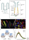
Maintenance of the mouse intestinal crypt. (A) The columnar epithelium of the small intestine is maintained by intestinal stem cells (ISCs), identified as crypt base columnar cells (CBCs), which localize around the base of glandular invaginations known as crypts. ISCs give rise to sublineage-restricted progenitors that differentiate into secretory and absorptive cells that, together, move in migration streams along the axis of the crypt and onto the villus, where they shed. Factors secreted by secretory (Paneth) cells as well as stromal cells provide a niche environment that maintain stem cell competence. As ISCs divide, some become displaced from the niche and enter into a differentiation program. (B) Lineage tracing using the multicolor R26R-Confetti reporter system induced at high (mosaic) labeling density at 8 wk (left) and 4 wk (right) postinduction. The left-hand image is a vertical section showing ribbons of lineage-labeled cells moving along the axis of the crypts (bottom) and onto the villi (top). The right-hand image is a horizontal section near the base of the crypt showing clusters of lineage-labeled cells expanding around the circumference of the crypt. (C) Following genetic labeling, ISC-derived clones undergo a process of neutral drift in which clones expand and contract around the crypt base circumference until the clone is lost or the crypt becomes monoclonally fixed. (D) Neutral drift dynamics of clone widths is evidenced by convergence of their size distribution onto statistical scaling behavior at intermediate times (see main text). This behavior masks a more refined organization in which ISCs positioned near the base of the crypt are biased toward duplication, whereas those at the niche border (defined by the range Lgr5 expression) are primed, but not committed, for differentiation (see main text). (A is based on data in Gehart and Clevers 2019; B is from Snippert et al. 2010; adapted, with permission, from Elsevier © 2010; C and D are from Lopez-Garcia et al. 2010; adapted, with permission, from The American Association for the Advancement of Science © 2010.)
Figure 4.
Figure 4.
Skin interfollicular epidermis, a squamous epithelial tissue. (A) The mouse skin interfollicular epidermis (IFE) comprises a stratified squamous epithelium interspersed with hair follicles, sweat glands, and sebaceous glands. All cell division in IFE takes place in the basal layer. Following commitment to terminal differentiation, cells delaminate from the basal layer and enter the suprabasal layers, where they mature into functional differentiated cell types before being shed from the skin surface. The mouse esophagus shows a similar organization as skin, but lacks most of the skin appendages. (B) Section through the basal layer of mouse esophagus showing typical clones induced using a ubiquitous promoter at a range of time points. Note that, despite expansion, clones remain roughly cohesive over time. (Cell nuclei marked by DAPI are blue. Scale bar, 10 μm.) In homeostasis, cells lost from the basal layer through differentiation are replenished by neighbors, leading to neutral drift dynamics of the clonal population. (CE) During this process, continual loss of basal clones (C) is compensated by a near-linear increase in the average size of surviving clones (D), whereas the size distribution (E) converges to the hallmark exponential scaling dependence. (A is from Jones and Simons 2008; adapted, with permission, from the authors who retained the copyright to reuse their own work; BE are from Doupé et al. 2012; adapted, with permission, from The American Association for the Advancement of Science © 2012.)
Figure 5.
Figure 5.
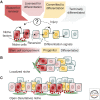
Niche-based model of stem cell regulation. (A) Schematic summarizing the regulation of stem cell density homeostasis based on the competition for niche factors. The reception of factors secreted from niche cells inhibits stem cell differentiation, leaving cells biased for renewal. When deprived of these factors, the up-regulation of differentiation licensing factors leaves stem cells primed, but not committed to differentiation. Reexposure to niche factors allows stem cells to reverse their fate bias. However, following exposure to secondary differentiation cues, released either by differentiating progeny or extrinsic signals, stem cells enter into a program that leaves them committed to differentiation. Following injury, progenitors may reprogram, reacquiring stem cell competence either directly or via some intermediate state. (B) In a discrete or localized niche, such as that found in the Drosophila germline or intestinal crypt, stem cells become spatially segregated from their differentiating progeny. (C) By contrast, in an open or facultative niche, such as that found in mouse testis, stem cells lie intermingled among their differentiating progenies.

Similar articles

Cited by

References

    1. Amoyel M, Bach EA. 2015. MT-nanotubes: lifelines for stem cells. Cell Stem Cell 17: 133–134. 10.1016/j.stem.2015.07.004 - DOI - PubMed
    1. Anderson PW. 1972. More is different. Science 177: 393–396. 10.1126/science.177.4047.393 - DOI - PubMed
    1. Ayyaz A, Kumar S, Sangiorgi B, Ghoshal B, Gosio J, Ouladan S, Fink M, Barutcu S, Trcka D, Shen J, et al. 2019. Single-cell transcriptomes of the regenerating intestine reveal a revival stem cell. Nature 569: 121–125. 10.1038/s41586-019-1154-y - DOI - PubMed
    1. Barker N, van Es JH, Kuipers J, Kujala P, van den Born M, Cozijnsen M, Haegebarth A, Korving J, Begthel H, Peters PJ, et al. 2007. Identification of stem cells in small intestine and colon by marker gene Lgr5. Nature 449: 1003–1007. 10.1038/nature06196 - DOI - PubMed
    1. Barrandon Y, Green H. 1987. Three clonal types of keratinocyte with different capacities for multiplication. Proc Natl Acad Sci 84: 2302–2306. 10.1073/pnas.84.8.2302 - DOI - PMC - PubMed

Publication types

LinkOut - more resources