

Porous bio-click microgel scaffolds control hMSC interactions and promote their secretory properties
- PMID: 31918222
- PMCID: PMC7047645
- DOI: 10.1016/j.biomaterials.2019.119725
Porous bio-click microgel scaffolds control hMSC interactions and promote their secretory properties
Abstract
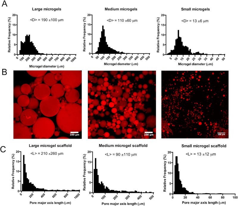
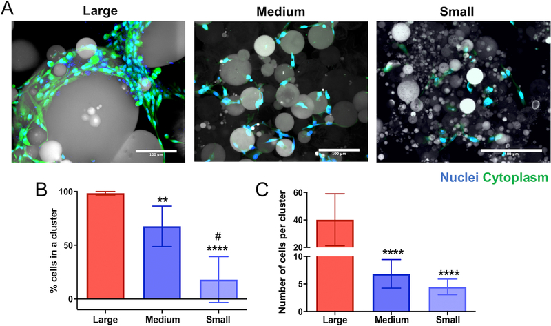
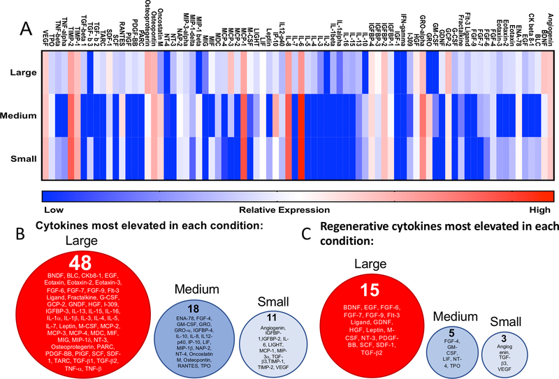
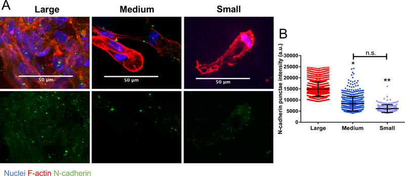
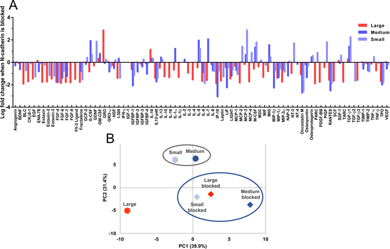
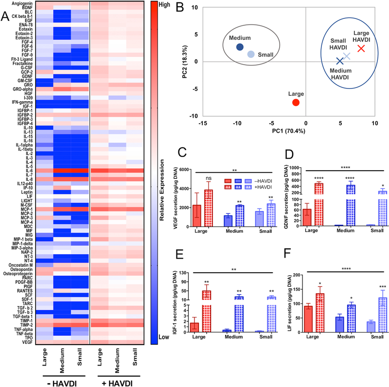
Human mesenchymal stem/stromal cells (hMSCs) are known to secrete numerous cytokines that signal to endogenous cells and aid in tissue regeneration. However, the role that biomaterial scaffolds can play in controlling hMSC secretory properties has been less explored. Here, microgels were co-assembled with hMSCs using three different microgel populations, with large (190 ± 100 μm), medium (110 ± 60 μm), and small (13 ± 6 μm) diameters, to create distinct porous environments that influenced hMSC clustering. Cells embedded in large diameter microgel networks resided in large clusters (~40 cells), compared to small clusters (~6 cells) observed in networks using medium diameter microgels and primarily single cells in small diameter microgel networks. Using a cytokine microarray, an overall increase in secretion was observed in scaffolds that promoted hMSC clustering, with over 60% of the measured cytokines most elevated in the large diameter microgel networks. N-cadherin interactions were identified as partially mediating these differences, so the microgel formulations were modified with an N-cadherin epitope, HAVDI, to mimic cell-cell interactions. Results revealed increased secretory properties for hMSCs in HAVDI functionalized scaffolds, even the non-clustered cells in small diameter microgel networks. Together, these results demonstrate opportunities for microgel-based scaffold systems for hMSC delivery and tailoring of porous materials properties to promote their secretory potential.
Keywords: Bio-click; HAVDI peptide; Mesenchymal stem/stromal cell; Microgels; Porous scaffolds; Secretome.
Copyright © 2019 Elsevier Ltd. All rights reserved.
Figures
Similar articles
-
Assembly of PEG Microgels into Porous Cell-Instructive 3D Scaffolds via Thiol-Ene Click Chemistry.Adv Healthc Mater. 2018 Jun;7(11):e1800160. doi: 10.1002/adhm.201800160. Epub 2018 Apr 16. Adv Healthc Mater. 2018. PMID: 29663702 Free PMC article.
-
Interplay between degradability and integrin signaling on mesenchymal stem cell function within poly(ethylene glycol) based microporous annealed particle hydrogels.Acta Biomater. 2020 Jan 1;101:227-236. doi: 10.1016/j.actbio.2019.11.009. Epub 2019 Nov 8. Acta Biomater. 2020. PMID: 31711899 Free PMC article.
-
Injectable degradable PVA microgels prepared by microfluidic technology for controlled osteogenic differentiation of mesenchymal stem cells.Acta Biomater. 2018 Sep 1;77:28-37. doi: 10.1016/j.actbio.2018.07.003. Epub 2018 Jul 5. Acta Biomater. 2018. PMID: 29981495
-
Supramolecular assemblies of multifunctional microgels for biomedical applications.J Mater Chem B. 2023 Jul 12;11(27):6265-6289. doi: 10.1039/d3tb00346a. J Mater Chem B. 2023. PMID: 37318041 Review.
-
Microgels: From responsive polymer colloids to biomaterials.Adv Colloid Interface Sci. 2009 Mar-Jun;147-148:251-62. doi: 10.1016/j.cis.2008.08.008. Epub 2008 Aug 24. Adv Colloid Interface Sci. 2009. PMID: 18809173 Review.
Cited by
-
Acoustofluidic Interfaces for the Mechanobiological Secretome of MSCs.Nat Commun. 2023 Nov 22;14(1):7639. doi: 10.1038/s41467-023-43239-6. Nat Commun. 2023. PMID: 37993431 Free PMC article.
-
Engineering the MSC Secretome: A Hydrogel Focused Approach.Adv Healthc Mater. 2021 Apr;10(7):e2001948. doi: 10.1002/adhm.202001948. Epub 2021 Feb 17. Adv Healthc Mater. 2021. PMID: 33594836 Free PMC article. Review.
-
Controlling Structure with Injectable Biomaterials to Better Mimic Tissue Heterogeneity and Anisotropy.Adv Healthc Mater. 2021 Jun;10(11):e2002221. doi: 10.1002/adhm.202002221. Epub 2021 May 5. Adv Healthc Mater. 2021. PMID: 33951341 Free PMC article. Review.
-
Phycocyanin-Loaded Alginate-Based Hydrogel Synthesis and Characterization.Mar Drugs. 2024 Sep 25;22(10):434. doi: 10.3390/md22100434. Mar Drugs. 2024. PMID: 39452842 Free PMC article.
-
Mesenchymal stem cell-inspired microgel scaffolds to control macrophage polarization.Bioeng Transl Med. 2021 Mar 21;6(2):e10217. doi: 10.1002/btm2.10217. eCollection 2021 May. Bioeng Transl Med. 2021. PMID: 34027099 Free PMC article.
References
-
- Trounson A. & Mcdonald C. Cell Stem Cell Stem Cell Therapies in Clinical Trials: Progress and Challenges. Stem Cell 17, 11–22 (2015). - PubMed
-
- da Silva Meirelles L, Fontes AM, Covas DT & Caplan AI Mechanisms involved in the therapeutic properties of mesenchymal stem cells. Cytokine Growth Factor Rev. 20, 419–427 (2009). - PubMed
-
- Engler AJ, Sen S, Sweeney HL & Discher DE Matrix Elasticity Directs Stem Cell Lineage Specification. Cell 126, 677–689 (2006). - PubMed
Publication types
MeSH terms
Substances
Grants and funding
LinkOut - more resources
Full Text Sources
Research Materials
