. 2020 Mar 1;318(3):C675-C694.
doi: 10.1152/ajpcell.00492.2019. Epub 2020 Jan 8.

The septate junction protein Mesh is required for epithelial morphogenesis, ion transport, and paracellular permeability in the Drosophila Malpighian tubule

Affiliations

The septate junction protein Mesh is required for epithelial morphogenesis, ion transport, and paracellular permeability in the Drosophila Malpighian tubule

Sima Jonusaite et al. Am J Physiol Cell Physiol. .

Abstract

Septate junctions (SJs) are occluding cell-cell junctions that have roles in paracellular permeability and barrier function in the epithelia of invertebrates. Arthropods have two types of SJs, pleated SJs and smooth SJs (sSJs). In Drosophila melanogaster, sSJs are found in the midgut and Malpighian tubules, but the functions of sSJs and their protein components in the tubule epithelium are unknown. Here we examined the role of the previously identified integral sSJ component, Mesh, in the Malpighian tubule. We genetically manipulated mesh specifically in the principal cells of the tubule at different life stages. Tubules of flies with developmental mesh knockdown revealed defects in epithelial architecture, sSJ molecular and structural organization, and lack of urine production in basal and kinin-stimulated conditions, resulting in edema and early adult lethality. Knockdown of mesh during adulthood did not disrupt tubule epithelial and sSJ integrity but decreased the transepithelial potential, diminished transepithelial fluid and ion transport, and decreased paracellular permeability to 4-kDa dextran. Drosophila kinin decreased transepithelial potential and increased chloride permeability, and it stimulated fluid secretion in both control and adult mesh knockdown tubules but had no effect on 4-kDa dextran flux. Together, these data indicate roles for Mesh in the developmental maturation of the Drosophila Malpighian tubule and in ion and macromolecular transport in the adult tubule.

Keywords: Drosophila; Malpighian tubule; Mesh; drosokinin; paracellular permeability; smooth septate junctions.


Conflict of interest statement

No conflicts of interest, financial or otherwise, are declared by the authors.

Figures

Fig. 1.
Fig. 1.
Diagram depicting mesh transcripts and target sites of the two different RNA interference lines used in this study. The introns are shown as solid lines, and the exons are shown as rectangular boxes. The arrows indicate the direction of transcription. The target regions of the RNAi lines are shown as dashed boxes for National Institute of Genetics (N) and Vienna Drosophila Resource Center (V). The Malpighian tubules express three splice variants of mesh as predicted by FlyAtlas 2 (40).
Fig. 2.
Fig. 2.
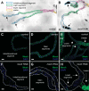
Developmental principal cell mesh knockdown in the Drosophila Malpighian tubule using mesh RNA interference (RNAi) line 12074-R1 results in morphological defects in adult tubules. A and B: bright-field images with depiction of the major segments of the anterior Malpighian tubules from a 1-day-old adult control female fly (w;c42-GAL4/+; A) and developmental principal cell mesh knockdown fly (w;UAS-meshRNAi/+;c42-GAL4/+; B). The mesh knockdown tubules appear dilated compared with control tubules and accumulate opaque concretions in the initial segments. CH: Mesh immunolocalizes to the smooth septate junctions (sSJs) between the epithelial cells throughout all segments in control tubules (C–E), but little to no Mesh immunoreactivity is observed in the initial/transitional, main, and lower segments and upper ureter of mesh knockdown tubules (F-H). Nuclei were counterstained blue with DAPI in CH. Ten samples per genotype were examined. Scale bars, 100 µm (A and B) and 50 μm (C–H). The mesh RNAi line 12074-R1 was used in subsequent figures unless otherwise noted.
Fig. 3.
Fig. 3.
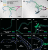
Developmental principal cell mesh knockdown in the Drosophila Malpighian tubule using mesh RNA interference (RNAi) line 6867 causes morphological defects in adult fly tubules. A and B: bright-field images with depiction of major morphological segments of the anterior Malpighian tubules from a 1-day-old adult control female fly [w;UAS-meshRNAi (II)/+; A] and developmental principal cell mesh knockdown fly [w;UAS-meshRNAi (II)/+;c42-GAL4/+; B]. The mesh knockdown tubules appear enlarged compared with control tubules, and their initial segments contain opaque concretions (A and B, arrows). CE: in control tubules, Mesh immunolocalizes to the smooth septate junctions between the epithelial cells throughout all segments. FH: in mesh knockdown tubules, only trace levels of Mesh immunofluorescence are observed in the initial/transitional, main, and lower segments. Nuclei were counterstained blue with DAPI in CH. Ten samples per genotype were examined. Scale bars, 100 µm (A and B) and 50 μm (CH).
Fig. 4.
Fig. 4.
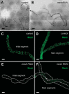
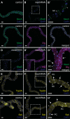
Developmental principal cell mesh knockdown in the larval Drosophila Malpighian tubule. A and B: bright-field images of the anterior Malpighian tubules from a nonwandering third-instar control larva (w;c42-GAL4/+; A) and developmental principal cell mesh knockdown larva (w;UAS-meshRNAi/+;c42-GAL4/+; B). The mesh knockdown tubules reveal some dilation compared with control tubules. C and D: Mesh immunolocalizes to the smooth septate junctions between the epithelial cells in control tubules. E and F: decreased Mesh immunofluorescence is observed in mesh knockdown tubules. Five samples per genotype were examined. Scale bars, 100 µm (A and B) and 50 μm (C–F).
Fig. 5.
Fig. 5.
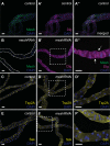
Developmental principal cell mesh knockdown in the Drosophila Malpighian tubule results in mislocalization of other smooth septate junction (sSJ) proteins. In the main segment of 1-day-old adult control female fly anterior tubules (w;c42-GAL4/+), Mesh colocalizes with Discs large (Dlg; A–A′′), tetraspanin 2A (Tsp2A; C), and Snakeskin (Ssk; E) at sSJs. In the main segment of principal cell mesh knockdown tubules (w;UAS-meshRNAi/+;c42-GAL4/+), in which Mesh signal is not detected (B), Dlg (B′ and B′′), Tsp2A (D and D′), and Ssk (F and F′) are distributed diffusely throughout the tubule epithelial cells. Some Dlg expression is still seen at sSJs (arrows in B′′). B′′, D′, and F′ show higher-magnification images of dashed-box areas in B′, D, and F, respectively. Nuclei were counterstained blue with DAPI. RNAi, RNA interference. Ten samples per genotype were examined. Scale bars, 50 μm.
Fig. 6.
Fig. 6.
Developmental principal cell mesh knockdown in the Drosophila Malpighian tubule does not alter the localization of Na+/K+-ATPase (NKA) or the organization of the actin cytoskeleton. The main segment of 1-day-old adult control female fly anterior tubules (w;c42-GAL4/+; A–A′′′ and C–C′′′) shows high expression of Mesh at bicellular contacts and NKA at the principal cells’ basolateral membrane and F-actin bundles that lie perpendicularly to the tubule long axis. A′′′ and C′′′ show single-confocal plane images indicated by dashed-box areas in A′′ and C′′, respectively. In principal cell mesh knockdown tubule main segments (w;UAS-meshRNAi/+;c42-GAL4/+; B–B′′′ and D–D′′′), NKA expression appears reduced but remains basolateral, and the expression and distribution of F-actin appear largely intact. B′′′ and D′′′ show single-confocal plane images indicated by dashed-box areas in B′′ and D′′, respectively. Nuclei were counterstained blue with DAPI. PC, principal cell; RNAi, RNA interference; SC, stellate cell. Ten samples per genotype were examined. Scale bars, 50 μm.
Fig. 7.
Fig. 7.
Developmental principal cell mesh knockdown in the Drosophila Malpighian tubule results in disruption of epithelial architecture and smooth septate junction (sSJ) organization. A–H: transmission electron micrographs of 1-day-old adult female fly anterior tubule main segment epithelial cells. Compared with a control tubule main segment (w;c42-GAL4/+; A and B), the epithelial cells of a principal cell mesh knockdown tubule main segment (w;UAS-meshRNAi/+;c42-GAL4/+; D–H) reveal a cytoplasm containing empty vacuoles (*), reduced or absent apical membrane microvilli (Amv) and associated mitochondria (mt), and reduced basal membrane infoldings (Bi). The sSJs between the principal cells (PCs) in a control tubule main segment (C and C′) show parallel plasma membranes and ladderlike septa (open arrowhead). C′ shows a higher-magnification image of dashed-box area in C. In mesh knockdown tubule main segment sSJs (F–H), the plasma membranes of adjacent cells are less parallel, and frequent large intercellular gaps are observed (*). F′ shows a higher-magnification image of dashed-box area in F. I–I′′ and J–J′′: confocal images of a 1-day-old adult female fly anterior tubule main segment expressing teashirt (tsh)-lacZ without (control; w;tsh04319/+;c42-GAL4/+) or with upstream activation sequence (UAS)-mesh RNA interference (mesh RNAi; w;tsh04319/+;c42-GAL4/UAS-meshRNAi) and stained for β-galactosidase (magenta) and DNA (blue, TOPO-3). Both control and mesh knockdown tubules express Tsh-LacZ restricted to the stellate cells with smaller nuclei (open arrowheads in I′ and J′). K–K′′ and L–L′′: confocal images of a 1-day-old adult female fly anterior tubule main segment stained for secretory chloride channel (secCl, green) and DNA (blue, DAPI). Control (w;c42-GAL4/+; K–K′′) and mesh knockdown (w;UAS-meshRNAi/+;c42-GAL4/+; L–L′′) tubules show secCl expression in the stellate cells with smaller nuclei (arrows and open arrowheads in K, K′, L, and L′). However, compared with control tubule main segment, the stellate cells of mesh knockdown tubules are abnormally shaped. Nc, nucleus; SC, stellate cell. Three samples per genotype were examined in A–J; 10 samples per genotype were examined in K and L. Scale bars, 100 µm (I–I′′ and J–J′′), 50 µm (K–K′′ and L–L′′), 5 μm (A, B, D, E, and E′), 1 μm (C, F, G, and H), 500 nm (F′), and 200 nm (C′).
Fig. 8.
Fig. 8.
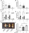
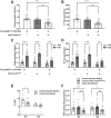
Developmental principal cell mesh knockdown in the Drosophila Malpighian tubule abolishes transepithelial fluid and K+ transport and response to drosokinin and causes a bloated abdomen phenotype. A and B: main segment fluid secretion (A) and K+ transport (B) is detected in 1-day-old adult control female fly tubules (w;c42-GAL4/+ and w;UAS-meshRNAi/+), but not the developmental principal cell mesh knockdown tubules (w;UAS-meshRNAi/+;c42-GAL4/+). Flies were reared at 28°C. Here, n = 22–30 tubules per genotype, one-way ANOVA P < 0.0001. C and D: Drosophila kinin (DK, 1 µM) treatment resulted in increased transepithelial fluid secretion and K+ flux in control tubules but had no effect on mesh knockdown tubules. Flies were reared at 18°C. Here, n = 33–34 control tubules per condition (100% of analyzed tubules secreting), and n = 5–7 mesh knockdown tubules per condition (~18% of analyzed tubules secreting); two-way ANOVA P < 0.0001 for the effects of genotype and DK and P = 0.0055 (C) and 0.0028 (D) for the interaction. ****P < 0.0001. E and F: mesh knockdown flies have distended abdomens with significantly larger abdominal volume. Flies were reared at 18°C. Here, n = 21 flies per genotype, one-way ANOVA P < 0.0001. Data are expressed as means ± SE; ****P < 0.0001. RNAi, RNA interference; UAS, upstream activation sequence. Adjusted P values for all multiple comparisons testing are shown in Table 1.
Fig. 9.
Fig. 9.
Impaired transepithelial fluid and K+ transport in the developmental principal cell mesh knockdown tubule using mesh RNA interference (RNAi) line 6867. A and B: transepithelial basal fluid secretion (A) and K+ transport (B) are significantly reduced in developmental mesh knockdown tubules from 3–4-day-old adult female flies [w;UAS-meshRNAi (II)/+;c42-GAL4/+] compared with control groups [w;c42-GAL4/+ and w;UAS-meshRNAi (II)/+]. Here, n = 17 control tubules per genotype (100% of analyzed tubules secreting), and n = 8 mesh knockdown tubules (~44% of analyzed tubules secreting); one-way ANOVA P < 0.0001 (A) and 0.0003 (B). ***P ≤ 0.001, ****P < 0.0001. P values for Tukey’s multiple comparisons test are shown in Table 2. C and D: compared with increased control tubule fluid and K+ transport, Drosophila kinin (DK, 1 µM) treatment has no effect on mesh knockdown tubule transport. Here, n = 20–24 control tubules per condition (100% of analyzed tubules secreting), and n = 7–12 mesh knockdown tubules per condition (~29% of analyzed tubules secreting in −DK and 50% of analyzed tubules secreting in +DK); two-way ANOVA P < 0.0001 for the effects of genotype, DK treatment, and interaction. ****P < 0.0001. P values for Sidak’s multiple comparisons test are shown in Table 2. Data are expressed as means ± SE.
Fig. 10.
Fig. 10.
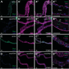
Principal cell mesh knockdown in the adult Drosophila Malpighian tubule results in mislocalized expression of other smooth septate junction (sSJ) proteins. The main segment of a 14-day-old adult control female fly anterior tubule (w;tub-GAL80ts20/+;c42-GAL4/+) shows Mesh expression at sSJs between all cells (A). Mesh colocalizes with Discs large (Dlg; C–C′′), tetraspanin 2A (Tsp2A; E), and Snakeskin (Ssk; G) at sSJs in control tubules. In the main segment of tubules subjected to 14-day adult principal cell mesh knockdown (w;tub-GAL80ts20/UAS-meshRNAi;c42-GAL4/+), Mesh immunoreactivity at sSJs between the principal cells is either greatly reduced or undetectable and is mostly confined to the borders of the stellate cells (B, arrowheads in B′). B′ shows a higher-magnification image of dashed-box area in B. The mesh knockdown tubules also show reduced levels of Dlg at sSJs and its spread into the cytoplasm of the principal cells (D–D′′, arrows in D′′). D′′ shows a higher-magnification image of merged dashed-box areas in D and D′. Tsp2A (F and F′) and Ssk (H and H′) are mostly expressed along the edges of the stellate cells (arrowheads in F′ and H′) and inside the principal cells of mesh knockdown tubules (arrows in F’ and H’). F′ and H′ show higher-magnification images of the dashed-box areas in F and H, respectively. Nuclei were counterstained blue with DAPI. PC, principal cell; RNAi, RNA interference; SC, stellate cell. Ten samples per genotype were examined. Scale bars, 50 μm.
Fig. 11.
Fig. 11.
Principal cell mesh knockdown in the adult Drosophila Malpighian tubule does not alter Na+/K+-ATPase (NKA) expression and actin cytoskeleton. NKA is expressed in the basolateral membrane of the principal cells of a 14-day-old adult control female fly tubule main segment (w;tub-GAL80ts20/+;c42-GAL4/+; A′′ and A′′′) and in the main segment of the tubule subjected to adult onset of principal cell mesh knockdown (w;tub-GAL80ts20/UAS-meshRNAi;c42-GAL4/+; B′′ and B′′′) where Mesh expression at smooth septate junctions (sSJs) between the principal cells is greatly reduced and is mostly restricted to the stellate cells (B and B′′′). A′′′ and B′′′ show single-confocal plane images indicated by dashed-box areas in A′′ and B′′, respectively. F-actin staining in a control tubule main segment (C′–C′′′) is also similar to its staining in a mesh knockdown tubule main segment (D′–D′′′). C′′′ and D′′′ show single-confocal plane images indicated by dashed-box areas in C′′ and D′′, respectively. Nuclei were counterstained blue with DAPI. RNAi, RNA interference; SC, stellate cell. Ten samples per genotype were examined. Scale bars, 50 μm.
Fig. 12.
Fig. 12.
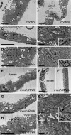
Principal cell mesh knockdown in the adult Drosophila Malpighian tubule does not affect epithelial integrity and smooth septate junction (sSJ) organization. Transmission electron micrographs of 14-day-old adult female fly anterior tubule main segment epithelial cells. Compared with control tubule main segment (w;tub-GAL80ts20/+;c42-GAL4/+; A and B), the morphology of the main segment tubule epithelium of 14-day adult principal cell mesh knockdown (w;tub-GAL80ts20/UAS-meshRNAi;c42-GAL4/+) is intact (E and F). Similarly, the sSJs between the principal cells of mesh knockdown tubule main segments (G, G′, H, and H′) appear normal with parallel plasma membranes and ladderlike septa (triangles in insets in G′ and H′) compared with control tubule sSJs (C, C′, D, and D′, triangles in insets in C′ and D′). Amv, apical microvilli; Bi, basal infoldings; mt, mitochondria; Nc, nucleus; PC, principal cell; RNAi, RNA interference; SC, stellate cell. Three samples per genotype were examined. Scale bars, 5 μm (AC, D, E, F, F′, G, and H), 500 nm (C′, D′, G′, and H′).
Fig. 13.
Fig. 13.
Principal cell mesh knockdown in the adult Drosophila Malpighian tubule decreases transepithelial fluid and ion transport, transepithelial potential (Vt), and paracellular permeability. A and B: transepithelial basal fluid secretion (A) and K+ transport (B) are significantly lower in the tubules subjected to 14-day adult-onset principal cell mesh knockdown (w;tub-GAL80ts20/UAS-meshRNAi;c42-GAL4/+) compared with control female fly tubules (w;tub-GAL80ts20/+;c42-GAL4/+ and w;UAS-meshRNAi/+). Here, n = 33–34 tubules per genotype, one-way ANOVA P < 0.0001. ***P < 0.001, **** P < 0.0001. Adjusted P values in multiple comparisons testing are shown in Table 3. C and D: fluid and K+ secretion is stimulated by Drosophila kinin (DK, 1 µM) in control and mesh knockdown tubules. Here, n = 33–38 tubules per genotype/condition, two-way ANOVA P < 0.0001 for the effects of genotype, DK treatment, and interaction. ***P < 0.001, ****P < 0.0001. Adjusted P values in multiple comparisons testing are shown in Table 3. E: Vt is diminished in mesh knockdown tubules compared with control tubules (w;tub-GAL80ts20/+;c42-GAL4/+) and is further reduced in both groups in the presence of DK. Here, n = 9–20 tubules per genotype, repeated measures two-way ANOVA P = 0.0204 for the effect of genotype, P < 0.0001 for the effect of DK, and P = 0.2686 for the interaction. *P < 0.05. Adjusted P values in multiple comparisons testing are shown in Table 4. F: Mesh knockdown tubules have reduced paracellular flux of FITC-labeled 3–5-kDa dextran (FITC-dextran). FITC-dextran flux in control and mesh knockdown tubules is not affected by DK. Here, n = 31–34 tubules per genotype, two-way ANOVA P < 0.0001 for the effect of genotype, P = 0.4122 for the effect of DK, and P = 0.8692 for the interaction. *P < 0.05, ***P < 0.001, ****P < 0.0001. Adjusted P values in multiple comparisons testing are shown in Table 5. RNAi, RNA interference; UAS, upstream activation sequence. All data are expressed as means ± SE.

Similar articles

Cited by

References

    1. Anderson JM, Van Itallie CM. Physiology and function of the tight junction. Cold Spring Harb Perspect Biol 1: a002584, 2009. doi:10.1101/cshperspect.a002584. - DOI - PMC - PubMed
    1. Beyenbach KW. Voltages and resistances of the anterior Malpighian tubule of Drosophila melanogaster. J Exp Biol 222: jeb201574, 2019. doi:10.1242/jeb.201574. - DOI - PubMed
    1. Beyenbach KW, Piermarini PM. Transcellular and paracellular pathways of transepithelial fluid secretion in Malpighian (renal) tubules of the yellow fever mosquito Aedes aegypti. Acta Physiol (Oxf) 202: 387–407, 2011. doi:10.1111/j.1748-1716.2010.02195.x. - DOI - PMC - PubMed
    1. Beyenbach KW, Skaer H, Dow JA. The developmental, molecular, and transport biology of Malpighian tubules. Annu Rev Entomol 55: 351–374, 2010. doi:10.1146/annurev-ento-112408-085512. - DOI - PubMed
    1. Bilder D, Li M, Perrimon N. Cooperative regulation of cell polarity and growth by Drosophila tumor suppressors. Science 289: 113–116, 2000. doi:10.1126/science.289.5476.113. - DOI - PubMed

Publication types

MeSH terms

LinkOut - more resources