. 2019 Dec 10:2019:8786396.
doi: 10.1155/2019/8786396. eCollection 2019.

Islet1 and Brn3 Expression Pattern Study in Human Retina and hiPSC-Derived Retinal Organoid

Affiliations

Islet1 and Brn3 Expression Pattern Study in Human Retina and hiPSC-Derived Retinal Organoid

Ziming Luo et al. Stem Cells Int. .

Abstract

This study was conducted to determine the dynamic Islet1 and Brn3 (POU4F) expression pattern in the human fetal retina and human-induced pluripotent stem cell- (hiPSC-) derived retinal organoid. Human fetal eyes from 8 to 27 fetal weeks (Fwks), human adult retina, hiPSC-derived retinal organoid from 7 to 31 differentiation weeks (Dwks), and rhesus adult retina were collected for cyrosectioning. Immunofluorescence analysis showed that Islet1 was expressed in retinal ganglion cells in the fetal retina, human adult retina, and retinal organoids. Unexpectedly, after Fwk 20, Brn3 expression gradually decreased in the fetal retina. In the midstage of development, Islet1 was detected in bipolar and developing horizontal cells. As the photoreceptor developed, the Islet1-positive cone precursors gradually became Islet1-negative/S-opsin-positive cones. This study highlights the distinguishing characteristics of Islet1 dynamic expression in human fetal retina development and proposes more concerns which should be taken regarding Brn3 as a cell-identifying marker in mature primate retina.


Conflict of interest statement

The authors declare no competing interests.

Figures

Figure 1
Figure 1
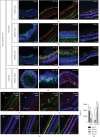
Lamination of human fetal retina and Islet1 dynamic expression in human fetal retina and hiPSC-derived retinal organoid. (a–f) Ganglion cell layer (GCL) first developed in the human fetal retina. Next, the neuroblast layer gradually thickened and divided into an inner nucleus layer (INL) and outer nucleus layer (ONL). (a′–f′) Islet1 was expressed in the GCL in the early stage and then appeared as a monolayer nucleus on the most outer side. Later, sporadic positive nuclei were observed in the thickening neuroblast layer and finally collected on the outer side of the INL. (a–f) Consistent with the human fetal retina, Islet1 was expressed in the GCL first in the retinal organoid. A monolayer of Islet1-positive cells was located on the apical side. In the midperiod, rosettes were detected in the organoid, disturbing lamination. Until late stages, retinal organoid was laminated and Islet1-positive cells were collected in the INL.
Figure 2
Figure 2
Islet1 coexpressed with Brn3 in retinal ganglion cells. (a–f) Islet1 and Brn3 colabeled retinal ganglion cells in the human fetal retina. From Fwk 20, Brn3 expression gradually decreased and was negative in Fwk 27. (g–i) Islet1-/Brn3-positive retinal ganglion cells were located on the basal side of the retinal organoid but were not laminated well. (j–l) Only sporadic Islet1-positive cells were on the basal side and were Brn3 negative. (m) Most Brn3-positive retinal ganglion cells exited the cell cycle in Fwk 9 retina. The high magnification showed one cell with Brn3 expression but still in the cell cycle. (n1) Triple staining with Islet1, Brn3, and progenitor marker MCM2 in Fwk 9 retina. (n2–n5) Showing high magnification of the white square in (n1) (scale bar = 100 μm).
Figure 3
Figure 3
Double immunolabeling of Islet1 and HuD in human fetal retinal and retinal organoid. (a–f) HuD coexpressed with Islet1 in GCL during the early stage of fetal retina development. From Fwk 16, a subpopulation of HuD-positive and Islet1-negative amacrine cells appeared in the INL. (g–l) HuD showed a similar expression pattern in retinal organoids. From Dwk 24, double-positive cells disappeared, and only HuD-single-positive amacrine cells organized as rosettes in the retinal organoid (scale bar = 100 μm).
Figure 4
Figure 4
Colabeling RGC with RBPMS, ISL, and Brn3 in human fetal retina, retinal organoid, and rhesus retina. (a) Colabeling RGC with RBPMS and Islet1 in fetal retina. (b) Costaining fetal retina with RBPMS and Brn3. (c) Double staining of HuD and RBPMS in fetal retina. (d) RPBMS expression shared the same pattern in retinal organoids. (e1, e2) In the human adult retina, Islet1 was expressed in retinal ganglion cells (white triangle) but these cells were Brn3 negative. (f1, f2) Human adult ganglion cells were HuD positive (white triangle) but Brn3 negative. (g1) No Brn3-positive cells were detected in rhesus retina. (g2) Retinal ganglion cells were RPBMS positive but could not been stained by Brn3 antibody. (g3) HuD antibody stained retinal ganglion cells along with RBPMS. (g4) Islet1 was positive in both GCL and INL, but RPBMS only labeled retinal ganglion cells. (h) Brn3 immunofluorescence intensity on human fetal retina, along with RBPMS. Scale bar = 100 μm.
Figure 5
Figure 5
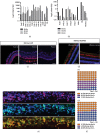
Brn3a expression in human fetal retina, retinal organoid, and rhesus retina. (a) Real-time PCR demonstrated the various expression of Brn3a, b, and c among retinal organoid, fetal retina, adult retina, rhesus retina, and Rb tissue. (b) Considering the rapidly decrease of RGC population in later developmental stage, Brn3a, b, and c expression levels were normalized with RBPMS expression. (c) Brn3a staining in human fetal retina. (d) Costaining of Brn3a and RBPMS in adult rhesus retina. (e–e) Immunostaining of Brn3a and Brn3 in Fwk 16 fetal retina. (f and f′) Cell counting of central (f) and peripheral (f′) Fwk 16 fetal retina.
Figure 6
Figure 6
Islet1 expression and postmitotic amacrine marker, AP2α. (a–f) Amacrine cell developed as early as Fwk 9 and then organized in the inner side of the INL, without coexpression of Islet1. (g–l) In the retinal organoid, AP2α-positive amacrine cell was observed at Dwk 13, later than in the fetal retina. Until Dwk 16, the number of amacrine cells obviously increased but was organized as rosettes. Until Dwk 31, the AP2α-positive cells showed a disorderly arrangement (scale bar = 100 μm).
Figure 7
Figure 7
Coimmunolabeling of Islet1 and CHX10 in human fetal retinal and retinal organoid. (a–c) CHX10 did not colocalize with Islet1 in the early stage, indicating CHX10 is a retinal progenitor marker during this period. (d, e) Layer of double-positive bipolar cells settled in the INL in the fetal retina. (f–i) CHX10-labeled retinal progenitors in the neuroblast layer in retinal organoids. (j) Some CHX10-positive cells colabeled with Islet1. (j, k) Another neural progenitor marker, MCM2, revealed undifferentiated cells in the fetal retina. (m–o) MCM2 and cell cycle marker, Ki67, confirmed that CHX10 was expressed in progenitors in this period rather than in bipolar cells. (p) To Dwk 26, CHX10-positive bipolar cells were MCM2 negative (scale bar = 100 μm).
Figure 8
Figure 8
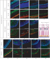
Isl1 expression in developing photoreceptor cells. (a–d) A monolayer of Islet1 immunoreactive nuclei was observed from Fwk 15; the cells were Recoverin positive. However, as indicated by the white triangle, some Recoverin-positive cells were Islet1 negative, and their nuclei were located on the inner side to Islet1-positive cells. (e) In Dwk 15, Recoverin-positive developing photoreceptors were migrating from the basal side to the apical side. (f) Similar to in the fetal retina, some developing photoreceptors with inner arranged nuclei were Recoverin positive but Islet1 negative. (g–h) Developing photoreceptors in the retinal organoid. (i) In Fwk 27, cells with inner arranged nuclei were Rhodopsin-positive rods, and the Islet1-nuclei were between the soma and inner segment of rods as the schematic diagram (h). (j–m) Rod differentiation in retinal organoids (scale bar = 100 μm).

Similar articles

Cited by

References

    1. Quaranta R., Fell J., Rühle F., et al. Revised roles of ISL1 in a hES cell-based model of human heart chamber specification. eLife. 2018;7, article e31706 doi: 10.7554/eLife.31706. - DOI - PMC - PubMed
    1. Kaku Y., Ohmori T., Kudo K., et al. Islet1 deletion causes kidney agenesis and hydroureter resembling CAKUT. Journal of the American Society of Nephrology. 2013;24(8):1242–1249. doi: 10.1681/ASN.2012050528. - DOI - PMC - PubMed
    1. Shao W., Szeto V., Song Z., et al. The LIM homeodomain protein ISL1 mediates the function of TCF7L2 in pancreatic beta cells. Journal of Molecular Endocrinology. 2018;61(1):1–12. doi: 10.1530/JME-17-0181. - DOI - PubMed
    1. Zhang Q., Huang R., Ye Y., et al. Temporal requirements for ISL1 in sympathetic neuron proliferation, differentiation, and diversification. Cell Death & Disease. 2018;9(2, article 283):p. 247. doi: 10.1038/s41419-018-0283-9. - DOI - PMC - PubMed
    1. Li L., Sun F., Chen X., Zhang M. ISL1 is upregulated in breast cancer and promotes cell proliferation, invasion, and angiogenesis. Onco Targets and Therapy. 2018;11:781–789. doi: 10.2147/OTT.S144241. - DOI - PMC - PubMed