. 2019 Dec 27;9(1):19950.
doi: 10.1038/s41598-019-56276-3.

Up-regulated fibronectin in 3D culture facilitates spreading of triple negative breast cancer cells on 2D through integrin β-5 and Src

Affiliations

Up-regulated fibronectin in 3D culture facilitates spreading of triple negative breast cancer cells on 2D through integrin β-5 and Src

Hwa -Jeong Park et al. Sci Rep. .

Abstract

Using MDA-MB-231 cells as a model of triple negative breast cancer (TNBC) and its metastatic sub-cell lines that preferentially metastasize to lung, bone or brain, we found that the mRNA and protein levels of fibronectin (FN) are increased in MDA-MB-231 cells and its lung metastatic derivative, when cultivated in three-dimensional (3D) suspension cultures. The increase of FN expression in 3D was dependent on p38 mitogen-activated protein kinase (MAPK) because it was prevented by treatment of cells with SB203580, an inhibitor of p38MAPK. The up-regulated FN was converted into fibrils, and it enhanced cell spreading when cells cultured in 3D were transferred to two-dimensional (2D) culture. The arginine-glycine-aspartate (RGD) peptides and siRNAs targeting of integrin β-5 inhibited spreading of cells regardless of the presence of FN on 2D culture dishes. In addition, the levels of phosphorylated Src were found to be increased in 3D and the treatment of cells with SU6656, an inhibitor of Src, decreased the rate of cell spreading on FN. Collectively, these studies demonstrate that increased cellular FN in 3D suspension culture facilitates cancer cell attachment and spreading via integrin β-5 and Src, suggesting that the increased FN promotes initial attachment of cancer cells to secondary organs after circulation during metastasis.


Conflict of interest statement

The authors declare no competing interests.

Figures
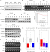
Figure 1
Figure 1
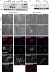
Changes in fibronectin expression of TNBCs in 3D and in vivo gene set analysis. (A) MDA-MB-231 parental, LM2, BoM2, and BrM2 were cultured in 2D or 3D condition for 48 hours, and the level of fibronectin protein was detected and the blots were cropped from different parts of the same gel. The mRNA levels (B) and protein levels (C) were monitored at 24, 48, and 72 hour in MDA-MB-231 parental and LM2 cells grown in 2D or 3D condition using electrophoretic gels and immunoblots, respectively. And the bands of blots were cropped from the same gel for each cell line (parental and LM2). MDA-MB-231 parental and LM2 cells cultured in 3D suspension conditions were transferred in 2D culture plates, and changes in fibronectin protein (D) and mRNA (E) expression were detected and the PCR products were observed using electrophoretic gels. (F) Kaplan-Meier survival analysis showing the relationship between FN1 gene expression and 5-year distant metastasis free survival outcomes in patients with ER-negative breast cancer. (G) A box plot exhibiting variation of FN1 gene expression according to breast cancer subtypes; basal-A, basal-B and luminal. (H) A box plot of FN1 gene expression in different breast cancer cell lines using GOBO gene-set analysis. TN; triple negative breast cancer cell lines, HER2; HER2-positive breast cancer cell lines, and HR; Hormone receptor positive breast cancer cell lines.
Figure 2
Figure 2
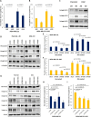
The effect of SB203580 treatment in fibronectin expression in 3D-grown cells. (A) Phosphorylation of p38MAPK and MAPKMAP2 increases in MDA-MB-231 parental and organotropic metastasis derivatives in 3D suspension culture for 48 hours. The same volume of the set of cell lysates were loaded into different SDS-page gels. The blots of phosphorylated MAPKAPK and p38MAPK were cropped from the same gel and the other blots were cropped from different gels. (B) SB203580 was added as serial concentrations in 3D culture media and changes in fibronectin protein expression were monitored after cells were cultivated for 48 hours in 3D. The same volume of the set of cell lysates was loaded into different gels and p-p38 MAPK and MAPKAPK were cropped from the same gel. The other blots were cropped from different gels. (C) MDA-MB-231 parental and LM2 cells were treated with the p38 MAPK inhibitor, SB203580 for 48 hours and formation of cell-spheroids was observed. Scale bar: 100 μm.
Figure 3
Figure 3
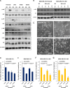
Increased fibronectin of MDA-MB-231 parental and LM2 cells in 3D facilitates cell attachment on 2D. Cells grown in 2D or 3D suspension cultures for 48 hours were re-cultivated in serum-free culture media on 2D culture plates and subjected to immunoblot after being harvested at 2.5 hr and 5 hr from 2D re-cultures and DOC-insoluble and DOC-soluble fibronectin was detected (A). DOC-soluble fibronectin used as internal loading control for comparison. DOC-soluble and DOC-insoluble lysates were loaded into different gels and cropped for immunoblots. (B) Protein expression changes were observed after FN1 siRNAs were treated in MDA-MA-231 parental and LM2 cells. The mixture of two different kinds of siRNAs against FN1 were transfected into cells in a serial manner in which 25 nM siRNAs were treated each time, and totally 50 nM siRNAs were treated. The blots for fibronectin and α-tubulin were cropped from the same gel. (C) Cells cultured in 2D or 3D condition for 48 hr were re-cultivated in 2D culture plates with serum-free media, and the spreading patterns of cells in each condition were observed. Scale bar: 50 μm. (D) FN1 or control siRNA treated cells were re-cultivated on 2D culture glasses for 2.5 hours after 3D incubation for 48 hours and fixed with 4% formalin. Fixed cells were not permeabilized before antibody staining to detect only the extracellular fibronectin. DAPI was used to stain nuclei. Blue: DAPI and Red: extracellular fibronectin. Scale bar: 10 μM. The below panels show extracellular fibronectin (White). (E) After 3D culture for 48hours,cells transfected with control or FN1 siRNA were cultivated for 2.5 hours and fixed, and permeabilized with PBS containing 1% deocycholate. Fixed cells were treated with antibodies against FN and phopho-paxillin, indicated squares(white line) were magnified in a black and white version. Blue:DAPI, Green:FN, and Red: phspho-paxillin. Scale bar: 20 μm.
Figure 4
Figure 4
Down-regulation of fibronectin in 3D suspension culture reduces attachment of MDA-MB-231 parental and LM2 cells to HUVECs. (A,B) MDA-MB-231 parental and LM2 cells were re-cultivated for 2.5 hours on uncoated or fibronectin VI-coated culture glasses, fixed with 4% formalin and stained with phalloidin (green) and DAPI (blue) and (C,D) the areas covered with cells that were attached to 2D culture glasses were measured. Scale bar: 100 μm. (G) MDA-MB-231 parental and LM2 cells were re-plated on a HUVEC monolayer for 2 hours, after being cultured in 3D suspension condition for 48 hours. Breast cancer cells were stained with green CMFDA, and the nuclei of breast cancer cells and HUVECs were stained DAPI (blue). Scale bar: 200 μm. (E,F) The relative ratio of attached MDA-MB-231 parental and LM2 cells on HUVECs was measured, based on CMFDA. For the experiments the mixture of two kinds of FN1 siRNA were transfected to parental and LM2 cells. (means ± s.e.m; Student’s t-test).
Figure 5
Figure 5
RGD peptides and integrin β-5 silencing disturb cell attachment from 3D to 2D. (A,B) 500 μM RGD peptides were added into 2D serum-free culture media before cells grown in 3D were transferred on uncoated- or fibronectin coated-2D culture glasses and the cells attached on 2D surfaces were fixed after 2.5 hours. And the areas occupied by the attached cells were measured. (C) Changes in protein levels of each integrin β isoform in different culture conditions were detected, and (D) the protein levels of fibronectin, integrin β-1, integrin β-3, and integrin β-5 of the respective siRNAs treated cells were examined by western blot. The same volume of the set of cell lysates were loaded in different gels to conduct immunoblots for each integrin β isoforms, fibronectin, β-actin, and α-tubulin and the blots were cropped from the gels. (E,F) The siRNA transfected cells were transferred to 2D culture plates uncoated or coated with fibronectin after 3D incubation for 48 hours and the areas covered by attached cells were measured. The mixtures of two different kinds of siRNAs against ITGB1 and ITGB5 and a siRNA against ITGB3 were used for siRNA transfection, respectively. Cells were transfected with siRNAs at concentration of 25 nM in a serial manner, total 50 nM. (G) siRNAs targeting different integrin isoforms were transfected to parental and LM2 cells in three different combinations, and 25 nM siRNA were treated per gene; ITGA5, ITGAV, ITGB1, ITGB3, and ITGB5 (totally 50 nM siRNA for a combination). The mixture of two different kinds of siRNA for ITGA5 and ITGAV were used. (H and I) siRNA transfected cells with each combination were spread on fibronectin coated culture glasses for 2.5 hours after being cultured in 3D suspension condition. And the areas covered by adherent cells were compared with those of control siRNA treated cells. (means ± s.e.m; Student’s t-test).
Figure 6
Figure 6
Pharmacological inhibition of SFK reduces the rate of cell-attachment from 3D to 2D. (A) Cells grown in 2D or 3D cultures for 48 hours were harvested and subjected to immunoblot analysis and phosphorylation of signaling proteins were detected. The same volumes of the set of cell lysates were loaded in different gels to carry out immunoblots for p-SFK, Src, Erk, JNK, AKT and α-tubulin, and the blots were cropped from different gels. (B) The changed levels of phosphorylated SFK were observed by western blot. The cells were transferred to 2D culture plate containing serum-free media after 3D suspension culture for 48 hours, and the cell lysates were harvested according to the order of time. The same volume of the set of cell lysates were loaded in different gel to carry out immunoblots for p-SFK, Src and β-actin. (C) SU6656 was added to culture media 30 min before transferring cells from 3D to 2D and the patterns of cell spreading were observed after 2.5 hours. Scale bar: 200 μm. (D–G) Control or fibroenctin down-regulated cells were re-cultivated on uncoated- or fibronectinVI coated-culture glasses after being cultured in 3D suspension condition. Before transferring the cells to 2D, 10 μM of SU6656 was added. The areas covered by attached cells were measured and compared. (means ± s.e.m; Student’s t-test).
Figure 7
Figure 7
Downregulation of fibronectin induces retarded collective migration of cellular monolayer derived from 3D cell-aggregates. (A) FN1 or control siRNA transfected MDA-MB-231 parental and LM2 cells (5,000 cells/well) were cultured in ultra-low attachment 96-well round bottom plates to form aggregates for 48 hours. Scale bar: 500 μm. (C,D) MDA-MB-231 parental cell-aggregates were transferred to uncoated or fibronectin VI-coated culture plates and the areas covered by cellular monolayer derived from the aggregates were measured by using ImageJ. The covered area with cellular monolayer at 24 hour was deducted from that at 48 hour. (B) A cartoon explains migration of cellular monolayer derived from cell-aggregates. (means ± s.e.m; Student’s t-test).

Similar articles

Cited by

References

    1. Dolznig H, et al. Modeling colon adenocarcinomas in vitro a 3D co-culture system induces cancer-relevant pathways upon tumor cell and stromal fibroblast interaction. Am J Pathol. 2011;179:487–501. doi: 10.1016/j.ajpath.2011.03.015. - DOI - PMC - PubMed
    1. Ekert Jason E., Johnson Kjell, Strake Brandy, Pardinas Jose, Jarantow Stephen, Perkinson Robert, Colter David C. Three-Dimensional Lung Tumor Microenvironment Modulates Therapeutic Compound Responsiveness In Vitro – Implication for Drug Development. PLoS ONE. 2014;9(3):e92248. doi: 10.1371/journal.pone.0092248. - DOI - PMC - PubMed
    1. Pickl M, Ries CH. Comparison of 3D and 2D tumor models reveals enhanced HER2 activation in 3D associated with an increased response to trastuzumab. Oncogene. 2009;28:461–8. doi: 10.1038/onc.2008.394. - DOI - PubMed
    1. Luca Anna C., Mersch Sabrina, Deenen René, Schmidt Stephan, Messner Isabelle, Schäfer Karl-Ludwig, Baldus Stephan E., Huckenbeck Wolfgang, Piekorz Roland P., Knoefel Wolfram T., Krieg Andreas, Stoecklein Nikolas H. Impact of the 3D Microenvironment on Phenotype, Gene Expression, and EGFR Inhibition of Colorectal Cancer Cell Lines. PLoS ONE. 2013;8(3):e59689. doi: 10.1371/journal.pone.0059689. - DOI - PMC - PubMed
    1. Sastry SK, Burridge K. Focal adhesions: a nexus for intracellular signaling and cytoskeletal dynamics. Exp Cell Res. 2000;261:25–36. doi: 10.1006/excr.2000.5043. - DOI - PubMed

Publication types

MeSH terms