. 2020 May;22(5):e13150.
doi: 10.1111/cmi.13150. Epub 2019 Dec 26.

Human microRNA-30 inhibits influenza virus infection by suppressing the expression of SOCS1, SOCS3, and NEDD4

Affiliations

Human microRNA-30 inhibits influenza virus infection by suppressing the expression of SOCS1, SOCS3, and NEDD4

Xian Lin et al. Cell Microbiol. 2020 May.

Abstract

Influenza A virus (IAV) has evolved multiple mechanisms to compromise type I interferon (IFN) responses. The antiviral function of IFN is mainly exerted by activating the JAK/STAT signalling and subsequently inducing IFN-stimulated gene (ISG) production. However, the mechanism by which IAV combat the type I IFN signalling pathway is not fully elucidated. In this study, we explored the roles of human microRNAs modulated by IAV infection in type I IFN responses. We demonstrated that microRNA-30 (miR-30) family members were downregulated by IAV infection. Our data showed that the forced expression of miR-30 family members inhibited IAV proliferation, while miR-30 family member inhibitors promoted IAV proliferation. Mechanistically, we found that miR-30 family members targeted and reduced SOCS1 and SOCS3 expression, and thus relieved their inhibiting effects on IFN/JAK/STAT signalling pathway. In addition, miR-30 family members inhibited the expression of NEDD4, a negative regulator of IFITM3, which is important for host defence against influenza viruses. Our findings suggest that IAV utilises a novel strategy to restrain host type I IFN-mediated antiviral immune responses by decreasing the expression of miR-30 family members, and add a new way to understand the mechanism of immune escape caused by influenza viruses.

Keywords: NEDD4; SOCS1; SOCS3; influenza virus; microRNA-30.


Conflict of interest statement

The authors declare no conflict of interest.

Figures
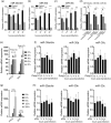
Figure 1
Figure 1
Influenza virus infection downregulates miR‐30 expression. A549 cells were infected with 0.1 MOI of H5N1 influenza virus, and the expression levels of miR‐30a/d/e (a), miR‐30b (b), and miR‐30c (c) were detected at 12, 24, 36, and 48 hpi by qRT‐PCR. (d) A549 cells were infected with heat‐inactivated H5N1, and after 36 hr, miR‐30 expression was detected by qRT‐PCR. A549 cells were transfected with 200 ng poly(I:C), IFN‐β and MX1 (e), and miR‐30 (f) were tested at the indicated time points post‐transfection by qRT‐PCR. A549 cells were infected by VSV with an MOI of 0.1, IFN‐β and MX1 (g), and miR‐30 (h) were detected at the indicated time points post‐infection by qRT‐PCR. The values are shown as the mean and SD and are representative of three independent experiments. The data in (a–c) were analysed using two‐way ANOVA; data in (d) were analysed using Student's t test. The values are shown as the mean and SD and are representative of three independent experiments. #: non‐significant, ***p < .001; **p < .01; *p < .05
Figure 2
Figure 2
MiR‐30 suppresses influenza virus replication. A549 cells were transfected with 80 nm miR‐30a/b/c mimics (a–c) or 100 nm inhibitors (d–f); 24 hr later, cells were infected with 0.2 MOI of H5N1 influenza virus (HM/H5N1). At the indicated time post‐infection, the RNA, total protein, and the supernatant were collected for NP mRNA (a and d), NP protein (b and e), and viral titre (c and f) detection by qRT‐PCR with GAPDH as housekeeping gene, western blot, and plaque assay analyses, respectively. NC, negative control; IN, inhibitor. The values are shown as the mean and SD and are representative of three independent experiments. Data were analysed using two‐way ANOVA; ***p < .001; **p < .01; *p < .05
Figure 3
Figure 3
Influenza virus genomic RNA is not the target of miR‐30. (a) Predicted interactions between miR‐30b/c and the influenza virus M gene using RNA22 V2. (b) Effects of miR‐30a/b/c/d/e mimics on the expression of the firefly luciferase gene from the pmirGLO reporter constructs containing the putative miR‐30b/c binding sites from the M gene (pmirGLO‐M) and 3′UTR of human p53 (pmirGLO‐p53). First, 293T cells were co‐transfected with reporter constructs together with miR‐30a/b/c/d/e mimics or NC. After 24 hr, cells were lysed, and luciferase activities were measured. The luciferase activity was normalised to the Renilla luciferase activity, and the data are expressed relative to that of the NC. The values are shown as the mean and SD and are representative of three independent experiments. Data were analysed using Student's t test. ***p < .001; **p < .01; *p < .05
Figure 4
Figure 4
MiR‐30 targets the 3′UTRs of SOCS1 and SOCS3. (a) Predicted target sites of miR‐30a/b/c in the 3′UTRs of SOCS1 and SOCS3. Nucleotides in blue are seed regions of miR‐30a/b/c; nucleotides in red are complementary to the miR‐30a/b/c seed region. (b and c) Effects of miR‐30a/b/c mimics on expression of the firefly luciferase gene from reporter constructs containing the SOCS1 and SOCS3 3′UTRs or SOCS1 and SOCS3 mutated 3′UTRs. First, 293T cells were transfected with NC or miR‐30a/b/c, together with the SOCS1 or SOCS3 3′UTR (3′UTR WT) or 3′UTR mutant (3′UTR Mut), and 24 hr after transfection, cells were harvested to assess luciferase activity. (d and e) MiR‐30a/b/c inhibit the SOCS1 and SOCS3 3′UTRs in a dose‐dependent manner. First, 293T cells were transfected with 30, 60, or 90 nM miR‐30a/b/c mimics, together with SOCS1 or SOCS3 3′UTR, and 24 hr after transfection, cells were lysed for luciferase activity analysis. (f and g) The 293T cells were transfected with 100 nM miR‐30a/b/c inhibitor or NC inhibitor (NC‐inhibitor), together with SOCS1 or SOCS3 3′UTR. Twenty‐four hours later, the cells were lysed for luciferase activity analysis. A549 cells were transfected with 80 nm miR‐30a/b/c mimics or 100 nM miR‐30a/b/c inhibitors, and 36 hr after transfection, the mRNA and protein levels of SOCS1 and SOCS3 were determined by qRT‐PCR (h and i) and western blot analyses (j and k), respectively. The values are shown as the mean and SD and are representative of at least three independent experiments. Data were analysed using Student's t test. ***p < .001; **p < .01; *p < .05
Figure 5
Figure 5
MiR‐30 targets NEDD4. (a) Analysis of NEDD4 3′UTR potential binding sites for miR‐30c; three putative binding sites were predicted (942–949, 960–967, and 1116–1122 in the 3′UTR of NEDD4 mRNA), and nucleotides in red show mutations of the 3′UTR of NEDD4, which is complementary to the seed region of miR‐30c. (b) Effects of miR‐30a/b/c on firefly luciferase activity from reporter constructs containing NEDD3 3′UTR WT or sequences with mutations in three putative positions (NEDD4‐Mut1, Mut2, and Mut3). First, 293T cells were co‐transfected with miR‐30a/b/c mimics and NEDD4 3′UTR or 3′UTR mutants, and 24 hr later, the cells were lysed for luciferase activity analysis. (c) Dose‐dependent inhibitory effects of miR‐30a/b/c on the NEDD4 3′UTR. First, 293T cells were transfected with 30, 60, or 90 nM miR‐30a/b/c, together with the NEDD4 3′UTR. Twenty‐four hours later, luciferase activity was analysed. (d) MiR‐30a/b/c inhibitors increased the NEDD4 3′UTR activity. First, 293T cells were transfected with 100 nM miR‐30a/b/c inhibitors and NEDD4 3′UTR. Twenty‐four hours later, luciferase activity was analysed. A549 cells were transfected with 80 nM miR‐30a/b/c mimics or 100 nM miR‐30a/b/c inhibitors, and 36 hr later, the mRNA and protein levels of NEDD4 were determined by qRT‐PCR (e and f) and western blot analyses (g). The values are shown as the mean and SD and are representative of at least three independent experiments. Data were analysed using Student's t test. ***p < .001; **p < .01; *p < .05
Figure 6
Figure 6
MiR‐30c promotes JAK–STAT signalling pathway activation. A549 cells were transfected with 80 nM NC or miR‐30c mimics, and 36 hr later, cells were treated with 500 U/ml human rIFN‐β for 30 min (a), transfected with 200 ng poly(I:C) for 4 hr (b), or infected with 5 MOI of H5N1 influenza virus for 4 hr (c). Cells were lysed, and cell extracts were analysed by western blot analysis. A549 cells were transfected with miR‐30c mimics; 36 hr later, cells were treated with 100 U/ml rIFN‐β for 3 hr, transfected with 100 ng/ml poly(I:C) for 5 hr, or infected with 3 MOI of H5N1 influenza virus for 8 hr. RNA was extracted for mRNA analyses of IFN‐β, IL‐6 (d), MX1, IFIT3, and CXCL10 (e) by qRT‐PCR. Data are shown as the mean and SD and as one representative of three independent experiments. Data were analysed using Student's t test. #: non‐significant; ***p < .001; **p < .01; *p < .05
Figure 7
Figure 7
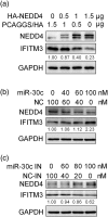
MiR‐30c is positively correlated with IFITM3 protein levels. (a) A549 cells were transfected with control vector (pCAGGS/HA) or 0.5, 1, and 1.5 μg PCAGGS/HA‐NEDD4, and 24 hr after transfection, cells were lysed for IFITM3 detection by western blot analysis. A549 cells were transfected with 0, 40, 60, and 100 nM miR‐30c mimics or negative control mimics (NC) (b) or 0, 60, 80, and 100 nM miR‐30c inhibitors (miR‐30c IN) or control inhibitor (NC‐IN) (c), and 24 hr later, the cells were lysed for IFITM3 and NEDD4 detection by western blot analysis. Data are shown as the mean and SD and as one representative of three independent experiments
Figure 8
Figure 8
SOCS1, SOCS3, and NEDD4 facilitate influenza virus replication. A549 cells were transfected with SOCS1 (a), SOCS3 (b), or NEDD4 (c) overexpression vector, and 24 hr after transfection, the cells were infected with 0.2 MOI of H5N1 influenza virus (HM/H5N1). After 36 hr, the supernatant was collected, and the cells were lysed for viral titre and NP protein determination by plaque and western blot analyses, respectively. A549 cells were transfected with 60 nM SOCS1 or SOCS3 siRNAs (d) or NEDD4 siRNAs (e); 24 hr later, A549 cells were infected with 0.2 MOI of HM/H5N1, and after 36 hr, the supernatant was collected, and the cells were lysed for viral titre and NP protein determination. The western blot results are shown as one representative of three independent experiments, and viral titres data are expressed as the mean and SD of three independent experiments. Data were analysed using Student's t test. **p < .01; *p < .05
Figure 9
Figure 9
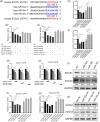
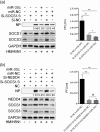
Antiviral function of miR‐30c is mainly dependent on SOCS1, SOCS3, and NEDD4 expression. A549 cells were transfected with 50 nm siRNA of SOCS1 and 50 nm siRNA of SOCS3 at the same time (a) or transfected with 50 nm siRNA of SOCS1, 50 nm siRNA of SOCS3, and 50 nm siRNA of NEDD4 at the same time (b). After 12 hr, 80 nm miR‐30c mimics or miRNA control (miR‐NC) was transfected; 24 hr later, A549 cells were infected with 0.2 MOI of HM/H5N1, and after 30 hr, the supernatant were collected for viral titres determination, and cells were lysed for NP test by western blot. Data are shown as the mean and SD and as one representative of three independent experiments. Data were analysed using Student's t test. #: non‐significant; **p < .01

Similar articles

Cited by

References

    1. Agrawal, R. , Tran, U. , & Wessely, O. (2009). The miR‐30 miRNA family regulates Xenopus pronephros development and targets the transcription factor Xlim1/Lhx1. Development, 136(23), 3927–3936. 10.1242/dev.037432 - DOI - PMC - PubMed
    1. Alexander, W. S. (2002). Suppressors of cytokine signalling (SOCS) in the immune system. Nature Reviews. Immunology, 2(6), 410–416. 10.1038/nri818 - DOI - PubMed
    1. Alexander, W. S. , & Hilton, D. J. (2004). The role of suppressors of cytokine signaling (SOCS) proteins in regulation of the immune response. Annual Review of Immunology, 22, 503–529. 10.1146/annurev.immunol.22.091003.090312 - DOI - PubMed
    1. Alexiou, P. , Maragkakis, M. , Papadopoulos, G. L. , Simmosis, V. A. , Zhang, L. , & Hatzigeorgiou, A. G. (2010). The DIANA‐mirExTra web server: From gene expression data to microRNA function. PLoS One, 5(2), e9171 10.1371/journal.pone.0009171 - DOI - PMC - PubMed
    1. Ambros, V. (2004). The functions of animal microRNAs. Nature, 431(7006), 350–355. 10.1038/nature02871 - DOI - PubMed

Publication types

MeSH terms