. 2020 Jan 7;117(1):717-726.
doi: 10.1073/pnas.1911243117. Epub 2019 Dec 23.

Shear force sensing of epithelial Na+ channel (ENaC) relies on N-glycosylated asparagines in the palm and knuckle domains of αENaC

Affiliations

Shear force sensing of epithelial Na+ channel (ENaC) relies on N-glycosylated asparagines in the palm and knuckle domains of αENaC

Fenja Knoepp et al. Proc Natl Acad Sci U S A. .

Abstract

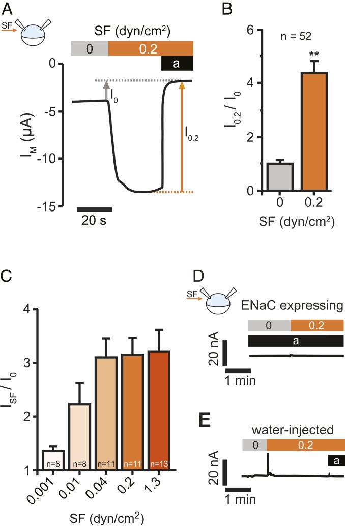
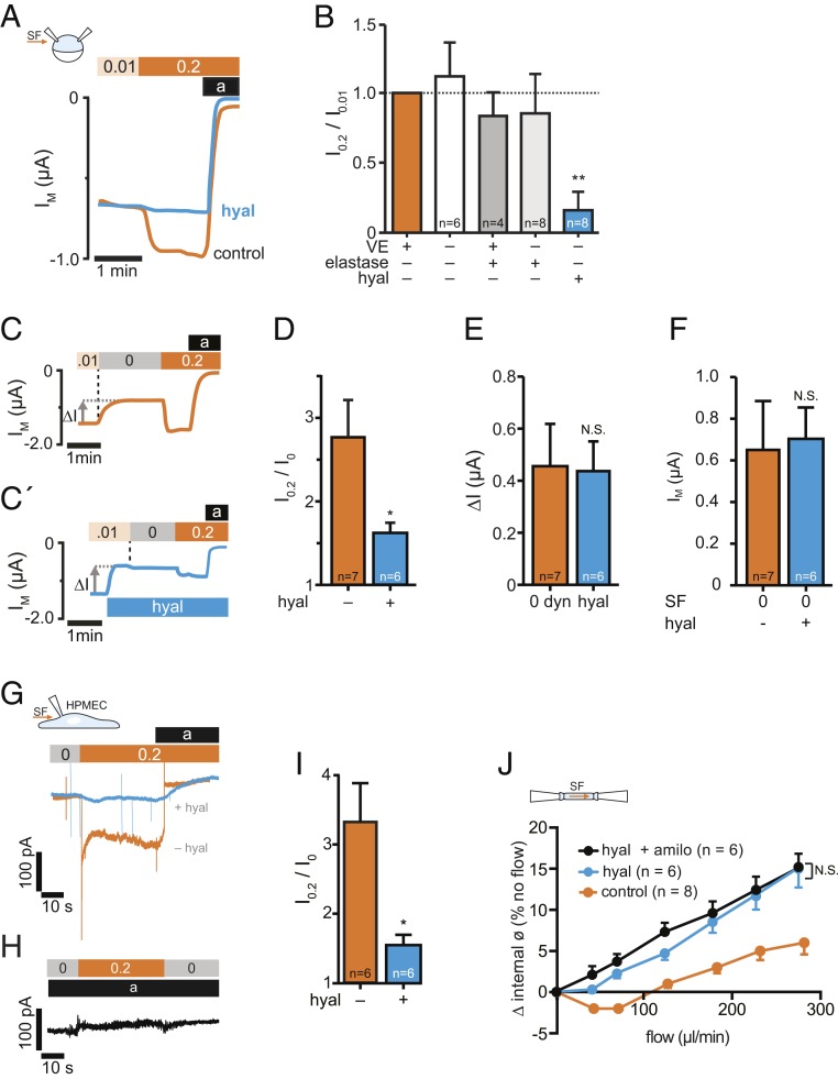
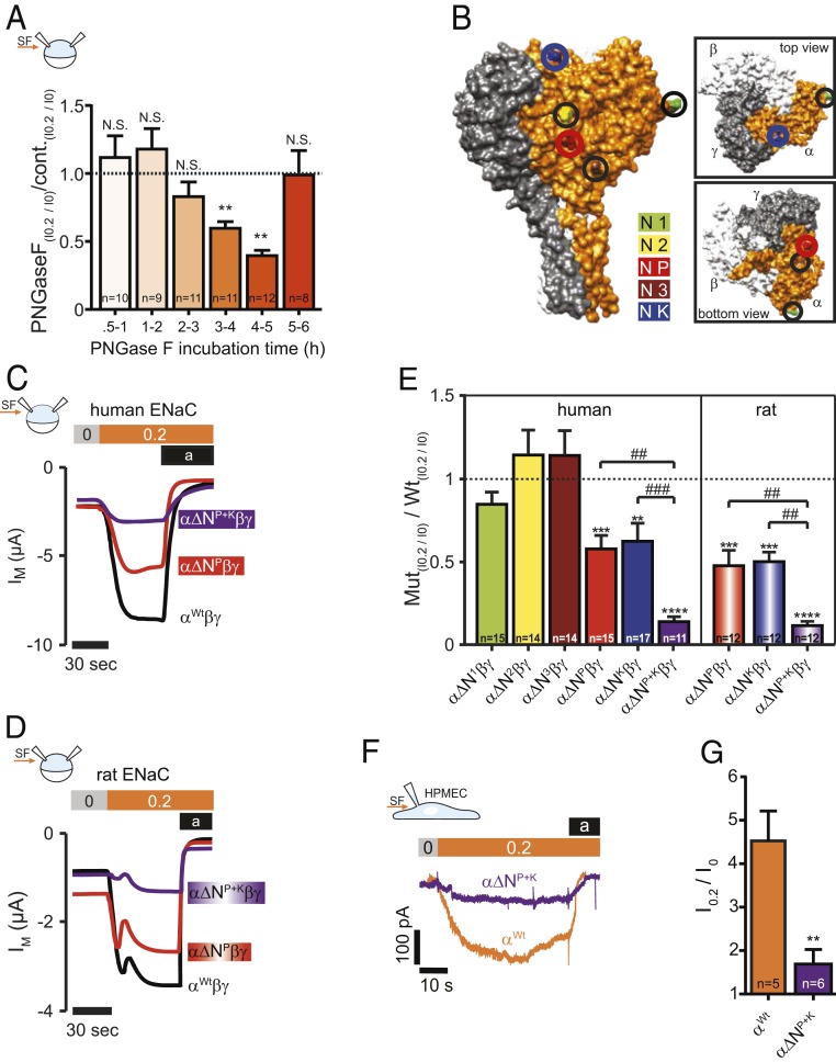
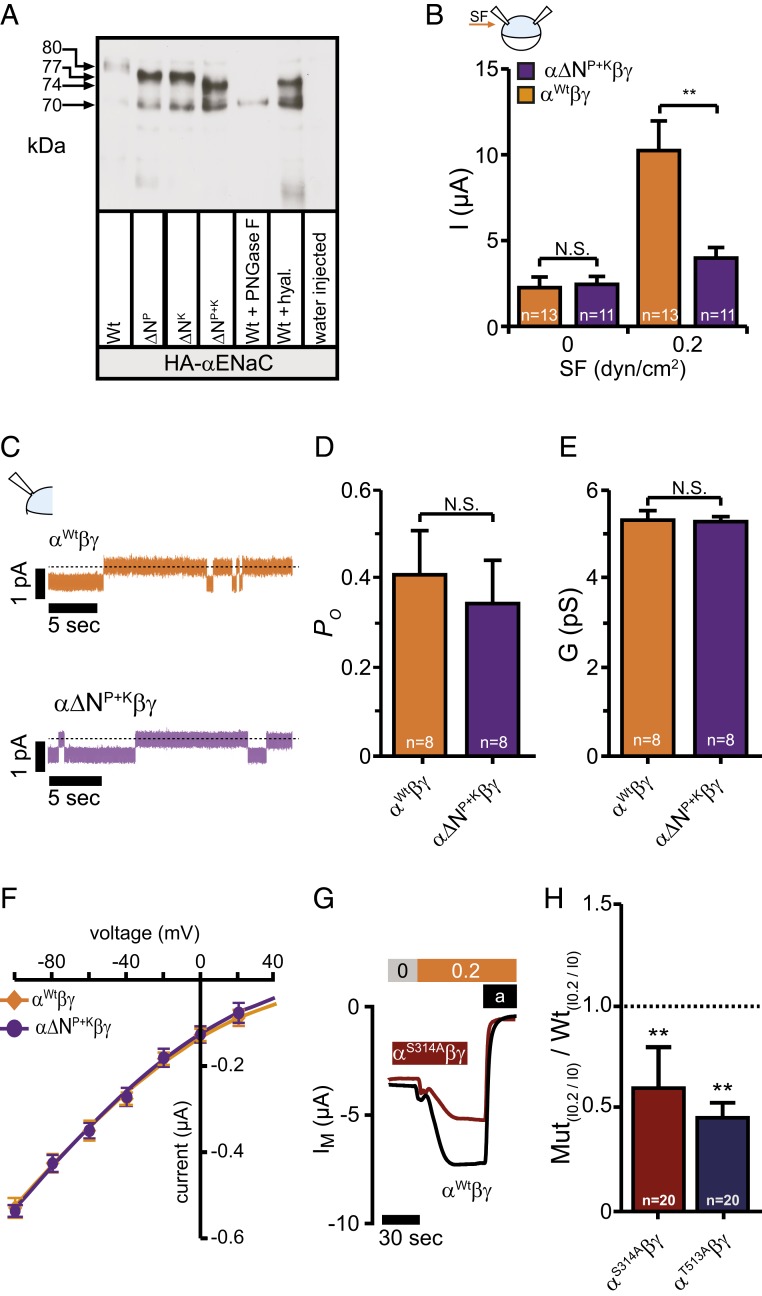
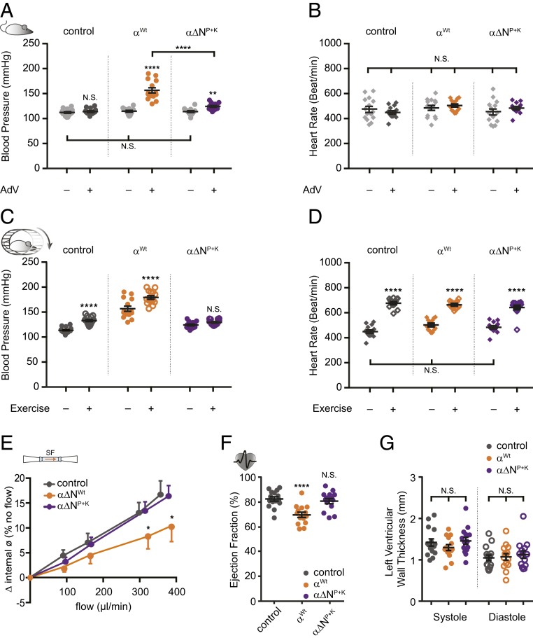
Mechanosensitive ion channels are crucial for normal cell function and facilitate physiological function, such as blood pressure regulation. So far little is known about the molecular mechanisms of how channels sense mechanical force. Canonical vertebrate epithelial Na+ channel (ENaC) formed by α-, β-, and γ-subunits is a shear force (SF) sensor and a member of the ENaC/degenerin protein family. ENaC activity in epithelial cells contributes to electrolyte/fluid-homeostasis and blood pressure regulation. Furthermore, ENaC in endothelial cells mediates vascular responsiveness to regulate blood pressure. Here, we provide evidence that ENaC's ability to mediate SF responsiveness relies on the "force-from-filament" principle involving extracellular tethers and the extracellular matrix (ECM). Two glycosylated asparagines, respectively their N-glycans localized in the palm and knuckle domains of αENaC, were identified as potential tethers. Decreased SF-induced ENaC currents were observed following removal of the ECM/glycocalyx, replacement of these glycosylated asparagines, or removal of N-glycans. Endothelial-specific overexpression of αENaC in mice induced hypertension. In contrast, expression of αENaC lacking these glycosylated asparagines blunted this effect. In summary, glycosylated asparagines in the palm and knuckle domains of αENaC are important for SF sensing. In accordance with the force-from-filament principle, they may provide a connection to the ECM that facilitates vascular responsiveness contributing to blood pressure regulation.

Keywords: N-glycosylation; epithelial Na+ channel (ENaC); extracellular tether; mechanotransduction; shear force.


Conflict of interest statement

The authors declare no competing interest.

Figures

Fig. 1.
Fig. 1.
ENaC is activated by SF. (A) Representative recording of SF application on αβγENaC currents in Xenopus oocytes. Activating the bath perfusion (0.2 dyn/cm2, orange bar) induced a rapid increase of the transmembrane current (IM). Amiloride (a, black bar) was applied to estimate ENaC-mediated current in the absence (I0) and presence (I0.2) of SF. (B) Current values with SF were normalized with respect to the values before SF was applied (I0.2/I0). (mean ± SEM; **P < 0.01, one-sample t test, two-tailed). (C) The SF-response (ISF) was augmented by increases in SF. (D) SF has no effect in the presence of amiloride (a, black bar, 10 µM), when ENaCs are blocked. (E) Neither SF nor amiloride affect endogenous ion channels of Xenopus oocytes. Oocytes were treated identical although no cRNA was dissolved in the water used for injection (representative traces of at least 12 recordings using oocytes from at least three different animals).
Fig. 2.
Fig. 2.
SF-mediated activation of ENaC is impaired by degradation of hyaluronic acid. (A) Hyaluronidase treatment before expression of ENaC impaired the SF response (hyal, blue line) in comparison to untreated oocytes (control, orange line). (B) Averaged SF-responses (I0.2/I0.01) from oocytes following removal/degradation of ECM components (VE, vitelline envelope). SF-responses were normalized to corresponding control oocytes (intact ECM, orange bar) using cells from the same animals (mean ± SEM; **P < 0.01, one-sample t test, two-tailed). (C) Stopping the bath perfusion (0.01 → 0 dyn/cm2, indicated by a vertical dashed line) induced a current decrease (∆I) and the subsequent application of SF (0 → 0.2 dyn/cm2) resulted in a current increase. (C′) The application of hyaluronidase (blue bar) in the presence of SF induced a similar effect on ∆I than stopping the bath perfusion (E), whereas the normalized SF-effect was reduced (D). (F) Membrane currents recorded in the absence of SF (0 dyn/cm2) were similar between oocytes that were untreated (−) or treated (+) with hyaluronidase (hyal, mean ± SEM; N.S., not significant, unpaired t test, two-tailed). (G) Current-traces of HPMECs, exposed to SF (0.2 dyn/cm2, orange bar). Amiloride (a, black bar) was used to estimate ENaC-mediated currents. Preincubation with hyaluronidase (hyal, blue trace), blunted the SF-induced increase of ENaC currents. (H) No SF-response was observed in the presence of amiloride (10 µM, black bar). (I) Statistical analysis from experiments depicted in G. *P < 0.05, unpaired t test, two-tailed. (J) Effect of intraluminal flow on isolated pressurized carotid arteries from mice at mean arterial pressure of 60 mmHg. Increased intraluminal flow affected the internal diameter of the arteries under control conditions (orange). Hyaluronidase augmented the SF-induced increase of the vessel diameter (blue) and amiloride in combination with hyaluronidase (black) had no additional effect (mean ± SEM; *P < 0.05, N.S., not significant, repeated-measures two-way ANOVA).
Fig. 3.
Fig. 3.
N-linked glycans and glycosylated asparagines contribute to the SF-effect. (A) Injection of PNGase F into the oocytes resulted in a time-dependent reduction of the SF-mediated response (mean ± SEM; **P < 0.01, N.S., not significant, one-sample t test, two-tailed). (B) Surface structure of a human α-, β-, γENaC-heterotrimer displaying the localization of glycosylated asparagines of αENaC. (C) Current-traces of the SF-effects in oocytes that express human or (D) αβγENaC Oocytes expressed either a wild-type (Wt) αENaC (black trace) or αENaC that had a single asparagine replaced in the palm domain (α∆NP, red trace) or two asparagines replaced (in palm and knuckle domain: α∆NP+K, purple trace). (E) Replacement of asparagines of human and rat αENaC in the palm and knuckle domains decreased the SF response. Dashed line represents SF responses from corresponding control experiments using either human or rat wild-type αβγENaC (mean ± SEM; ****P < 0.0001, ***P < 0.001, **P < 0.01, one-sample t test, two-tailed; ###P < 0.001. ##P < 0.01 and as indicated, one-way ANOVA with Bonferroni´s multiple comparison test). (F) Representative current-traces of HPMECs exposed to SF (0.2 dyn/cm2, orange bar). Amiloride (a, black bar) was applied to estimate ENaC-mediated currents. Compared to wild-type HPMECs (αWt, orange current trace), CRISPR/Cas9-mediated replacement of the asparagines in the palm and knuckle domains of αENaC (α∆NP+K, purple trace) blunted the SF-induced increase in current in these cells. (G) Statistical analysis from experiments depicted in F. **P < 0.01, unpaired t test, two-tailed.
Fig. 4.
Fig. 4.
N-linked glycans contribute to the SF-mediated effect but do not affect basic channel properties. (A) Immunoblotting of whole-cell lysates from oocytes expressing either HA-tagged human or wild-type αENaC (Wt) subunits with the asparagine in the palm or knuckle domains replaced or both. The shift in relative molecular mass (MR) is observed between the wild-type (∼80 kDa) and the modified subunits (∼77 kDa for ∆NP and ∆NK and ∼74 kDa for ∆NP+K) corresponding to a lack of N-linked glycosylation. Total removal of N-linked glycosylation by PNGase F resulted in the complete loss of the upper (glycosylated) band and shifted the band to ∼70 kDa (Wt+PNGase F). Treatment of wild-type with hyaluronidase resulted in a band of ∼74 kDa (Wt+hyal). Cell lysates of water-injected control-oocytes served as a control for antibody specificity. The depicted blot is representative for n = 6. (B) Amiloride sensitive currents at 0 dyn are similar, but increased in wild-type when exposed to 0.2 dyn/cm2 SF (mean ± SEM; **P < 0.01, N.S., not significant, 2-way ANOVA followed by Sidak’s multiple comparison test). (C) Current-traces of cell attached single-channel recordings at a membrane-potential of -100 mV using devitellinized oocytes. (DF) Neither the open probability (PO), the conductance (G), nor the single-channel current voltage relationship was affected by replacement of the asparagines. Means were fitted by the Goldman–Hodgkin–Katz equation. The calculated permeability for Na+ was wild-type: 1.72 × 10⁻¹² cm−3 s−1; α∆NP+KβγENaC: 1.73 × 10⁻¹² cm−3 s−1 (mean ± SEM; N.S., not significant, unpaired t test, two-tailed). (G and H) Disruption of the glycosylation motif in the palm and knuckle domains did reduce the SF-mediated effect (mean ± SEM; **P < 0.01, one-sample t test, two-tailed).
Fig. 5.
Fig. 5.
Blood pressure increase after viral transduction relies on glycosylated asparagines. (A) Systolic blood pressure from three groups recorded before and after viral transduction. The increase in blood pressure was smaller in animals that received an αENaC subunit lacking asparagines in the palm and knuckle domains (α∆NP+K, purple; mean ± SEM; ****P < 0.0001, **P < 0.01, N.S., not significant, 2-way ANOVA followed by Tukey’s multiple comparison test). (B) The heart rate of the animals was not affected by the viral transduction. (C) Replacement of asparagines prevented the increase in blood pressure in response to exercise. (D) Changes in heart rate during exercise were not affected (mean ± SEM; ****P < 0.0001, **P < 0.01, N.S., not significant, two-way ANOVA followed by Tukey´s multiple comparison test). (E) Flow-mediated vasodilation was blunted in carotid arteries isolated from animals that received the wild-type αENaC subunit (n = 5; orange), whereas those of α∆NP+K were protected (n = 7; purple; control: n = 5; dark gray; mean ± SEM; *P < 0.05, two-way ANOVA with Newman Keul’s multiple comparison test). (F) The increased blood pressure by αENaC transduction was accompanied by changes of the heart function including reduced ejection fraction (mean ± SEM; N.S., not significant, ****P < 0.0001, one-way ANOVA with Tukey’s multiple comparison test). (G) Changes of the heart structure (ventricular wall thickness) were not observed (mean ± SEM; N.S., not significant, one-way ANOVA with Tukey’s multiple comparison test).

Similar articles

Cited by

References

    1. Hamill O. P., Martinac B., Molecular basis of mechanotransduction in living cells. Physiol. Rev. 81, 685–740 (2001). - PubMed
    1. Jaalouk D. E., Lammerding J., Mechanotransduction gone awry. Nat. Rev. Mol. Cell Biol. 10, 63–73 (2009). - PMC - PubMed
    1. Teng J., Loukin S., Anishkin A., Kung C., The force-from-lipid (FFL) principle of mechanosensitivity, at large and in elements. Pflugers Arch. 467, 27–37 (2015). - PMC - PubMed
    1. Martinac B., Adler J., Kung C., Mechanosensitive ion channels of E. coli activated by amphipaths. Nature 348, 261–263 (1990). - PubMed
    1. Katta S., Krieg M., Goodman M. B., Feeling force: Physical and physiological principles enabling sensory mechanotransduction. Annu. Rev. Cell Dev. Biol. 31, 347–371 (2015). - PubMed

Publication types

MeSH terms

LinkOut - more resources