Developments, applications, and prospects of cryo-electron microscopy
- PMID: 31854478
- PMCID: PMC7096719
- DOI: 10.1002/pro.3805
Developments, applications, and prospects of cryo-electron microscopy
Abstract
Cryo-electron microscopy (cryo-EM) is a structural biological method that is used to determine the 3D structures of biomacromolecules. After years of development, cryo-EM has made great achievements, which has led to a revolution in structural biology. In this article, the principle, characteristics, history, current situation, workflow, and common problems of cryo-EM are systematically reviewed. In addition, the new development direction of cryo-EM-cryo-electron tomography (cryo-ET), is discussed in detail. Also, cryo-EM is prospected from the following aspects: the structural analysis of small proteins, the improvement of resolution and efficiency, and the relationship between cryo-EM and drug development. This review is dedicated to giving readers a comprehensive understanding of the development and application of cryo-EM, and to bringing them new insights.
Keywords: 3D reconstruction; 3D structure; X-ray crystallography; cryo-electron microscopy; structural biology.
© 2019 The Protein Society.
Conflict of interest statement
The authors declare no potential conflict of interest.
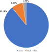
Figures


Similar articles
-
Introduction to high-resolution cryo-electron microscopy.Postepy Biochem. 2016;62(3):383-394. Postepy Biochem. 2016. PMID: 28132494 Review. English.
-
Structure Determination by Single-Particle Cryo-Electron Microscopy: Only the Sky (and Intrinsic Disorder) is the Limit.Int J Mol Sci. 2019 Aug 27;20(17):4186. doi: 10.3390/ijms20174186. Int J Mol Sci. 2019. PMID: 31461845 Free PMC article. Review.
-
The integrative role of cryo electron microscopy in molecular and cellular structural biology.Biol Cell. 2017 Feb;109(2):81-93. doi: 10.1111/boc.201600042. Epub 2016 Nov 25. Biol Cell. 2017. PMID: 27730650 Review.
-
Cryo-Electron Microscopy Methodology: Current Aspects and Future Directions.Trends Biochem Sci. 2019 Oct;44(10):837-848. doi: 10.1016/j.tibs.2019.04.008. Epub 2019 May 8. Trends Biochem Sci. 2019. PMID: 31078399 Review.
-
Progress in spatial resolution of structural analysis by cryo-EM.Microscopy (Oxf). 2023 Apr 6;72(2):135-143. doi: 10.1093/jmicro/dfac053. Microscopy (Oxf). 2023. PMID: 36269102 Review.
Cited by
-
Using in vivo intact structure for system-wide quantitative analysis of changes in proteins.Nat Commun. 2024 Oct 29;15(1):9310. doi: 10.1038/s41467-024-53582-x. Nat Commun. 2024. PMID: 39468068 Free PMC article.
-
AI-Driven Deep Learning Techniques in Protein Structure Prediction.Int J Mol Sci. 2024 Aug 1;25(15):8426. doi: 10.3390/ijms25158426. Int J Mol Sci. 2024. PMID: 39125995 Free PMC article. Review.
-
Toward the analysis of functional proteoforms using mass spectrometry-based stability proteomics.Front Anal Sci. 2023;3:1186623. doi: 10.3389/frans.2023.1186623. Epub 2023 Jun 21. Front Anal Sci. 2023. PMID: 39072225 Free PMC article.
-
Exploring HIV-1 Maturation: A New Frontier in Antiviral Development.Viruses. 2024 Sep 6;16(9):1423. doi: 10.3390/v16091423. Viruses. 2024. PMID: 39339899 Free PMC article. Review.
-
Metal-Based Drug-DNA Interactions and Analytical Determination Methods.Molecules. 2024 Sep 13;29(18):4361. doi: 10.3390/molecules29184361. Molecules. 2024. PMID: 39339356 Free PMC article. Review.
References
-
- Yin C‐C. Structural biology revolution led by technical breakthroughs in cryo‐electron microscopy. Chinese J Biochem Mol Biol. 2018;34:1–12.
-
- De Rosier DJ, Klug A. Reconstruction of three dimensional structures from electron micrographs. Nature. 1968;217:130–134. - PubMed
-
- Rubinstein JL. Cryo‐EM captures the dynamics of ion channel opening. Cell. 2017;168:341–343. - PubMed
-
- Renaud JP, Chari A, Ciferri C, et al. Cryo‐EM in drug discovery: Achievements, limitations and prospects. Nat Rev Drug Discov. 2018;17:471–492. - PubMed
Publication types
MeSH terms
Substances
LinkOut - more resources
Full Text Sources
Other Literature Sources

