. 2020 Feb;214(2):381-395.
doi: 10.1534/genetics.119.302939. Epub 2019 Dec 18.

Antioxidant CoQ10 Restores Fertility by Rescuing Bisphenol A-Induced Oxidative DNA Damage in the Caenorhabditis elegans Germline

Affiliations

Antioxidant CoQ10 Restores Fertility by Rescuing Bisphenol A-Induced Oxidative DNA Damage in the Caenorhabditis elegans Germline

Maria Fernanda Hornos Carneiro et al. Genetics. 2020 Feb.

Abstract

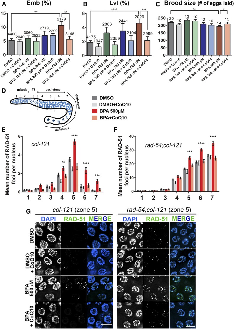
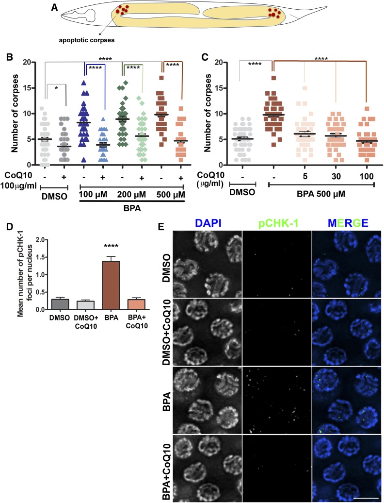
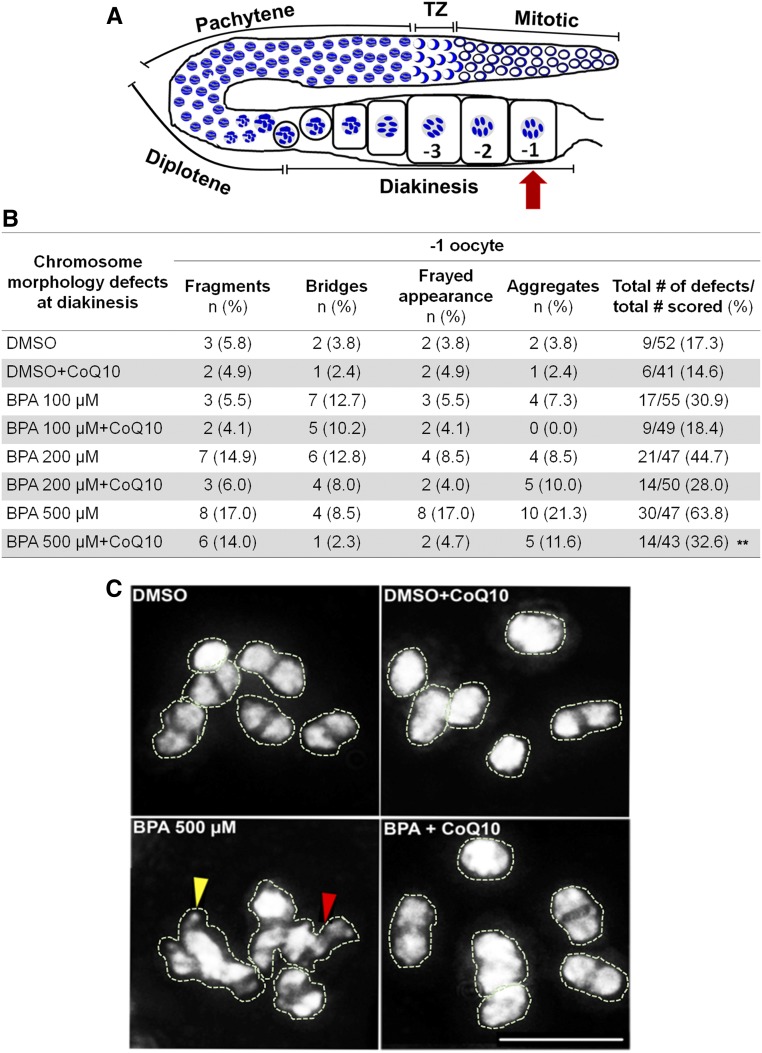
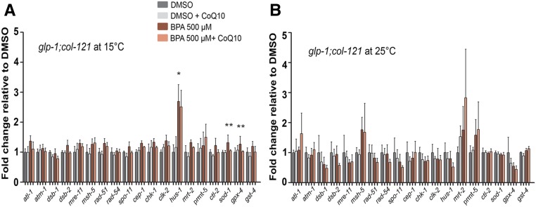
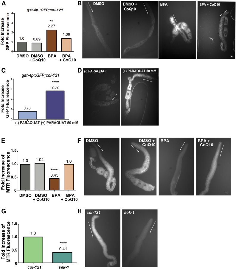
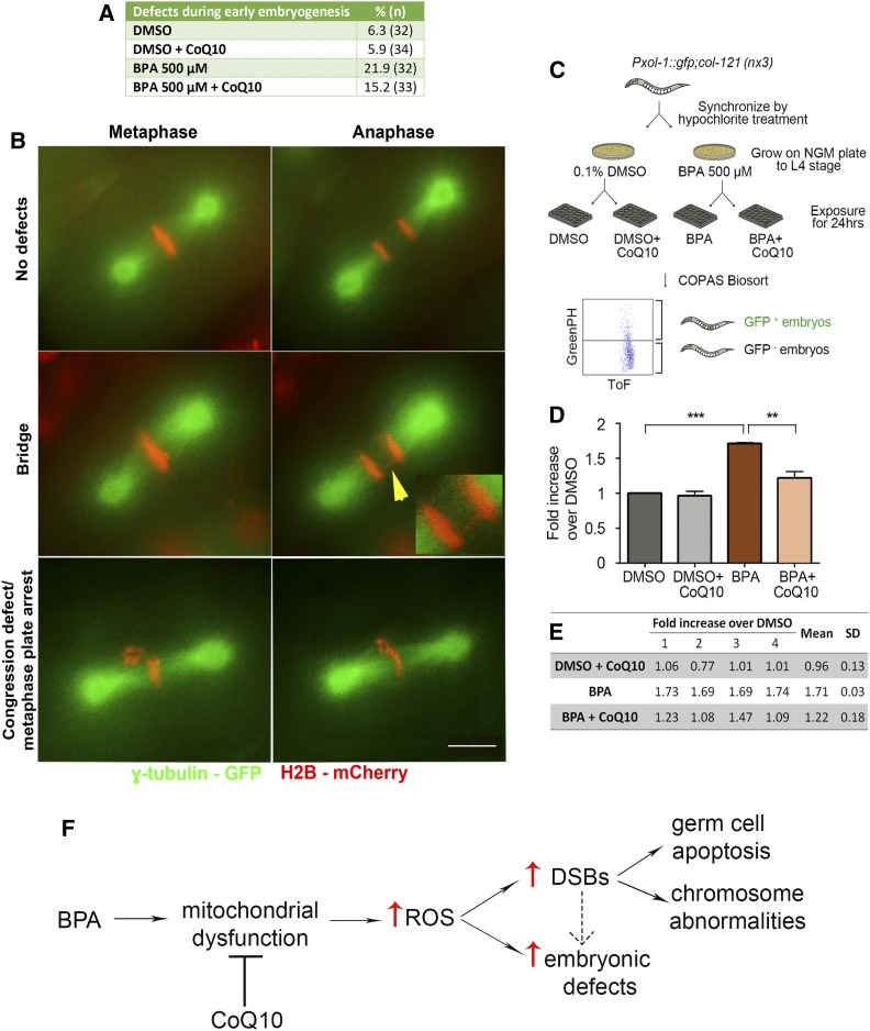
Endocrine-disrupting chemicals are ubiquitously present in our environment, but the mechanisms by which they adversely affect human reproductive health and strategies to circumvent their effects remain largely unknown. Here, we show in Caenorhabditis elegans that supplementation with the antioxidant Coenzyme Q10 (CoQ10) rescues the reprotoxicity induced by the widely used plasticizer and endocrine disruptor bisphenol A (BPA), in part by neutralizing DNA damage resulting from oxidative stress. CoQ10 significantly reduces BPA-induced elevated levels of germ cell apoptosis, phosphorylated checkpoint kinase 1 (CHK-1), double-strand breaks (DSBs), and chromosome defects in diakinesis oocytes. BPA-induced oxidative stress, mitochondrial dysfunction, and increased gene expression of antioxidant enzymes in the germline are counteracted by CoQ10. Finally, CoQ10 treatment also reduced the levels of aneuploid embryos and BPA-induced defects observed in early embryonic divisions. We propose that CoQ10 may counteract BPA-induced reprotoxicity through the scavenging of reactive oxygen species and free radicals, and that this natural antioxidant could constitute a low-risk and low-cost strategy to attenuate the impact on fertility by BPA.

Keywords: Bisphenol A; C. elegans; Coenzyme Q10; germline; meiosis.


Figures

Figure 1
Figure 1
CoQ10 rescues BPA-induced embryonic lethality (Emb), larval lethality (Lvl), and decreased brood size, and it protects from BPA-induced defects in DNA double-strand break formation and repair. (A) Embryonic lethality observed among the progeny of worms exposed to vehicle alone (0.1% DMSO) or the indicated doses of BPA, either with or without subsequent supplementation with 100 µg/ml CoQ10. Indicated above the error bars are the total numbers of eggs laid by the exposed hermaphrodites. (B) Larval lethality observed among the hatched progeny of exposed worms. Indicated above the error bars are the total numbers of larvae hatched from the eggs laid by the exposed hermaphrodites. (C) Mean number of eggs laid by worms exposed to the indicated conditions. Indicated above the error bars are the total numbers of hermaphrodites exposed from L1 to 24 hr post-L4, for which entire brood sizes were scored. Error bars represent SEM. P values calculated by the two-tailed Mann-Whitney test, 95% C.I. *P ≤ 0.05; **P ≤ 0.01; ***P ≤ 0.001; ****P ≤ 0.0001. (D) Diagram of the C. elegans germline indicating the position of the seven zones scored for RAD-51 foci. TZ, transition zone, corresponds to leptotene/zygotene stages of meiosis. (E and F) Mean number of RAD-51 foci scored per nucleus (y-axis) for each zone along the germline axis (x-axis) for the indicated genotypes. (E) Histogram indicates a significant increase in levels of RAD-51 foci in col-121 worms exposed to BPA-only compared to other cases. (F) Histogram indicates that the mean number of RAD-51 foci detected per nucleus is increased starting in midpachytene (zone 5) in rad-54;col-121 worms exposed to BPA-only compared to other cases. (G) Representative images of the immunostaining for RAD-51 on midpachytene nuclei (zone 5) from dissected gonads of col-121 (left) and rad-54;col-121 (right) treated worms. Error bars in (E and F) represent SEM. Two-tailed Mann-Whitney test, 95% C.I. Levels of RAD-51 foci in worms exposed to BPA only are statistically different from other cases with the following P values: **P ≤ 0.01; ***P ≤ 0.001; ****P ≤ 0.0001. DMSO was used at 0.1%; CoQ10 at 100 µg/ml, and BPA at 500 µM. Bar, 5 μm. N = 6 gonads.
Figure 2
Figure 2
CoQ10 rescues BPA-induced elevated germ cell apoptosis and pCHK-1 foci levels detected in pachytene nuclei. (A) Schematic representation of the U-shaped gonad arms of C. elegans indicating where the acridine orange stained corpses are scored. (B) Dose-response curve showing the quantification of apoptotic corpses for the indicated doses of BPA with and without CoQ10 (100 µg/ml) supplementation. (C) Lower doses of CoQ10 were also able to counteract the elevated BPA-induced (500 µM) germ cell apoptosis. Two-tailed Mann-Whitney test, 95% C.I. *P ≤ 0.05; ***P ≤ 0.001; ****P ≤ 0.0001. DMSO was used at 0.1%. N ≥ 32, number of gonads scored per condition. (D) Mean number of pCHK-1 foci per nucleus ± SEM scored in mid to late pachytene for the indicated conditions. (E) Representative images of the immunostaining for pCHK-1 on midpachytene (zone 5) nuclei from dissected gonads. Two-tailed Mann-Whitney test, 95% C.I. ****P ≤ 0.0001 compared to all other conditions. DMSO was used at 0.1%; CoQ10 at 100 µg/ml and BPA at 500 µM. Bar, 5 µm. N = 6, number of gonads scored per condition.
Figure 3
Figure 3
CoQ10 ameliorates the deleterious effects of BPA on chromosome morphology observed at diakinesis (−1 oocyte). (A) Schematic representation of one gonad arm of C. elegans indicating the location of the −1 oocyte (red arrow). (B) Quantification of the chromosome morphology defects observed in bivalents at diakinesis. n = number of −1 oocytes in which each indicated type of defect was observed. DMSO was used at 0.1%; CoQ10 at 100 µg/ml. Fisher’s exact test pairwise comparisons for the total numbers of defects/total number of oocytes scored revealed **P = 0.0035 for BPA 500 µM vs. BPA 500 µM + CoQ10, P = 0.0043 for DMSO vs. BPA 200 µM and P < 0.0001 for DMSO vs. BPA 500 µM. (C) Representative images of the DAPI-stained bodies in the −1 oocyte. Six intact bivalents are shown for control and either control or BPA 500 µM supplemented with CoQ10. In contrast, higher frequencies of oocytes carrying chromosomes with a frayed appearance, aggregates, chromatin bridges (red arrowhead), and fragments (yellow arrowhead) are observed upon BPA-treatment. Bivalents and aggregates have been traced to facilitate visualization of the DAPI-stained bodies being scored, given that, once the 3D data stacks encompassing whole nuclei are “flattened,” some of the bivalents look superimposed onto each other in the final projections. Bar, 5 µm.
Figure 4
Figure 4
CoQ10 counteracts BPA-induced altered gene expression of antioxidant enzymes in the germline. (A and B) Expression levels of a panel of genes implicated in DSBR, DNA damage response, and oxidative stress scavenging were assayed by quantitative RT-PCR. Analysis of glp-1;col-121 worms at 15°C (A) reports expression in both the germline and the soma, while only expression in the soma can be scored at 25°C (B). Error bars represent SEM. BPA is statistically different from DMSO at *P = 0.0286 (hus-1); BPA is statistically different from BPA + CoQ10 at **P = 0.006 and **P = 0.0017 for sod-1 and gpx-4, respectively, by the unpaired two-tailed t-test, 95% C.I. DMSO was used at 0.1%; CoQ10 at 100 µg/ml.
Figure 5
Figure 5
BPA-induced oxidative stress and mitochondrial dysfunction in the germline are rescued by CoQ10 supplementation. (A) Fold increase of gonadal GFP fluorescence showing CoQ10 rescues the elevated signal in gonads from worms exposed to BPA. gst-4p::GFP;col-121 transgenic worms were examined. (B) Dissected gonads from transgenic gst-4p::GFP; col-121 worms exposed to the indicated chemicals. GFP signal is elevated in the germlines of BPA-exposed worms, and that signal is decreased in the gonads of BPA-exposed worms supplemented with CoQ10. Gonads from worms exposed to either DMSO-only or DMSO + CoQ10 are shown as controls. (C) Fold increase of gonadal GFP fluorescence showing paraquat treatment (50 mM) significantly augments the signal in gst-4p::GFP;col-121 worms. (D) Dissected gonads from transgenic gst-4p::GFP; col-121 worms either exposed (+) or not (−) to paraquat 50 mM. (E–H) Staining of mitochondria using Mitotracker Red was examined in the gonads. (E) Fold increase of gonadal Mitotracker Red (MTR) fluorescence showing CoQ10 rescues mitochondrial function in BPA-exposed worms. (F) Dissected gonads from col-121 worms exposed to the indicated chemicals. Mitotracker Red signal is significantly decreased in the germlines of BPA-exposed worms and increased in the gonads of BPA-exposed worms supplemented with CoQ10. Gonads from worms exposed to either DMSO-only or DMSO + CoQ10 are shown as controls. (G) Quantification of gonadal Mitotracker Red (MTR) signal detected in untreated sek-1 mutants showing a significant decrease compared to untreated col-121 (control). (H) Dissected gonads from col-121 and sek-1 worms were examined after Mitotracker staining. White arrows are positioned adjacent to the premeiotic region indicating the direction of germline progression. Dissected gonads are shown to facilitate visualization and quantitation of signal specifically in the germline, which otherwise is confounded by signal in other regions of the worms’ bodies. Statistically different from DMSO and DMSO + CoQ10 at P ≤ 0.0001, statistically different from BPA + CoQ10 at **P ≤ 0.01 (A); statistically different from (−) paraquat at ****P ≤ 0.0001 (C); statistically different from DMSO and BPA at ****P < 0.0001, statistically different from BPA + CoQ10 at ****P < 0.0001 (E); statistically different from col-121 and sek-1 at ****P < 0.0001 (G) by the two-tailed Mann-Whitney test, 95% C.I. DMSO used at 0.1%; CoQ10 at 100 µg/ml and BPA at 500 µM. Bar, 5 µm. N ≥ 18, number of gonads scored per treatment.
Figure 6
Figure 6
CoQ10 supplementation rescues BPA-induced defects during the first embryonic division. (A and B) A higher frequency of chromosome segregation defects during the first embryonic cell division is seen in H2B::mCherry; γ-tubulin::GFP; col-121 embryos after treatment with BPA compared to vehicle (DMSO). CoQ10 partially rescues this effect. (A) Quantification of defects detected during the first embryonic cell division. (B) Time-lapse images of the first embryonic division. Shown are examples of normal chromosome alignment at metaphase and segregation at anaphase (top row), with chromosomes in red and spindle microtubules in green. Center and bottom rows show examples of chromatin bridges (yellow arrowhead; insert shows a magnified image of the chromatin bridge), congression defects, and metaphase arrest observed. A Pearson Chi-Square test of independence was calculated for a pairwise comparison of defective and nondefective first embryonic divisions. DMSO used at 0.1%; CoQ10 at 100 µg/ml. Bar, 5 µm. N ≥ 32 per condition. (C–E) High-throughput analysis with the COPAS Biosort shows CoQ10 rescues the elevated chromosome nondisjunction detected during embryogenesis following BPA exposure. (C) Flowchart for high-throughput analysis with the COPAS Biosort. Age-matched L1 stage animals were grown on either DMSO- or BPA-containing NGM plates until reaching the L4 stage. L4 stage animals from DMSO plates were dispensed into 24-well plates (300 worms/well) containing OP50 E. coli (OD600 = 24) in M9 buffer and either DMSO alone or DMSO with CoQ10, and L4 stage animals from BPA plates were dispensed into 24-well plates with either BPA or BPA with CoQ10. After a 24-hr incubation at 20°, worms were thoroughly washed with M9 and sorted with the COPAS Biosort based on fluorescence intensity. Time-of-flight (Tof) and GFP peak height were used as reading parameters. (D) Levels of GFP+ embryos (resulting from X chromosome nondisjunction) detected in the uterus of BPA-treated worms are significantly higher relative to DMSO (***P < 0.0001) and CoQ10 treatment results in a significant reduction in the elevation induced by BPA (**P = 0.0021). DMSO used at 0.1%; BPA at 500 µM; CoQ10 at 100 µg/ml. Statistical comparisons were performed using the unpaired two-tailed t-test, 95% C.I. Error bars represent SEM. (E) Readouts obtained with the COPAS Biosort for the indicated chemicals. Shown is the fold increase over DMSO alone calculated for each biological repeat (labeled as 1 through 4). SD, standard deviation. (F) Model for how CoQ10 restores fertility by rescuing BPA-induced oxidative DNA damage in the C. elegans germline. BPA exposure may lead to mitochondrial dysfunction in the germline resulting in increased reactive oxygen species (ROS) that in turn may account, at least in part, for the increased levels of DNA double-strand breaks (DSBs) and the embryonic defects observed. Unrepaired or aberrantly repaired meiotic DSBs lead to the activation of a late pachytene DNA damage checkpoint resulting in increased germ cell apoptosis, the increased chromosome abnormalities detected in oocytes at late diakinesis, and some of the observed embryonic defects (the latter is depicted by a dashed arrow, given that a direct effect on embryos may also be possible). Only the pathway/mode of action directly tested in this study is depicted, but other studies have shown additional effects such as alterations in gene expression (Allard and Colaiacovo 2010; Chen et al. 2016).

Similar articles

Cited by

References

    1. Allard P., and Colaiacovo M. P., 2010. Bisphenol A impairs the double-strand break repair machinery in the germline and causes chromosome abnormalities. Proc. Natl. Acad. Sci. USA 107: 20405–20410. 10.1073/pnas.1010386107 - DOI - PMC - PubMed
    1. Allard P., Kleinstreuer N. C., Knudsen T. B., and Colaiacovo M. P., 2013. A C. elegans screening platform for the rapid assessment of chemical disruption of germline function. Environ. Health Perspect. 121: 717–724. 10.1289/ehp.1206301 - DOI - PMC - PubMed
    1. Alpi A., Pasierbek P., Gartner A., and Loidl J., 2003. Genetic and cytological characterization of the recombination protein RAD-51 in Caenorhabditis elegans. Chromosoma 112: 6–16. 10.1007/s00412-003-0237-5 - DOI - PubMed
    1. Arany I., Carter A., Hall S., Fulop T., and Dixit M., 2017. Coenzyme Q10 protects renal proximal tubule cells against nicotine-induced apoptosis through induction of p66shc-dependent antioxidant responses. Apoptosis 22: 220–228. 10.1007/s10495-016-1309-3 - DOI - PubMed
    1. Asencio C., Navas P., Cabello J., Schnabel R., Cypser J. R. et al. , 2009. Coenzyme Q supports distinct developmental processes in Caenorhabditis elegans. Mech. Ageing Dev. 130: 145–153. 10.1016/j.mad.2008.10.004 - DOI - PubMed

Publication types

MeSH terms