Proposition of a novel animal model of systemic sclerosis induced by type V collagen in C57BL/6 mice that reproduces fibrosis, vasculopathy and autoimmunity
- PMID: 31829272
- PMCID: PMC6907238
- DOI: 10.1186/s13075-019-2052-2
Proposition of a novel animal model of systemic sclerosis induced by type V collagen in C57BL/6 mice that reproduces fibrosis, vasculopathy and autoimmunity
Abstract
Background: Type V collagen (Col V) has the potential to become an autoantigen and has been associated with the pathogenesis of systemic sclerosis (SSc). We characterized serological, functional, and histopathological features of the skin and lung in a novel SSc murine model induced by Col V immunization.
Methods: Female C57BL/6 mice (n = 19, IMU-COLV) were subcutaneously immunized with two doses of Col V (125 μg) emulsified in complete Freund adjuvant, followed by two intramuscular boosters. The control group (n = 19) did not receive Col V. After 120 days, we examined the respiratory mechanics, serum autoantibodies, and vascular manifestations of the mice. The skin and lung inflammatory processes and the collagen gene/protein expressions were analyzed.
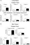
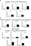
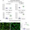
Results: Vascular manifestations were characterized by endothelial cell activity and apoptosis, as shown by the increased expression of VEGF, endothelin-1, and caspase-3 in endothelial cells. The IMU-COLV mice presented with increased tissue elastance and a nonspecific interstitial pneumonia (NSIP) histologic pattern in the lung, combined with the thickening of the small and medium intrapulmonary arteries, increased Col V fibers, and increased COL1A1, COL1A2, COL3A1, COL5A1, and COL5A2 gene expression. The skin of the IMU-COLV mice showed thickness, epidermal rectification, decreased papillary dermis, atrophied appendages, and increased collagen, COL5A1, and COL5A2 gene expression. Anti-collagen III and IV and ANA antibodies were detected in the sera of the IMU-COLV mice.
Conclusion: We demonstrated that cutaneous, vascular, and pulmonary remodeling are mimicked in the Col V-induced SSc mouse model, which thus represents a suitable preclinical model to study the mechanisms and therapeutic approaches for SSc.
Keywords: Autoantibodies; Fibrosis; Mouse model; Systemic sclerosis; Type V collagen; Vascular.
Conflict of interest statement
The authors declare that they have no competing interests.
Figures


Similar articles
-
Identification of Autoimmunity to Peptides of Collagen V α1 Chain as Newly Biomarkers of Early Stage of Systemic Sclerosis.Front Immunol. 2021 Feb 12;11:604602. doi: 10.3389/fimmu.2020.604602. eCollection 2020. Front Immunol. 2021. PMID: 33643291 Free PMC article.
-
Pathological pulmonary vascular remodeling is induced by type V collagen in a model of scleroderma.Pathol Res Pract. 2021 Apr;220:153382. doi: 10.1016/j.prp.2021.153382. Epub 2021 Feb 19. Pathol Res Pract. 2021. PMID: 33647866
-
Autoantibody profile in the experimental model of scleroderma induced by type V human collagen.Immunology. 2007 Sep;122(1):38-46. doi: 10.1111/j.1365-2567.2007.02610.x. Epub 2007 Apr 18. Immunology. 2007. PMID: 17442023 Free PMC article.
-
Abnormal collagen V deposition in dermis correlates with skin thickening and disease activity in systemic sclerosis.Autoimmun Rev. 2012 Sep;11(11):827-35. doi: 10.1016/j.autrev.2012.02.017. Epub 2012 Mar 2. Autoimmun Rev. 2012. PMID: 22406224 Review.
-
Pathogenesis of systemic sclerosis-current concept and emerging treatments.Immunol Res. 2017 Aug;65(4):790-797. doi: 10.1007/s12026-017-8926-y. Immunol Res. 2017. PMID: 28488090 Review.
Cited by
-
Recent Insights into Cellular and Molecular Mechanisms of Defective Angiogenesis in Systemic Sclerosis.Biomedicines. 2024 Jun 14;12(6):1331. doi: 10.3390/biomedicines12061331. Biomedicines. 2024. PMID: 38927538 Free PMC article. Review.
-
Molecular Mechanisms of Fetal Tendon Regeneration Versus Adult Fibrous Repair.Int J Mol Sci. 2021 May 25;22(11):5619. doi: 10.3390/ijms22115619. Int J Mol Sci. 2021. PMID: 34070692 Free PMC article.
-
Establishment of a humanized animal model of systemic sclerosis in which T helper-17 cells from patients with systemic sclerosis infiltrate and cause fibrosis in the lungs and skin.Exp Mol Med. 2022 Sep;54(9):1577-1585. doi: 10.1038/s12276-022-00860-7. Epub 2022 Sep 29. Exp Mol Med. 2022. PMID: 36175484 Free PMC article.
-
Identification of Autoimmunity to Peptides of Collagen V α1 Chain as Newly Biomarkers of Early Stage of Systemic Sclerosis.Front Immunol. 2021 Feb 12;11:604602. doi: 10.3389/fimmu.2020.604602. eCollection 2020. Front Immunol. 2021. PMID: 33643291 Free PMC article.
-
Collagen V α1 Chain Decrease in Papillary Dermis from Early Systemic Sclerosis: A New Proposal in Cutaneous Fibrosis Molecular Structure.Int J Mol Sci. 2022 Oct 21;23(20):12654. doi: 10.3390/ijms232012654. Int J Mol Sci. 2022. PMID: 36293511 Free PMC article.
References
Publication types
MeSH terms
Substances
LinkOut - more resources
Full Text Sources
Medical
Research Materials
Miscellaneous

