

Microscopic Examination of Nail Clippings from Patients with Palmoplantar Pustulosis
- PMID: 31828062
- PMCID: PMC6902250
- DOI: 10.1159/000503704
Microscopic Examination of Nail Clippings from Patients with Palmoplantar Pustulosis
Abstract
This study describes the clinical characteristics and microscopic findings of nails from 25 patients with palmoplantar pustulosis.
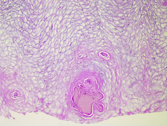
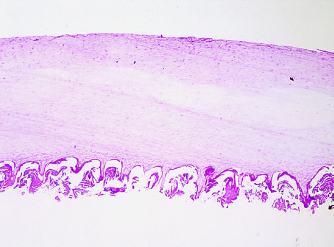
Methods: This is a cross-sectional study of adult patients with clear-cut palmoplantar pustulosis. Onychodystrophy severity was evaluated in fingernails using the nail psoriasis severity index (NAPSI). A fragment of the most dystrophic fingernail was collected from each patient and submitted to routine histotechnical processing. The following microscopic parameters were evaluated: nail plate and subungual region thickness, presence or absence of parakeratosis, number of layers of parakeratosis, and presence of neutrophils, serous lakes, bacteria, blood, and fungi.
Results: Twenty-one patients (84%) presented onychodystrophy with a mean NAPSI score of 12.67. The most common nail change was pitting (76.19% of patients). On average, nail plate thickness and subungual region thickness measured 0.42 and 0.14 mm, respectively. Neutrophils and fungi were not observed, but serous lakes were found in 4.7%, bacteria in 28.57%, blood in 4.76%, and parakeratosis in 19.05% of the patients.
Conclusions: although palmoplantar pustulosis is a disease with great amounts of neutrophils in the epidermis, those cells were not found in the nail clippings studied herein. Furthermore, when clinical aspects and microscopic findings of palmoplantar pustulosis are compared to those of similar studies in psoriasis vulgaris, they show different characteristics.
Keywords: Microscopy; Nail clipping; Nails; Onychodystrophy; Palmoplantar pustulosis; Psoriasis.
Copyright © 2019 by S. Karger AG, Basel.
Conflict of interest statement
The authors have no conflicts of interest to declare.
Figures
Similar articles
-
Microscopic nail clipping findings in patients with psoriasis.Am J Dermatopathol. 2015 Jun;37(6):429-39. doi: 10.1097/DAD.0000000000000197. Am J Dermatopathol. 2015. PMID: 25993403
-
Comparative microscopic analysis of nail clippings from patients with cutaneous psoriasis and psoriatic arthritis.An Bras Dermatol. 2017 Jan-Feb;92(1):21-25. doi: 10.1590/abd1806-4841.20175056. An Bras Dermatol. 2017. PMID: 28225951 Free PMC article.
-
Microscopic examination of normal nail clippings.Dermatol Pract Concept. 2013 Jul 31;3(3):9-14. doi: 10.5826/dpc.0303a04. eCollection 2013. Dermatol Pract Concept. 2013. PMID: 24106655 Free PMC article.
-
Similarity and difference between palmoplantar pustulosis and pustular psoriasis.J Dermatol. 2021 Jun;48(6):750-760. doi: 10.1111/1346-8138.15826. Epub 2021 Mar 2. J Dermatol. 2021. PMID: 33650702 Review.
-
The interleukin 1 receptor antagonist anakinra to reduce disease severity of palmoplantar pustulosis in adults: APRICOT RCT and PLUM mechanistic study.Southampton (UK): NIHR Journals Library; 2022 Mar. Southampton (UK): NIHR Journals Library; 2022 Mar. PMID: 35377574 Free Books & Documents. Review.
Cited by
-
Prevalence of onychomycosis among psoriasis patients: a clinico-mycological and dermoscopic comparative cross sectional study.Sci Rep. 2024 Sep 18;14(1):21743. doi: 10.1038/s41598-024-71321-6. Sci Rep. 2024. PMID: 39289407 Free PMC article.
References
-
- Mrowietz U, van de Kerkhof PC. Management of palmoplantar pustulosis: do we need to change? Br J Dermatol. 2011 May;164((5)):942–6. - PubMed
-
- Sugiura K. The genetic background of generalized pustular psoriasis: IL36RN mutations and CARD14 gain-of-function variants. J Dermatol Sci. 2014 Jun;74((3)):187–92. - PubMed
-
- Navarini AA, Burden AD, Capon F, Mrowietz U, Puig L, Köks S, et al. ERASPEN Network European consensus statement on phenotypes of pustular psoriasis. J Eur Acad Dermatol Venereol. 2017 Nov;31((11)):1792–9. - PubMed
-
- Farley E, Masrour S, McKey J, Menter A. Palmoplantar psoriasis: a phenotypical and clinical review with introduction of a new quality-of-life assessment tool. J Am Acad Dermatol. 2009 Jun;60((6)):1024–31. - PubMed
-
- Adişen E, Tekin O, Gülekon A, Gürer MA. A retrospective analysis of treatment responses of palmoplantar psoriasis in 114 patients. J Eur Acad Dermatol Venereol. 2009 Jul;23((7)):814–9. - PubMed
LinkOut - more resources
Full Text Sources
