. 2020 Jan 24;295(4):1077-1090.
doi: 10.1074/jbc.RA119.011424. Epub 2019 Dec 10.

A 49-residue sequence motif in the C terminus of Nav1.9 regulates trafficking of the channel to the plasma membrane

Affiliations

A 49-residue sequence motif in the C terminus of Nav1.9 regulates trafficking of the channel to the plasma membrane

Daria V Sizova et al. J Biol Chem. .

Abstract

Genetic and functional studies have confirmed an important role for the voltage-gated sodium channel Nav1.9 in human pain disorders. However, low functional expression of Nav1.9 in heterologous systems (e.g. in human embryonic kidney 293 (HEK293) cells) has hampered studies of its biophysical and pharmacological properties and the development of high-throughput assays for drug development targeting this channel. The mechanistic basis for the low level of Nav1.9 currents in heterologous expression systems is not understood. Here, we implemented a multidisciplinary approach to investigate the mechanisms that govern functional Nav1.9 expression. Recombinant expression of a series of Nav1.9-Nav1.7 C-terminal chimeras in HEK293 cells identified a 49-amino-acid-long motif in the C terminus of the two channels that regulates expression levels of these chimeras. We confirmed the critical role of this motif in the context of a full-length channel chimera, Nav1.9-Ct49aaNav1.7, which displayed significantly increased current density in HEK293 cells while largely retaining the characteristic Nav1.9-gating properties. High-resolution live microscopy indicated that the newly identified C-terminal motif dramatically increases the number of channels on the plasma membrane of HEK293 cells. Molecular modeling results suggested that this motif is exposed on the cytoplasmic face of the folded C terminus, where it might interact with other channel partners. These findings reveal that a 49-residue-long motif in Nav1.9 regulates channel trafficking to the plasma membrane.

Keywords: Nav1.9; channel trafficking; electrophysiology; functional expression; live imaging; nociception; pain; sensory neurons; sodium channel; structural model; trafficking; voltage clamp.


Conflict of interest statement

The authors declare that they have no conflicts of interest with the contents of this article

Figures

Figure 1.
Figure 1.
C terminus of Nav1.9 is expressed at low levels in transiently transfected HEK293 cells. A, schematic representation of the MS-CtX-SF construct showing location and composition of the myristoylation signal (green box, full sequence included) and streptavidin-FLAG tag (light blue box with stripes). The C terminus of a sodium channel (CtX) or a control full GFP sequence was inserted in-frame between the myristoylation and SF tags (yellow). B, representative Western blotting showing comparative expression levels of MS-CtNav1.9-SF, MS-CtNav1.7-SF, and MS-CtNav1.6-SF constructs as well as a control MS-GFP-SF. Total protein extracts were run on a 4–12% polyacrylamide gel, transferred to a nitrocellulose membrane, and probed with FLAG (top) or β-actin (bottom) antibodies. Lane E, a control transfection with an empty vector.
Figure 2.
Figure 2.
Chimeric construct with a 49-amino acid swap between Nav1.7 and Nav1.9 is sufficient for rescuing expression of the Nav1.9 C terminus. A, aa sequences of Nav1.7 and Nav1.9 C termini are aligned using DNASTAR Lasergene software; nonconserved residues in Nav1.9 are highlighted in red. Aligned sequences were divided into three parts that contained divergent sequences separated by stretches of identical residues (region Pr (proximal) in blue, region Md (middle) in pink, and region Ds (distal) in green). B, three chimeric constructs were created by replacing the three regions of the Nav1.9 C terminus (yellow) with the corresponding sequences from the Nav1.7 C terminus. The 1.7Pr sequence (proximal 52 aa residues from Nav1.7 C terminus) is represented by a blue rectangle; 1.7Md (middle 49 aa from Nav1.7 C terminus) is represented by a pink rectangle; and 1.7Ds (distal 116 aa from Nav1.7 C terminus) is represented by a green rectangle. C, representative Western blotting showing comparative expression of ChimPr, ChimMd, and ChimDs constructs. Total protein extracts were probed with FLAG (top) or β-actin (bottom) antibodies. D, three chimeric constructs named ChimMd#1, ChimMd#2, and ChimMd#3 were created by replacing three subregions of the 49-aa middle region of the Nav1.9 C terminus (yellow) with the corresponding sequences from Nav1.7 (Md#1, Md#2, and Md#3, red, purple, and orange, respectively). E, representative Western blotting showing protein levels of ChimMd#1, ChimMd#2, and ChimMd#3 chimera. Total protein extracts were probed with FLAG (top) or β-actin (bottom) antibodies.
Figure 3.
Figure 3.
The reciprocal swapping of the 49-amino acid motif between Nav1.7 and Nav1.9 has only a minor effect on steady-state protein level. A, schematic representation of the Nav1.9-Ct49aaNav1.7 and Nav1.7-Ct49aaNav1.9 chimeric constructs showing approximate location of the 49-aa residue swap between Nav1.9 and Nav1.7 in the context of full channel sequences (green for Nav1.9, purple for Nav1.7) as well as the N-terminal GFP-2A sequence (bright green rectangle). B, representative Western blotting image showing protein levels of Nav1.9, Nav1.9-CtNav1.7, Nav1.9-Ct49aaNav1.7, Nav1.7, and Nav1.7-Ct49aaNav1.9 in transiently transfected HEK293 cells. Total protein extracts were probed with pan-sodium channel (top) or β-actin (bottom) antibodies. C, protein expression levels from three independent transfections for each construct were quantified using ImageLab software and presented as mean ± S.D. (error bars). A 2.2-fold higher protein level of Nav1.7 channel compared with Nav1.9 channel was observed (Nav1.9: 6.24 ± 0.64 AU, n = 3; Nav1.7: 13.46 ± 2.62 AU, n = 3, p < 0.05), by one-way ANOVA (F(4, 10) = 5.220, p = 0.0156), followed by Tukey's post hoc analysis. Differences in protein levels of all of the other constructs were statistically insignificant.
Figure 4.
Figure 4.
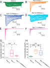
The 49-aa motif in the C terminus increases Nav1.9 current amplitude in HEK293 cells. A–F, representative current traces recorded from HEK293 cells expressing either Nav1.9 (A, green), Nav1.9-CtNav1.7 (B, orange), Nav1.9-Ct49aaNav1.7 (C, navy blue), Nav1.7 (E, red), or Nav1.7-Ct49aaNav1.9 (F, magenta). Current traces recorded from HEK293 cells expressing Nav1.9-Ct49aaNav1.7 and demonstrating ultra-slow inactivation kinetics and large persistent current (in ∼20% HEK293 cells) similar to the parent Nav1.9 current are shown separately (D, light blue). G, comparison of current density among Nav1.9 (n = 15), Nav1.7 (n = 11), and chimera Nav1.7-Ct49aaNav1.9 (n = 10). H, comparison of current density among Nav1.9 (n = 15), Nav1.9-CtNav1.7 (n = 8), and Nav1.9-Ct49aaNav1.7 (n = 7) channels. Error bars, S.E. Statistical significance is indicated by asterisks: *, p < 0.05; **, p < 0.01; ***, p < 0.001, by one-way ANOVA followed by Tukey's multiple-comparison test. ns, not significant.
Figure 5.
Figure 5.
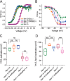
The C terminus of Nav1.9 modulates voltage-dependence of steady-state fast-inactivation but not activation. Boltzmann fits of voltage dependence of activation (A) and steady-state fast inactivation (B) for WT Nav1.9 and Nav1.7 channels and their chimeras. C, comparison of midpoint voltage of activation (V½Act) among all tested channels showing no difference in chimeric channels Nav1.9-CtNav1.7 (n = 8), Nav1.9-Ct49aaNav1.7 (n = 7), and Nav1.7-Ct49aaNav1.9 (n = 10), compared with parent channels Nav1.9 (n = 4) and Nav1.7 (n = 10). D, comparison of midpoint voltage of fast inactivation (V½Inact) among all tested channels (Nav1.9-CtNav1.7 (n = 8); Nav1.9-Ct49aaNav1.7 (n = 7); Nav1.7-Ct49aaNav1.9 (n = 10); Nav1.9 (n = 4); Nav1.7 (n = 10)). Error bars, S.E. Statistical significance is indicated by asterisks: *, p < 0.05; **, p < 0.01; ***, p < 0.001, by one-way ANOVA followed by Tukey's multiple-comparison test. ns, not significant.
Figure 6.
Figure 6.
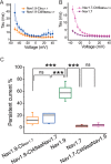
The effect of the 49-aa motif on kinetics of open-state fast inactivation and persistent current. A, time constants of open-state fast inactivation for Nav1.9-CtNav1.7 (orange, n = 8) and Nav1.9-Ct49aaNav1.7 (blue, n = 7). B, time constants of open-state fast inactivation were compared between Nav1.7 (n = 11) and Nav1.7-Ct49aaNav1.9 (n = 10). C, comparison of persistent current, normalized to the peak current, among all channels tested (Nav1.9-CtNav1.7, n = 8; Nav1.9-Ct49aaNav1.7, n = 7; Nav1.9 (n = 4); Nav1.7 (n = 11); Nav1.7-Ct49aaNav1.9 (n = 10)). Error bars, S.E. ***, statistical significance with p < 0.001. ns, not significant.
Figure 7.
Figure 7.
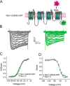
Nav1.9-BAD-GFP channels produce currents comparable with untagged Nav1.9 channels. A, schematic representation of the Nav1.9-BAD-GFP construct showing locations of the BAD (orange loop) and the C-terminal GFP tag (green star). The BAD domain is biotinylated by a co-transfected bacterial biotin ligase, BirA, which allows channel detection using a streptavidin-conjugated fluorophore CF-640R (red star). B, representative current traces recorded from superior cervical ganglion neurons expressing either Nav1.9 (green) or Nav1.9-BAD-GFP (gray). C, comparison of voltage dependence of activation for Nav1.9 (green, n = 9) and Nav1.9-BAD-GFP (gray, n = 8) channels. D, comparison of voltage dependence of steady-state fast inactivation for Nav1.9 (green, n = 9) or Nav1.9-BAD-GFP (gray, n = 8) channels. Error bars, S.E.
Figure 8.
Figure 8.
Steady-state protein levels of Nav1.9-BAD-GFP and Venus-Nav1.7-BAD in transiently transfected HEK293 cells. A, Western blotting of cell lysate of HEK293 cells transfected with either Nav1.9-BAD-GFP or Venus-Nav1.7-BAD. The blot was probed with pan-sodium channel (top) or β-actin (bottom) antibodies. Three independent transfections of different batches of HEK293 cells were analyzed: lanes 1–3 (Nav1.9-BAD-GFP) and lanes 4–6 (Venus-Nav1.7-BAD). B, protein expression levels were quantified using ImageLab software and presented as mean ± S.D. (error bars). A 3.1-fold difference was observed in protein expression between Nav1.9-BAD-GFP (0.32 ± 0.10 AU; n = 3) and Venus-Nav1.7-BAD constructs (1.00 ± 0.36 AU; n = 3), p < 0.05. Significance was determined using a two-tailed unpaired t test.
Figure 9.
Figure 9.
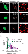
Imaging of Nav1.9 and Nav1.7 channels at the cell surface in HEK293 cells. A, representative TIRF images (green, 488-nm laser, first column of images; red, 647-nm laser, second column) of HEK293 cells transfected with any of the three constructs Venus-Nav1.7-BAD, Nav1.9-BAD-GFP, and Nav1.9-BAD-Ct49aaNav1.7-GFP. The third column shows ×10 magnified areas (insets marked by a square) within images from the second column. B, histograms of surface fluorescence intensity of SA-CF-640R–labeled Nav1.9-BAD-GFP (green), Nav1.9-BAD-Ct49aaNav1.7-GFP (blue), and Venus-Nav1.7-BAD (purple) channels on HEK293 cell surface. Solid green, blue, and purple lines are fits to the histogram distribution of the Nav1.9-BAD-GFP and Venus-Nav1.7-BAD constructs, respectively, with a mean value of 249 ± 38 AU, n = 30 for Nav1.9-BAD-GFP; 658 ± 64 AU, n = 30 for Nav1.9-BAD-Ct49aaNav1.7-GFP; and 1156 ± 100 AU, n = 30 for the Venus-Nav1.7-BAD construct. Differences between these values are statistically significant (one-way ANOVA (F(2, 87) = 9.769, p = 0.0001), followed by Tukey's multiple-comparison test).
Figure 10.
Figure 10.
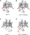
Structures for the C-terminal domains of Nav1.7 and Nav1.9 and their chimeric versions. All channel structures were created using the homology modeling tool in the MOE program using the recently published core structure of hNav1.7 (Protein Data Bank entry 6J8I) to create energetically stable conformations of the intracellular loops of the channel constructs used in the current study. The highly constrained structure of the core membrane-spanning segments are colored in gray. Although all of the intracellular sequences were modeled, only the C-terminal structure is shown for clarity. The default color scheme is red for α-helix and yellow for β-sheet. A, view of the Nav1.9 homology model illustrating the C-terminal domain. The 49-aa motif in Nav1.9 is colored green. B, view of the Nav1.9-Ct49aaNav1.7 chimera homology model illustrating the C-terminal domain. The 49-aa motif from Nav1.7 is colored cyan. C, view of the Nav1.7 homology model illustrating the C-terminal domain. The 49-aa motif in Nav1.7 is colored cyan. D, view of the Nav1.7-Ct49aaNav1.9 chimera homology model illustrating the C-terminal domain. The 49-aa motif from Nav1.9 is colored green.

Similar articles

Cited by

References

    1. Dib-Hajj S. D., Tyrrell L., Black J. A., and Waxman S. G. (1998) NaN, a novel voltage-gated Na channel, is expressed preferentially in peripheral sensory neurons and down-regulated after axotomy. Proc. Natl. Acad. Sci. U.S.A. 95, 8963–8968 10.1073/pnas.95.15.8963 - DOI - PMC - PubMed
    1. Cummins T. R., Dib-Hajj S. D., Black J. A., Akopian A. N., Wood J. N., and Waxman S. G. (1999) A novel persistent tetrodotoxin-resistant sodium current in SNS-null and wild-type small primary sensory neurons. J. Neurosci. 19, RC43 10.1523/JNEUROSCI.19-24-j0001.1999 - DOI - PMC - PubMed
    1. Rush A. M., Cummins T. R., and Waxman S. G. (2007) Multiple sodium channels and their roles in electrogenesis within dorsal root ganglion neurons. J. Physiol. 579, 1–14 10.1113/jphysiol.2006.121483 - DOI - PMC - PubMed
    1. Dib-Hajj S. D., Black J. A., and Waxman S. G. (2015) NaV1.9: a sodium channel linked to human pain. Nat. Rev. Neurosci. 16, 511–519 10.1038/nrn3977 - DOI - PubMed
    1. Bennett D. L., Clark A. J., Huang J., Waxman S. G., and Dib-Hajj S. D. (2019) The role of voltage-gated sodium channels in pain signaling. Physiol. Rev. 99, 1079–1151 10.1152/physrev.00052.2017 - DOI - PubMed

Publication types

MeSH terms

Substances

Associated data

LinkOut - more resources