. 2019 Dec 5;76(5):738-752.e7.
doi: 10.1016/j.molcel.2019.10.034.

The Integrator Complex Attenuates Promoter-Proximal Transcription at Protein-Coding Genes

Affiliations

The Integrator Complex Attenuates Promoter-Proximal Transcription at Protein-Coding Genes

Nathan D Elrod et al. Mol Cell. .

Abstract

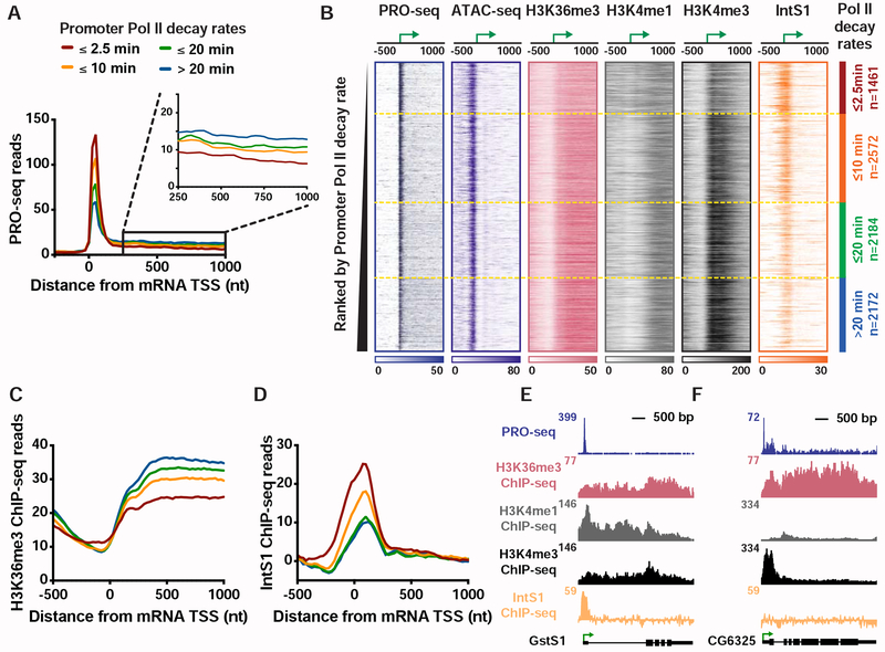
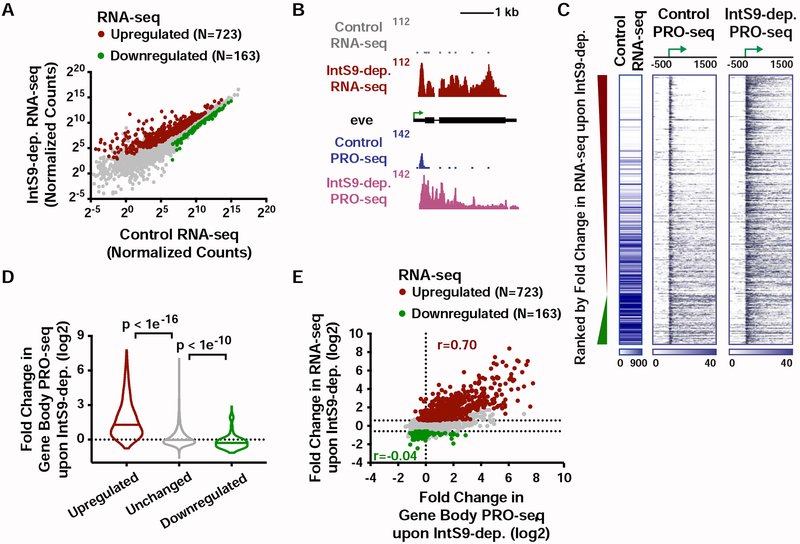
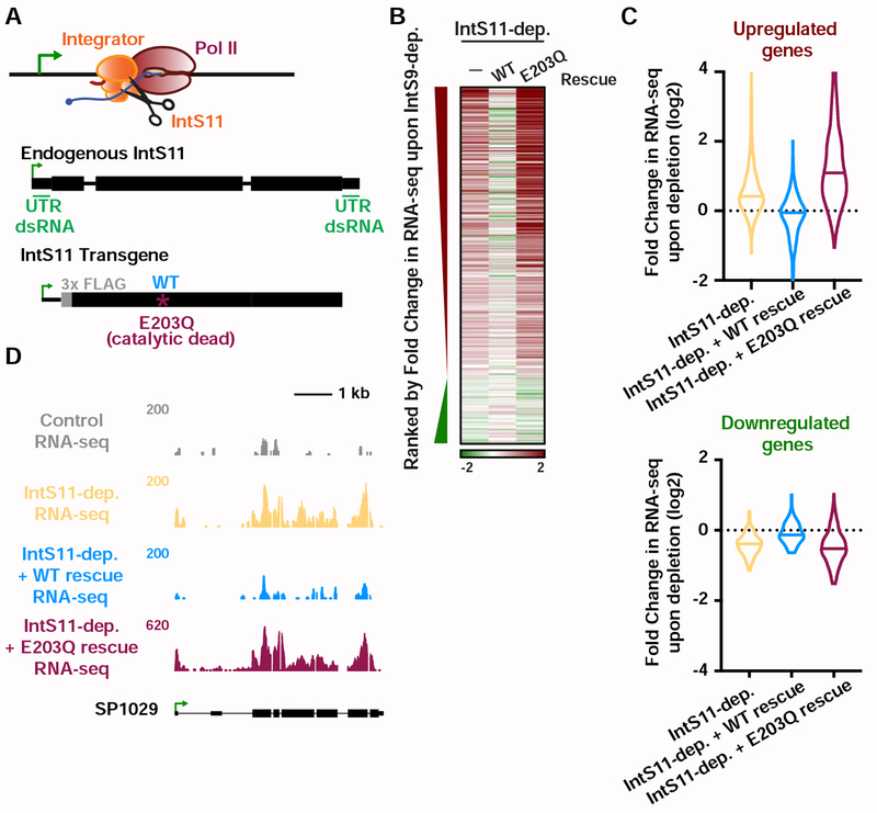
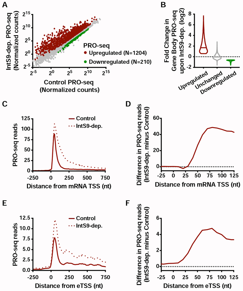
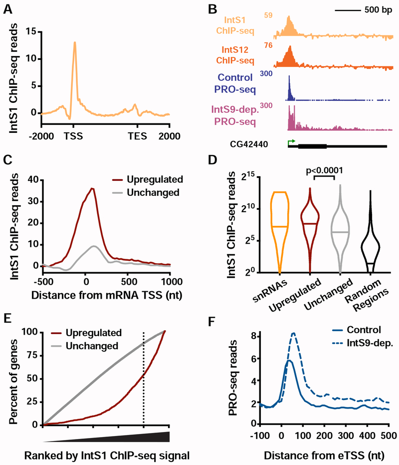
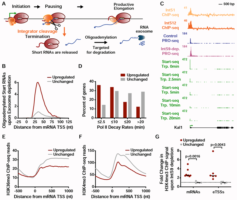
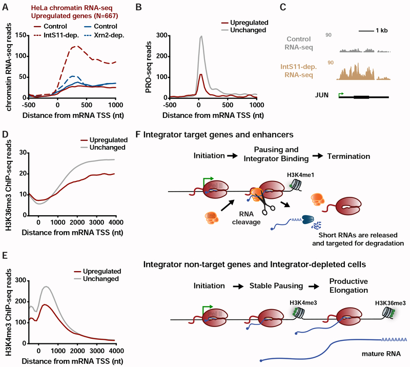
The transition of RNA polymerase II (Pol II) from initiation to productive elongation is a central, regulated step in metazoan gene expression. At many genes, Pol II pauses stably in early elongation, remaining engaged with the 25- to 60-nt-long nascent RNA for many minutes while awaiting signals for release into the gene body. However, 15%-20% of genes display highly unstable promoter Pol II, suggesting that paused polymerase might dissociate from template DNA at these promoters and release a short, non-productive mRNA. Here, we report that paused Pol II can be actively destabilized by the Integrator complex. Specifically, we present evidence that Integrator utilizes its RNA endonuclease activity to cleave nascent RNA and drive termination of paused Pol II. These findings uncover a previously unappreciated mechanism of metazoan gene repression, akin to bacterial transcription attenuation, wherein promoter-proximal Pol II is prevented from entering productive elongation through factor-regulated termination.

Keywords: Integrator complex; enhancer RNA; histone methylation; polymerase pausing; transcription regulation; transcription termination.


Conflict of interest statement

DECLARATION OF INTERESTS

The authors declare no competing interests.

Figures

Figure 1.
Figure 1.. Genes with unstable promoter Pol II are characterized by poor transcription elongation and enriched binding of Integrator.
(A) The average distribution of PRO-seq signal is shown at mRNA transcription start sites (TSSs), with genes divided into four groups based on Pol II promoter decay rates following Triptolide treatment (groups defined in Henriques et al., 2018). Inset shows the gene body region. Read counts are summed in 25-nt bins. (B) Heatmap representations of PRO-seq and ATAC-seq signal, along with ChIP-seq reads for H3K36me3, H3K4me1 and H3K4me3 histone modifications and the Integrator subunit 1 (IntS1). Data are aligned around mRNA TSSs, shown as a green arrow (n=8389). Data are ranked by Promoter Pol II decay rate, where promoters with fastest decay rates (≤2.5 min) are on top. Dotted line separates each group of genes. (C and D) Average distribution of (C) H3K36me3, and (D) IntS1, ChIP-seq signal is shown, aligned around TSSs and divided into groups based on Pol II decay rate, as in A. (E and F) Example gene loci representative of genes in the (E) fast, or (F) slow, Pol II promoter decay groups displaying profiles of PRO-seq and ChIP-seq signals, as indicated. See also Figure S1.
Figure 2.
Figure 2.. The Integrator complex attenuates expression of protein-coding genes.
(A) Cells were treated for 60 h with control dsRNA, or dsRNA targeting IntS9 (N=3). Normalized RNA-seq signal is shown, with significantly affected genes defined as P<0.0001 and fold change >1.5. (B) even skipped (eve, CG2328) locus displaying profiles of RNA-seq and PRO-seq in control and IntS9-depleted cells. (C) Heatmap representations of RNA-seq levels are shown, along with PRO-seq reads from control and IntS9-depleted cells (treated as in A). The location of mRNA TSSs is indicated by an arrow. Genes that are upregulated or downregulated upon IntS9-depletion in RNA-seq are shown, ranked from most upregulated to most downregulated. (D) Violin plots depict the change in gene body PRO-seq signal upon IntS9-depletion for each group of genes. IntS9-affected genes are defined as in A, as compared to 8613 unchanged genes. Plots show the range of values, with a line indicating median. P-values are calculated using a Mann-Whitney test. (E) Comparison of fold changes in RNA-seq and PRO-seq signals upon IntS9-depletion. Pearson correlations are shown separately for upregulated and downregulated genes, indicating good agreement between steady-state RNA-seq and nascent PRO-seq signals for upregulated genes, but little correspondence for downregulated genes. See also Figure S2.
Figure 3.
Figure 3.. IntS11 endonuclease activity is essential for gene repression.
(A) IntS11 harbors RNA endonuclease activity (depicted as scissors). Cells were depleted of IntS11 and rescued using a stably integrated transgene expressing WT IntS11, or IntS11 with a mutation that disrupts endonuclease activity (E203Q). To specifically deplete endogenous IntS11 from the rescue cell lines, a dsRNA targeting the untranslated (UTR) regions of endogenous IntS11 (green) was used. Cells were treated for 60 h with control or IntS11 UTR RNAi (N=3, see STAR Methods), and RNA harvested for RNA-seq. (B) Heatmap representations of RNA-seq fold changes in IntS11-depleted cells, as compared to cells rescued with WT or E203Q mutant. Genes shown are those affected upon IntS9-depletion, ranked by fold-change as in Figure 2C. (C) Fold Change in RNA-seq signal upon IntS11-depletion at genes (top) upregulated (N=723) or (bottom) downregulated by IntS9-depletion (N=163). Changes in RNA-seq levels as compared to the parental cell line are shown in IntS11-depleted cells, and those rescued by WT or E203Q mutant IntS11. Violin plots show range of values, with a line indicating median. (D) SP1029 (CG11956) locus showing an upregulated gene whose expression is rescued by WT IntS11, but not by the catalytic dead mutant (E203Q mutation). RNA-seq tracks are shown in control cells and each of the treatments. See also Figure S3.
Figure 4.
Figure 4.. Integrator represses productive elongation at genes and enhancers.
(A) Drosophila cells were treated for 60 h with control or IntS9 RNAi (N=3). Normalized PRO-seq signal across gene bodies is shown, with IntS9-affected genes defined as P<0.0001 and fold change >1.5. (B) Violin plots depict the change in gene body PRO-seq signal upon IntS9-depletion for each group of genes. IntS9-affected genes are defined as in A, as compared to unchanged genes (N=8085). Violin plots show range of values, with a line indicating median. (C) Average distribution of PRO-seq signal in control and IntS9-depleted cells is shown at upregulated genes. (D) The difference in PRO-seq signal between IntS9-depleted and control cells for upregulated genes is shown. Increased signal in IntS9-depleted cells is consistent with the position of Pol II pausing, from +25 to +60 nt downstream of the TSS. (E) Average distribution of PRO-seq reads from control and IntS9-depleted cells are displayed, centered on enhancer transcription start sites (eTSS) that are upregulated upon IntS9 RNAi (N=228). (F) Difference in PRO-seq signal between IntS9-depleted and control cells for IntS9-upregulated enhancer RNAs. Note that signal increases at enhancers in the same interval (+25-60 nt from TSS) as at coding loci. See also Figure S4.
Figure 5.
Figure 5.. Integrator binding is enriched at target genes and enhancers.
(A) Distribution of IntS1 ChIP-seq signal along the transcription units of all active mRNA genes (N=9499). Windows are from 2 kb upstream of the TSS to 2 kb downstream of the transcription end site (TES). Bin size within genes is scaled according to gene length. (B) Example locus (CG42440) of an upregulated gene upon IntS9-dep. showing PRO-seq and Integrator ChIP-seq. (C) Metagene analysis of average IntS1 ChIP-seq signal around promoters of upregulated (N=1204) and unchanged (N=8085) mRNA genes in IntS9-depleted cells. Data are shown in 25-nt bins. (D) Promoter-proximal IntS1 ChIP-seq reads for each group of sites: snRNAs (N=31), upregulated or unchanged genes, and randomly-selected intergenic regions (N=5000). Violin plots show range of values, with a line indicating median. P-values are calculated using a Mann-Whitney test. (E) All active genes (N=9499) were rank ordered by increasing IntS1 ChIP-seq signal around promoters (± 250bp), and the cumulative distribution of upregulated or unchanged genes across the range of IntS1 signal is shown. IntS1 levels at unchanged genes show no deviation from the null model, but upregulated genes display a significant bias towards elevated IntS1 ChIP-seq signal. (F) Average distribution of PRO-seq signal at eTSSs bound by Integrator (N=691) in control and IntS9-depleted cells is shown. See also Figure S5.
Figure 6.
Figure 6.. Integrator attenuates mRNA expression through promoter-proximal termination.
(A) Schematic of transcription cycle with possible fates of Pol II. Paused Pol II can enter into productive elongation or terminate and release a short RNA. A small fraction of released RNA is oligoadenylated to facilitate degradation by the RNA exosome. (B) The 3′ end locations of oligoadenylated RNAs identified in exosome-depleted cells are shown at mRNA genes that are upregulated or unchanged by IntS9-depletion. (C) Kal1 (CG6173) locus displaying profiles of ChIP-seq for Integrator subunits, PRO-seq, and Start-seq following a time course of Triptolide treatment. (D) Decay rates for promoter Pol II were determined using Start-seq over a Triptolide treatment time course, and the percentage of upregulated or unchanged genes in each group is shown. (E-F) Average distribution of (E) H3K36me3 and (F) H3K4me3 ChIP-seq signal is shown, aligned around mRNA TSSs. Genes shown are those upregulated or unchanged in the PRO-seq assay upon IntS9-depletion. (G) Quantification of the change in H3K4me3 ChIP signal around mRNA promoters or eTSSs upon IntS9-depletion. H3K4me3 signal was quantified relative to Control cells, using primer pairs centered near the +1 nucleosome for each site. P-values are from Mann-Whitney test. See also Figure S6.
Figure 7.
Figure 7.. The Integrator complex represses expression of mammalian mRNAs.
(A) Average distribution of chromatin RNA-seq reads in control and IntS11-depleted or Xrn2-depleted HeLa cells is shown for genes upregulated upon IntS11-depletion (data from Lai et al., 2015; Nojima et al., 2015). (B) Average distribution of PRO-seq signal in HeLa cells (data from Nilson et al. 2017) is shown at upregulated (N=667) and unchanged (N=15979) genes. (C) JUN locus showing upregulation of transcription upon IntS11-depletion. Shown are profiles of chromatin RNA-seq in control and IntS11-depleted HeLa cells (data from Lai et al., 2015). (D-E) Average distribution of (D) H3K36me3 and (E) H3K4me3 histone modifications (data from ENCODE) is shown around mRNA TSSs for upregulated (N=667) and unchanged (N=15979) genes. (F) Schematic representation of the effect of Integrator at protein-coding and enhancer loci. See also Figure S7.

Similar articles

Cited by

References

    1. Adelman K, and Lis JT (2012). Promoter-proximal pausing of RNA polymerase II: emerging roles in metazoans. Nat. Rev. Genet 13, 720–731. - PMC - PubMed
    1. Anders S, and Huber W (2010). Differential expression analysis for sequence count data. Genome Biol. 11, R106. - PMC - PubMed
    1. Andersson R, Sandelin A, and Danko CG (2015). A unified architecture of transcriptional regulatory elements. Trends in Genetics 31, 426–433. - PubMed
    1. Arnold CD, Gerlach D, Stelzer C, Boryń ŁM, Rath M, and Stark A (2013). Genome-wide quantitative enhancer activity maps identified by STARR-seq. Science 339, 1074–1077. - PubMed
    1. Austenaa LMI, Barozzi I, Simonatto M, Masella S, Chiara, Della G, Ghisletti S, Curina A, de Wit E, Bouwman BAM, de Pretis S, et al. (2015). Transcription of Mammalian cis-Regulatory Elements Is Restrained by Actively Enforced Early Termination. Molecular Cell 60, 460–474. - PubMed

Publication types

MeSH terms