Development of a diagnostic compatible BCG vaccine against Bovine tuberculosis
- PMID: 31780694
- PMCID: PMC6882907
- DOI: 10.1038/s41598-019-54108-y
Development of a diagnostic compatible BCG vaccine against Bovine tuberculosis
Erratum in
-
Author Correction: Development of a diagnostic compatible BCG vaccine against Bovine tuberculosis.Sci Rep. 2020 Oct 1;10(1):16654. doi: 10.1038/s41598-020-73832-4. Sci Rep. 2020. PMID: 33004994 Free PMC article.
Abstract
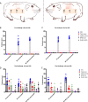
Bovine tuberculosis (BTB) caused by Mycobacterium bovis remains a major problem in both the developed and developing countries. Control of BTB in the UK is carried out by test and slaughter of infected animals, based primarily on the tuberculin skin test (PPD). Vaccination with the attenuated strain of the M. bovis pathogen, BCG, is not used to control bovine tuberculosis in cattle at present, due to its variable efficacy and because it interferes with the PPD test. Diagnostic tests capable of Differentiating Infected from Vaccinated Animals (DIVA) have been developed that detect immune responses to M. bovis antigens absent in BCG; but these are too expensive and insufficiently sensitive to be used for BTB control worldwide. To address these problems we aimed to generate a synergistic vaccine and diagnostic approach that would permit the vaccination of cattle without interfering with the conventional PPD-based surveillance. The approach was to widen the pool of M. bovis antigens that could be used as DIVA targets, by identifying antigenic proteins that could be deleted from BCG without affecting the persistence and protective efficacy of the vaccine in cattle. Using transposon mutagenesis we identified genes that were essential and those that were non-essential for persistence in bovine lymph nodes. We then inactivated selected immunogenic, but non-essential genes in BCG Danish to create a diagnostic-compatible triple knock-out ΔBCG TK strain. The protective efficacy of the ΔBCG TK was tested in guinea pigs experimentally infected with M. bovis by aerosol and found to be equivalent to wild-type BCG. A complementary diagnostic skin test was developed with the antigenic proteins encoded by the deleted genes which did not cross-react in vaccinated or in uninfected guinea pigs. This study demonstrates the functionality of a new and improved BCG strain which retains its protective efficacy but is diagnostically compatible with a novel DIVA skin test that could be implemented in control programmes.
Conflict of interest statement
The authors declare no competing interests.
Figures


Similar articles
-
Differentiation between Mycobacterium bovis BCG-vaccinated and M. bovis-infected cattle by using recombinant mycobacterial antigens.Clin Diagn Lab Immunol. 1999 Jan;6(1):1-5. doi: 10.1128/CDLI.6.1.1-5.1999. Clin Diagn Lab Immunol. 1999. PMID: 9874655 Free PMC article.
-
Mycobacterium bovis Δmce2 double deletion mutant protects cattle against challenge with virulent M. bovis.Tuberculosis (Edinb). 2013 May;93(3):363-72. doi: 10.1016/j.tube.2013.02.004. Epub 2013 Mar 19. Tuberculosis (Edinb). 2013. PMID: 23518075 Clinical Trial.
-
Field evaluation of specific mycobacterial protein-based skin test for the differentiation of Mycobacterium bovis-infected and Bacillus Calmette Guerin-vaccinated crossbred cattle in Ethiopia.Transbound Emerg Dis. 2022 Jul;69(4):e1-e9. doi: 10.1111/tbed.14252. Epub 2021 Aug 19. Transbound Emerg Dis. 2022. PMID: 34331511 Free PMC article.
-
Bovine TB and the development of new vaccines.Comp Immunol Microbiol Infect Dis. 2008 Mar;31(2-3):77-100. doi: 10.1016/j.cimid.2007.07.003. Epub 2007 Aug 30. Comp Immunol Microbiol Infect Dis. 2008. PMID: 17764740 Review.
-
Vaccination of cattle against Mycobacterium bovis.Tuberculosis (Edinb). 2001;81(1-2):125-32. doi: 10.1054/tube.2000.0254. Tuberculosis (Edinb). 2001. PMID: 11463233 Review.
Cited by
-
Evidence, Challenges, and Knowledge Gaps Regarding Latent Tuberculosis in Animals.Microorganisms. 2022 Sep 15;10(9):1845. doi: 10.3390/microorganisms10091845. Microorganisms. 2022. PMID: 36144447 Free PMC article. Review.
-
Review on Bovine Tuberculosis: An Emerging Disease Associated with Multidrug-Resistant Mycobacterium Species.Pathogens. 2022 Jun 21;11(7):715. doi: 10.3390/pathogens11070715. Pathogens. 2022. PMID: 35889961 Free PMC article. Review.
-
Prime Vaccination with Chitosan-Coated Phipps BCG and Boosting with CFP-PLGA against Tuberculosis in a Goat Model.Animals (Basel). 2021 Apr 8;11(4):1046. doi: 10.3390/ani11041046. Animals (Basel). 2021. PMID: 33917739 Free PMC article.
-
Bacillus Calmette-Guérin (BCG) Revaccination and Protection Against Tuberculosis: A Systematic Review.Cureus. 2024 Mar 21;16(3):e56643. doi: 10.7759/cureus.56643. eCollection 2024 Mar. Cureus. 2024. PMID: 38646352 Free PMC article. Review.
-
Differences in skin test reactions to official and defined antigens in guinea pigs exposed to non-tuberculous and tuberculous bacteria.Sci Rep. 2023 Feb 20;13(1):2936. doi: 10.1038/s41598-023-30147-4. Sci Rep. 2023. PMID: 36806813 Free PMC article.
References
-
- Perry, B. D. Investing in animal health research to alleviate poverty. (ILRI (aka ILCA and ILRAD), 2002).
-
- Organization, W. H. Global Tuberculosis Report 2017. (World Health Organization, 2017).
-
- Organization, W. H. Roadmap for zoonotic tuberculosis (2017).
Publication types
MeSH terms
Substances
Grants and funding
LinkOut - more resources
Full Text Sources
Other Literature Sources
Medical

