

CYR61, a potential biomarker of tumor inflammatory response in epithelial ovarian cancer microenvironment of tumor progress
- PMID: 31766991
- PMCID: PMC6878653
- DOI: 10.1186/s12885-019-6321-x
CYR61, a potential biomarker of tumor inflammatory response in epithelial ovarian cancer microenvironment of tumor progress
Abstract
Background: Recent studies have found that inflammatory response is involved in the pathogenesis of ovarian cancer. Advanced ovarian cancer is often presented with ascites that is rich in cytokines, inflammatory factors or cancer cells. Therefore, it is important to study the microenvironment of ascites in order to further clarify the occurrence and progression of ovarian cancer. As a pro-inflammatory factor, the Cyr61 expression patterns are inconsistent in human tumors. Although it has been reported that Cyr61 is related to the progression of ovarian cancer, its specific mechanism is not yet clear. This study sought to evaluate the Cyr61 levels of ascites, serum and different tissues of ovarian cancer to explore the potential association of Cyr61with the tumor-associated inflammatory microenvironment of EOC.
Methods: Tumor specimens were procured from patients with ovarian serous cystadenocarcinoma and ovarian serous cystadenoma. Cyr61 and IL-6 levels of serum or ascites were determined by ELISA (Enzyme-Linked ImmunoSorbent Assay), while Cyr61 expressions of different ovarian tumor tissues were evaluated by IHC (Immunohistochemistry). Then the correlation of Cyr61 level in ascites with clinicopathologic features was analyzed. And other laboratory data were obtained from medical records.
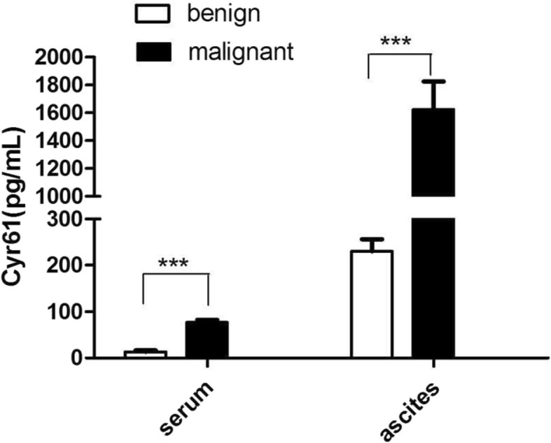
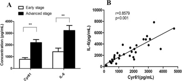
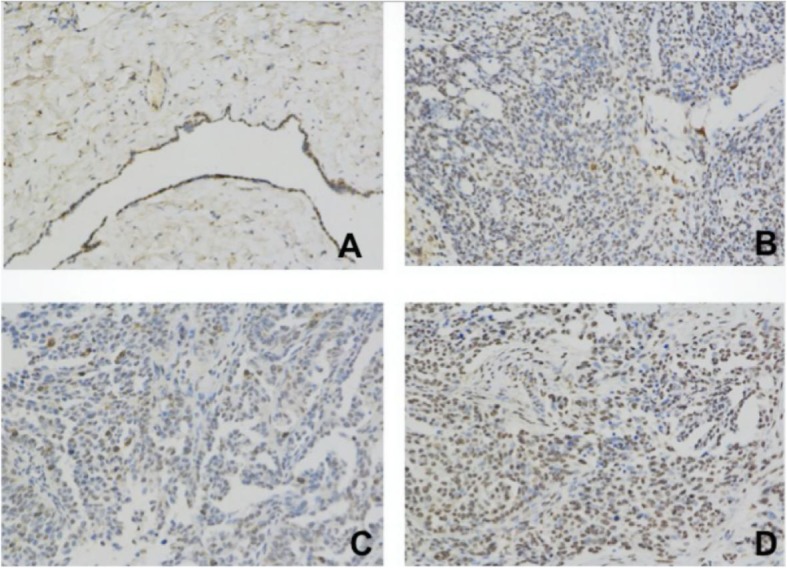
Results: Both in ascites and serum, significantly higher Cyr61 levels were found in ovarian serous cystadenocarcinoma. In malignant ascites, higher Cyr61 level of ovarian serous cystadenocarcinoma was more closely associated with FIGO stage, initial tumor size > 10 cm and the residual tumor size. And the increased IL-6 level was linearly related to Cyr61 level. Moreover, the serum levels of Cyr61, IL-6 and CRP in advanced stage of ovarian cancer were much higher than those in early stage. Lastly, the IHC data demonstrate that Cyr61 expression of ovarian serous adenocarcinoma was higher than that of ovarian serous cystadenoma, but it was lower than the paired metastatic lesions.
Conclusions: As a pro-inflammatory factor, increased ascites Cyr61 level is associated with FIGO stage, initial tumor size > 10 cm and the residual tumor size. Moreover, serum Cyr61 may be used as a potential marker for EOC inflammatory response. Finally, Cyr61 may be involved in the process of tumor metastasis and progression by producing IL-6 and CRP in the EOC inflammatory microenvironment.
Keywords: Cyr61; Epithelial ovarian cancer; Tumor progression; Tumor-associated inflammatory microenvironment.
Conflict of interest statement
The authors declare that they have no competing interests .
Figures
Similar articles
-
Inflammation-regulating factors in ascites as predictive biomarkers of drug resistance and progression-free survival in serous epithelial ovarian cancers.BMC Cancer. 2015 Jul 1;15:492. doi: 10.1186/s12885-015-1511-7. BMC Cancer. 2015. PMID: 26122176 Free PMC article.
-
Validity and prognostic significance of sperm protein 17 as a tumor biomarker for epithelial ovarian cancer: a retrospective study.BMC Cancer. 2018 Oct 11;18(1):970. doi: 10.1186/s12885-018-4880-x. BMC Cancer. 2018. PMID: 30309325 Free PMC article.
-
Correlation of CD44v6 expression with ovarian cancer progression and recurrence.BMC Cancer. 2013 Apr 8;13:182. doi: 10.1186/1471-2407-13-182. BMC Cancer. 2013. Retraction in: BMC Cancer. 2020 Mar 19;20(1):236. doi: 10.1186/s12885-020-06753-0 PMID: 23565736 Free PMC article. Retracted.
-
Immune Tumor Microenvironment in Ovarian Cancer Ascites.Int J Mol Sci. 2022 Sep 14;23(18):10692. doi: 10.3390/ijms231810692. Int J Mol Sci. 2022. PMID: 36142615 Free PMC article. Review.
-
Adipocytes: active facilitators in epithelial ovarian cancer progression?J Ovarian Res. 2020 Sep 23;13(1):115. doi: 10.1186/s13048-020-00718-4. J Ovarian Res. 2020. PMID: 32967712 Free PMC article. Review.
Cited by
-
Hypoxia-initiated Cysteine-rich protein 61 secretion promotes chemoresistance of acute B lymphoblastic leukemia cells.Am J Cancer Res. 2024 Jul 15;14(7):3388-3403. doi: 10.62347/CKMT4065. eCollection 2024. Am J Cancer Res. 2024. PMID: 39113880 Free PMC article.
-
Systematic analysis of the relationship between ovarian cancer prognosis and alternative splicing.J Ovarian Res. 2021 Sep 15;14(1):120. doi: 10.1186/s13048-021-00866-1. J Ovarian Res. 2021. PMID: 34526089 Free PMC article.
-
Tumor-Associated Microglia and Macrophages in the Glioblastoma Microenvironment and Their Implications for Therapy.Cancers (Basel). 2021 Aug 24;13(17):4255. doi: 10.3390/cancers13174255. Cancers (Basel). 2021. PMID: 34503065 Free PMC article. Review.
-
ZEB2 facilitates peritoneal metastasis by regulating the invasiveness and tumorigenesis of cancer stem-like cells in high-grade serous ovarian cancers.Oncogene. 2021 Aug;40(32):5131-5141. doi: 10.1038/s41388-021-01913-3. Epub 2021 Jul 1. Oncogene. 2021. PMID: 34211089 Free PMC article.
-
Immune environment and antigen specificity of the T cell receptor repertoire of malignant ascites in ovarian cancer.PLoS One. 2023 Jan 6;18(1):e0279590. doi: 10.1371/journal.pone.0279590. eCollection 2023. PLoS One. 2023. PMID: 36607962 Free PMC article.
References
MeSH terms
Substances
LinkOut - more resources
Full Text Sources
Medical
Research Materials
Miscellaneous
