. 2019 Nov 13;8(4):234.
doi: 10.3390/pathogens8040234.

Porcine Alveolar Macrophages' Nitric Oxide Synthase-Mediated Generation of Nitric Oxide Exerts Important Defensive Effects against Glaesserella parasuis Infection

Affiliations

Porcine Alveolar Macrophages' Nitric Oxide Synthase-Mediated Generation of Nitric Oxide Exerts Important Defensive Effects against Glaesserella parasuis Infection

Qi Cao et al. Pathogens. .

Abstract

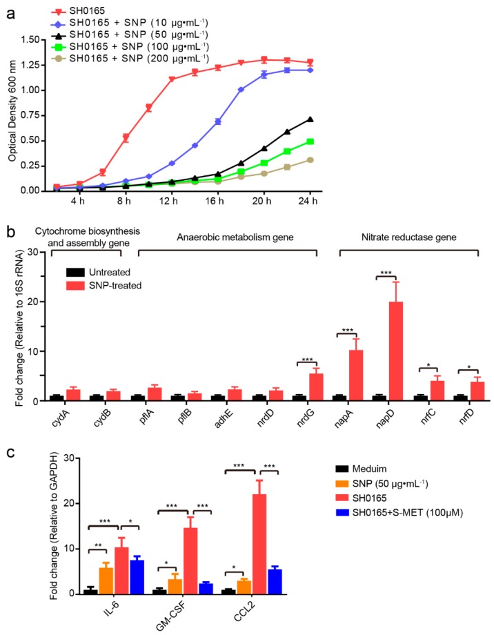
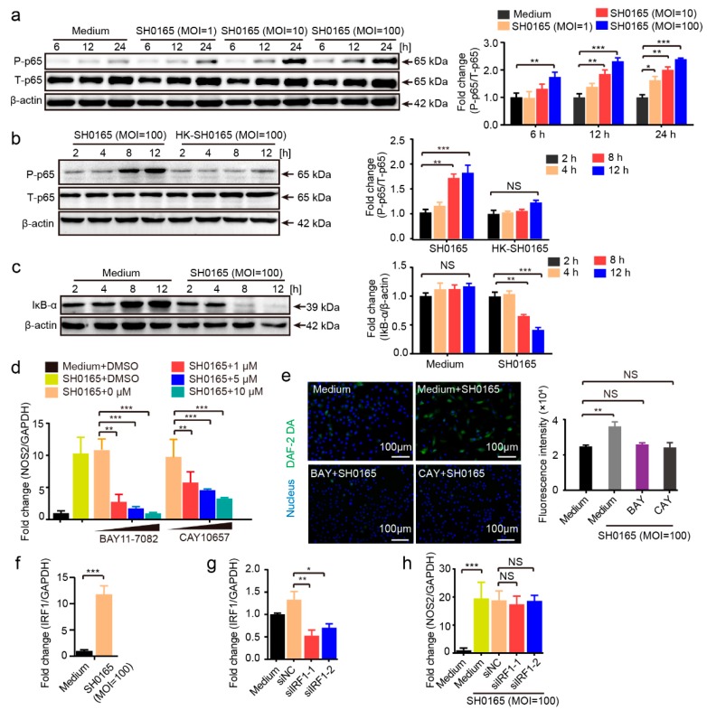
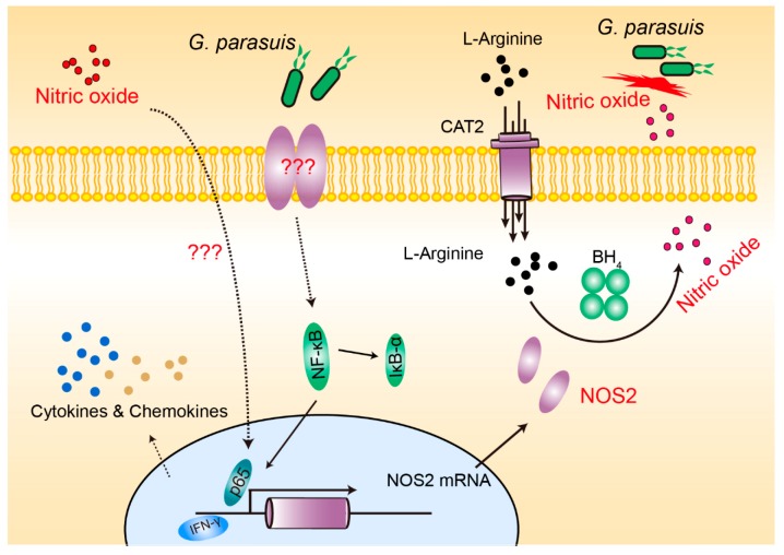
Glaesserella parasuis is a habitual bacterium of pigs' upper respiratory tracts. Its infection initiates with the invasion and colonization of the lower respiratory tracts of pigs, and develops as the bacteria survive host pulmonary defenses and clearance by alveolar macrophages. Alveolar macrophage-derived nitric oxide (NO) is recognized as an important mediator that exerts antimicrobial activity as well as immunomodulatory effects. In this study, we investigated the effects and the signaling pathway of NO generation in porcine alveolar macrophages 3D4/21 during G. parasuis infection. We demonstrated a time and dose-dependent generation of NO in 3D4/21 cells by G. parasuis, and showed that NO production required bacterial viability and nitric oxide synthase 2 upregulation, which was largely contributed by G. parasuis-induced nuclear factor-κB signaling's activation. Moreover, the porcine alveolar macrophage-derived NO exhibited prominent bacteriostatic effects against G. parasuis and positive host immunomodulation effects by inducing the production of cytokines and chemokines during infection. G. parasuis in turn, selectively upregulated several nitrate reductase genes to better survive this NO stress, revealing a battle of wits during the bacteria-host interactions. To our knowledge, this is the first direct demonstration of NO production and its anti-infection effects in alveolar macrophages with G. parasuis infection.

Keywords: Glaesserella parasuis; NF-κB signaling; NOS2; nitric oxide; porcine alveolar macrophages.


Conflict of interest statement

The authors declare no conflict of interest.

Figures

Figure 1
Figure 1
Glaesserella parasuis induced the time-dependent and multiplicity of infection (MOI)-dependent production of nitrite in 3D4/21 cells which required bacterial viability. (a) 3D4/21 cells were challenged with or without different doses (MOI of 1, 10 and 100) of SH0165 for 12 h. (b) 3D4/21 cells were infected with SH0165 (MOI 100) or HK-SH0165 at the same MOIs for 12 h. The supernatants were collected for nitrite measurement at 2 h intervals via the Griess reaction. Cells alone and bacteria alone at different colony forming units were used as controls. Data are shown as the means ± SDs from three replicates. Statistical differences were analyzed using the Student’s t-test (***, p < 0.001).
Figure 2
Figure 2
NOS2 and CAT2 facilitated G. parasuis-induced NO production. (a) Real-time PCR analyzing the expression levels of NOS isoforms in 3D4/21 cells in response to SH0165 infection for 12 h. (b) Real-time PCR analysis of NOS2 expression in 3D4/21 cells in response to different infection doses of SH0165 at indicated time points. (c) NOS2 expression in 3D4/21 cells challenged with SH0165 or HK-SH0165 at indicated time points were analyzed by real-time PCR. (d) CAT expression levels in 3D4/21 cells infected with or without SH0165 for 12 h. (e) Intracellular NO generation at 12 h of SH0165 infection at MOI of 100 was detected by the DAF-2 DA probe via fluorescence microscope capture and quantified with a fluorescence microplate reader. Scale bar, 100 μm. (f) Nitrite levels from culture supernatants of cells with or without SH0165 infection and other treatments at 12 h via the Griess reaction. (g) Real-time PCR analysis and comparison of NOS2 expression in SH0165-infected 3D4/21 cells receiving other treatments. All real-time PCR results were normalized to GAPDH and shown as means ± SDs from three independent assays. Statistical differences were calculated using two-way ANOVA (***, p < 0.001; **, p < 0.01; *, p < 0.05; NS: not significant). #, undetectable.
Figure 3
Figure 3
NO reduced the in vitro growth of G. parasuis and induced proinflammatory cytokine and chemokine expression in 3D4/21 cells. (a) The NO donor SNP exhibited a dose-dependent inhibition on the growth of G. parasuis SH0165. (b) Real-time PCR detection of multiple NO response-associated genes in G. parasuis SH0165, including cytochrome biosynthesis and assembly genes (cydAB), anaerobic metabolism genes (pflAB, adhE and nrdDG) and nitrate reductase genes (napAD and nrfCD). (c) Real-time PCR analysis of cytokine and chemokine expression in 3D4/21 cells receiving SNP (50 μg·mL−1), SH0165 or SH0165 with pretreatment of S-MET (100 μM) for 24 h. Cells with medium alone were used as the control group. Results were generated from three duplicate assays and were normalized to bacterial 16S rRNA or GAPDH of cells. Statistical differences were analyzed using two-way ANOVA (***, p < 0.001; **, p < 0.01; *, p < 0.05).
Figure 4
Figure 4
SH0165-induced activation of NF-κB signaling pathway-mediated NO generation in 3D4/21 cells. (a) Western blot showing time-dependent and bacterial MOI-dependent phosphorylation of the NF-κB p65 subunit in 3D4/21 cells in response to SH0165. (b) Western blot and comparison of p65 phosphorylation in response to SH0165 and HK-SH0165 (MOI 100) at the time points indicated. (c) Western blot analysis of IκB-α degradation in whole-cell lysates of 3D4/21 cells with or without SH0165 infection. (d) Real-time PCR analysis of NOS2 expression in 3D4/21 cells upon 12 h of SH0165 infection (MOI 100) with or without pretreatment of BAY11-7082 or CAY10657. (e) Intracellular NO generation at 12 h of infection at MOI of 100 was detected by the DAF-2 DA probe via fluorescence microscope and quantified with a fluorescence microplate reader. Scale bar, 100 μm. (f) Real-time PCR analysis of IRF1 expression in 3D4/21 cells upon SH0165 infection. (g) Cells were collected 24 h after siRNA transfection and real-time PCR analysis of IRF1 expression. NC, negative control. (h) IRF1 silencing by siRNA and was then infected with SH0165 or non-infected for 12 h. Real-time PCR analysis of IRF1 expression in 3D4/21 cells. Band densitometry was normalized to T-p65 or β-actin and shown as mean ± SD from three independent analyses. All real-time PCR data were normalized to GAPDH and shown as means ± SDs from three independent assays. Statistical differences were analyzed using Student’s t-tests (***, p < 0.001; **, p < 0.01; *, p < 0.05; NS: not significant).
Figure 5
Figure 5
The effects of NF-κB p65 binding sites on the transcriptional activity of the NOS2 promoter via luciferase reporter assays. (a) The potential p65 binding sites (red triangles) and IRF1 binding sites (blue triangles) in the NOS2 promoter region. (b) The NOS2 full-length promoter region and different truncations were co-transfected with p65 or IRF1 to analyze and compare luciferase activities. (c) A set of p65 binding site mutations was introduced into the NOS2 promoter region and transcriptional activities were analyzed by luciferase reporter assays with p65 co-transfection. Firefly luciferase activity was measured and normalized to Renilla luciferase activity. Results are expressed as means ± SDs from six well duplicates. The statistical differences were calculated using two-way ANOVA (***, p < 0.001; *, p < 0.05; NS: not significant).
Figure 6
Figure 6
Schematic diagram of the pathways and molecules involved in this NO process in response to G. parasuis infection.

Similar articles

References

    1. Costa-Hurtado M., Olvera A., Martinez-Moliner V., Galofre-Mila N., Martinez P., Dominguez J., Aragon V. Changes in macrophage phenotype after infection of pigs with Haemophilus parasuis strains with different levels of virulence. Infect. Immun. 2013;81:2327–2333. doi: 10.1128/IAI.00056-13. - DOI - PMC - PubMed
    1. Oliveira S., Pijoan C. Haemophilus parasuis: New trends on diagnosis, epidemiology and control. Vet. Microbiol. 2004;99:1–12. doi: 10.1016/j.vetmic.2003.12.001. - DOI - PubMed
    1. Cai X., Chen H., Blackall P.J., Yin Z., Wang L., Liu Z., Jin M. Serological characterization of Haemophilus parasuis isolates from China. Vet. Microbiol. 2005;111:231–236. doi: 10.1016/j.vetmic.2005.07.007. - DOI - PubMed
    1. Correa-Fiz F., Fraile L., Aragon V. Piglet nasal microbiota at weaning may influence the development of Glasser’s disease during the rearing period. BMC Genom. 2016;17:404. doi: 10.1186/s12864-016-2700-8. - DOI - PMC - PubMed
    1. Costa-Hurtado M., Aragon V. Advances in the quest for virulence factors of Haemophilus parasuis. Vet. J. 2013;198:571–576. doi: 10.1016/j.tvjl.2013.08.027. - DOI - PubMed

LinkOut - more resources