. 2019 Nov 15;11(11):2777.
doi: 10.3390/nu11112777.

Inhibitory Effects of Osthole on Human Breast Cancer Cell Progression via Induction of Cell Cycle Arrest, Mitochondrial Dysfunction, and ER Stress

Affiliations

Inhibitory Effects of Osthole on Human Breast Cancer Cell Progression via Induction of Cell Cycle Arrest, Mitochondrial Dysfunction, and ER Stress

Wonhyoung Park et al. Nutrients. .

Abstract

Background: Breast cancer is the most commonly diagnosed cancer and the second leading cause of cancer death in women. Although, recently, the number of pathological studies of breast cancer have increased, it is necessary to identify a novel compound that targets multiple signaling pathways involved in breast cancer.

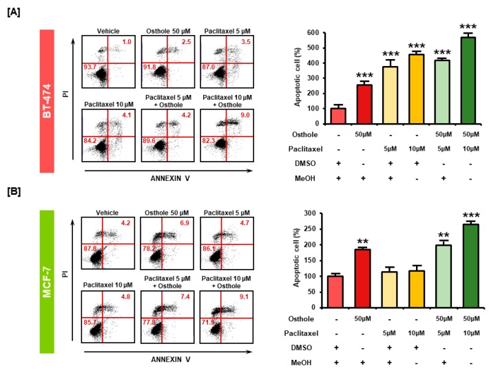
Methods: The effects of osthole on cell viability, apoptosis, mitochondria-mediated apoptosis, production of reactive oxygen species (ROS), and endoplasmic reticulum (ER) stress proteins of BT-474 and MCF-7 breast cancer cell lines were investigated. Signal transduction pathways in both cells in response to osthole were determined by western blot analyses.

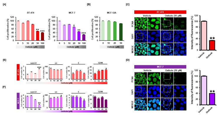
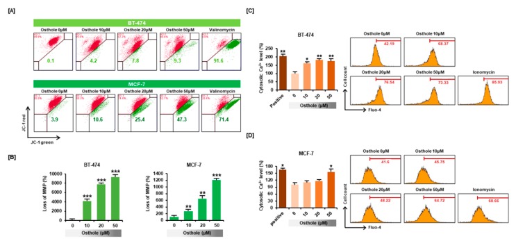
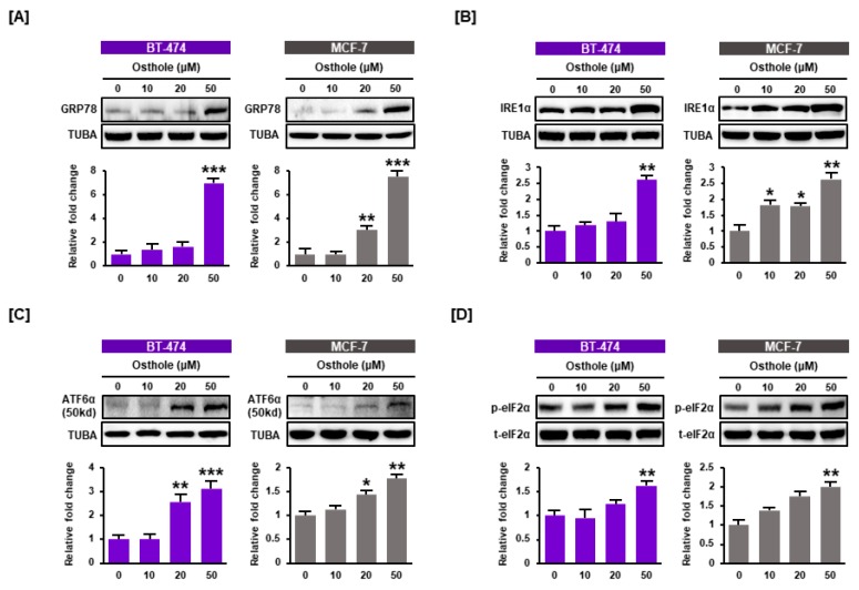
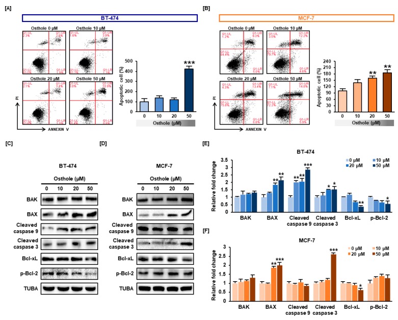
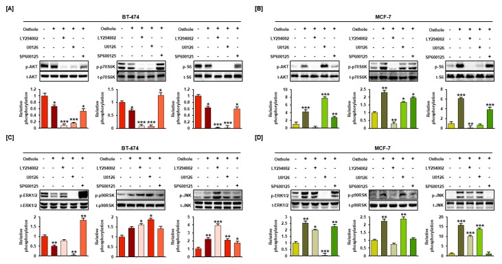
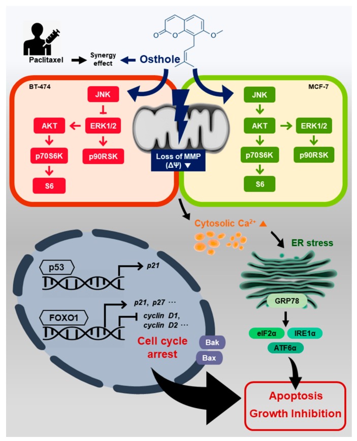
Results: Here, we demonstrated that osthole inhibited cellular proliferation and induced cell cycle arrest through modulation of cell cycle regulatory genes in BT-474 and MCF-7 cells. Additionally, osthole induced loss of mitochondrial membrane potential (MMP), intracellular calcium imbalance, and ER stress. Moreover, osthole induced apoptosis by activating the pro-apoptotic protein, Bax, in both cell lines. Osthole regulated phosphorylation of signaling proteins such as Akt and ERK1/2 in human breast cancer cells. Furthermore, osthole-induced activation of JNK protein-mediated apoptosis in both cell lines.

Conclusions: Collectively, the results of the present study indicated that osthole may ameliorate breast cancer and can be a promising therapeutic agent for treatment of breast cancer.

Keywords: ER stress; MMP depolarization; apoptosis; breast cancer; calcium imbalance; osthole.


Conflict of interest statement

The authors declare no conflict of interest.

Figures

Figure 1
Figure 1
Osthole inhibited cell proliferation in human breast cancer cells. (A) Proliferation of breast cancer cells was analyzed using cell proliferation assays. Osthole (0, 5, 10, 20, 50, and 100 μM) decreased the proliferation of BT-474 and MCF-7 cells in a dose-dependent manner. (B) Effect of osthole on MCF-12A normal mammary epithelial cell proliferation in a dose dependent manner (0, 10, 20, and 50 μM). (C,D) Proliferative cell nuclear antigen (PCNA) was detected using immunofluorescence analysis. The green signal associated with PCNA was attenuated in osthole-treated cells compared to vehicle-treated cells. Nuclei were counterstained with DAPI (blue). The graph shows the intensity of PCNA fluorescence. (E,F) Regulatory effect of osthole on cell cycle progression in breast cancer cells. The number of cells in each cell cycle phase was analyzed by propidium iodide (PI) staining of DNA contents using flow cytometry. All experiments were performed in biological triplicate and the asterisks represent statistically significant differences compared to control (DMSO-treated cells; 0 μM) (* p < 0.05, ** p < 0.01 and *** p < 0.001).
Figure 2
Figure 2
Relative mRNA expression of cell cycle-related genes including cyclin E1 (A), cyclin D1 (B), CDK2 (C), CDK4 (D), CDK6 (E), ERα (F), P21 (G), TP53 (H), and FOXO1 (I). Expression of cell cycle-related genes in breast cancer cell lines was analyzed by quantitative RT-PCR using cDNA templates based on RNA isolated from BT-474 and MCF-7 cells. The graphs indicate the relative mRNA expression in breast cancer cell lines treated with vehicle and osthole for 24 h. All experiments were performed in technical triplicate and the asterisks represent statistically significant differences compared to vehicle-treated cells (** p < 0.01 and *** p < 0.001).
Figure 3
Figure 3
Effects of osthole on mitochondrial dysfunction and cytosolic calcium influx in human breast cancer cells. (A,B) Mitochondrial membrane potential (MMP) was evaluated in JC-1 stained BT-474 and MCF-7 cells using flow cytometry. Depolarization of mitochondria was indicated by increased green fluorescence in the dot plot. The cells treated with Valinomycin (1 μg/mL) for 20 min were used as a positive control for mitochondrial depolarization. (C,D) Calcium levels in the cytoplasm were evaluated using the cytosolic calcium indicator, Fluo-4. The cells treated with Ionomycin (12 μM) for 5 min were used as a positive control for calcium flux. All experiments were performed in biological triplicate and the asterisks represent statistically significant differences compared to vehicle-treated cells (* p < 0.05, ** p < 0.01 and *** p < 0.001).
Figure 4
Figure 4
ER stress induced by osthole treatment in human breast cancer cells. The levels of the ER regulatory proteins GRP78 (A), IRE1α (B), ATF6α (C), and eIF2α (D) were analyzed using western blot analysis of BT-474 and MCF-7 cells. The levels of GRP78, IRE1α, and ATF6α were normalized to α-tubulin (TUBA), and phosphor eIF2α was normalized to total eIF2α protein. All experiments were performed in biological triplicate and the asterisks represent statistically significant differences (* p < 0.05, ** p < 0.01 and *** p < 0.001).
Figure 5
Figure 5
The apoptotic effects of osthole on human breast cancer cells. (A,B) Apoptotic cells were detected by flow cytometry using Annexin V/PI staining. The graph represents increased numbers of cells in the late apoptotic phase (upper right side of dot plot) in response to osthole (0, 10, 20, and 50 μM). Data were analyzed relative to the DMSO control (0 μM). (C,D) The expressions of the pro-apoptotic proteins including Bax, Bak, cleaved caspase 3 and cleaved caspase 9, and anti-apoptotic proteins including Bcl-xL and p-BcL-2 were analyzed by western blot in BT-474 and MCF-7 cells. The expression levels of apoptotic proteins in BT-474 (E) and MCF-7 (F) were represented as bar graphs. The expression levels of proteins were normalized to α-tubulin (TUBA). All experiments were performed in biological triplicate and the asterisks indicate statistically significant differences compared to control (DMSO-treated cells; 0 μM) (* p < 0.05, ** p < 0.01 and *** p < 0.001).
Figure 6
Figure 6
Effect of osthole on phosphorylation of PI3K/Akt and MAPK pathway proteins in human breast cancer cells. The levels of Akt, p70S6K, S6 (A,B), ERK1/2, p90RSK, and JNK (C,D) were detected by western blot analysis. Each phosphorylated protein was normalized to the corresponding total protein. Band intensity is shown in the graph as relative value compared to non-treated control (0 μM). All experiments were performed in biological triplicate and the asterisks indicate statistically significant differences (* p < 0.05, ** p < 0.01 and *** p < 0.001).
Figure 7
Figure 7
Combinational effect of pharmacological inhibitors on osthole-mediated signaling pathways in human breast cancer cells. After incubation with LY294002 (Akt inhibitor), U0126 (ERK1/2 inhibitor), and SP600125 (JNK inhibitor) for 2 h, BT-474 and MCF-7 cells were treated with 50 μM osthole for 1 h. Changes in phosphorylation of Akt, p70S6K, S6 (A,B), ERK1/2, p90RSK, and JNK (C,D) were analyzed by western blotting. The graph shows relative phosphorylation of each protein compared to that in non-treated control (0 μM). All experiments were performed in biological triplicate and the asterisks indicate statistically significant differences compared to non-treated control (* p < 0.05, ** p < 0.01 and *** p < 0.001).
Figure 8
Figure 8
Combinational apoptotic effect of osthole and conventional chemotherapeutic agents. The proportion of apoptotic cells following treatment with osthole with and without paclitaxel was detected using Annexin V/PI staining in BT-474 (A) and MCF-7 (B). BT-474 and MCF-7 cells were incubated with osthole, paclitaxel, or a combination of both for 48 h. Data are represented in the graph as the percentage of apoptotic cells compared with vehicle-treated control (100%). All experiments were performed in biological triplicate and the asterisks indicate statistically significant differences compared to Vehicle-treated control (** p < 0.01 and *** p < 0.001).
Figure 9
Figure 9
Hypothetical mechanism of action of osthole in human breast cancer cell lines. Osthole induced loss of the MMP and increased cytoplasmic calcium levels. ER stress-response proteins and pro-apoptotic proteins were increased in BT-474 and MCF-7 cells. Osthole upregulated JNK in both cell lines, downregulated the Akt and ERK1/2 signaling pathways in BT-474 cells, and upregulated these pathways in MCF-7 cells. Osthole induced growth inhibition and apoptosis in breast cancer cell lines.

Similar articles

Cited by

References

    1. Siegel R.L., Miller K.D., Jemal A. Cancer statistics, 2018. CA A Cancer J. Clin. 2018;68:7–30. doi: 10.3322/caac.21442. - DOI - PubMed
    1. Winters S., Martin C., Murphy D., Shokar N.K. Breast Cancer Epidemiology, Prevention, and Screening. Prog. Mol. Biol. Transl. Sci. 2017;151:1–32. doi: 10.1016/bs.pmbts.2017.07.002. - DOI - PubMed
    1. Winters S., Martin C., Murphy D., Shokar N.K. Chapter One—Breast Cancer Epidemiology, Prevention, and Screening. In: Lakshmanaswamy R., editor. Progress in Molecular Biology and Translational Science. Volume 151. Academic Press; Cambridge, MA, USA: 2017. pp. 1–32. - PubMed
    1. Redig A.J., McAllister S.S. Breast cancer as a systemic disease: A view of metastasis. J. Intern. Med. 2013;274:113–126. doi: 10.1111/joim.12084. - DOI - PMC - PubMed
    1. Sinha D., Sarkar N., Biswas J., Bishayee A. Seminars in Cancer Biology. Volume 40. Academic Press; Cambridge, MA, USA: 2016. Resveratrol for breast cancer prevention and therapy: Preclinical evidence and molecular mechanisms; pp. 209–232. - DOI - PubMed

MeSH terms