. 2020 Jan 17;94(3):e01168-19.
doi: 10.1128/JVI.01168-19. Print 2020 Jan 17.

Puumala and Andes Orthohantaviruses Cause Transient Protein Kinase R-Dependent Formation of Stress Granules

Affiliations

Puumala and Andes Orthohantaviruses Cause Transient Protein Kinase R-Dependent Formation of Stress Granules

Wanda Christ et al. J Virol. .

Abstract

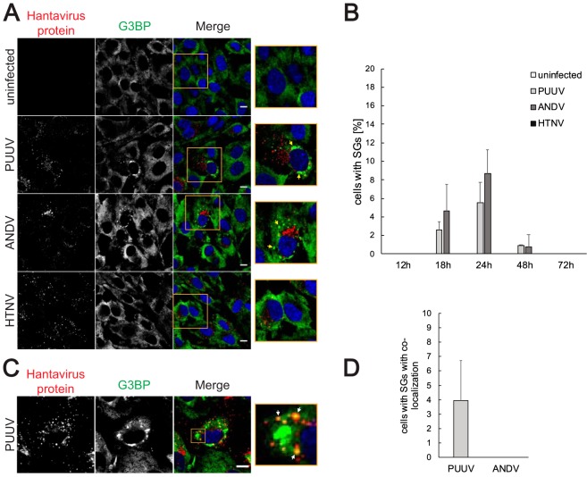
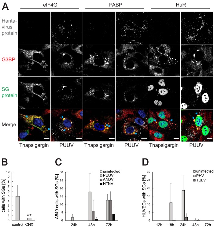
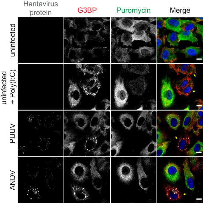
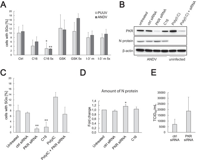
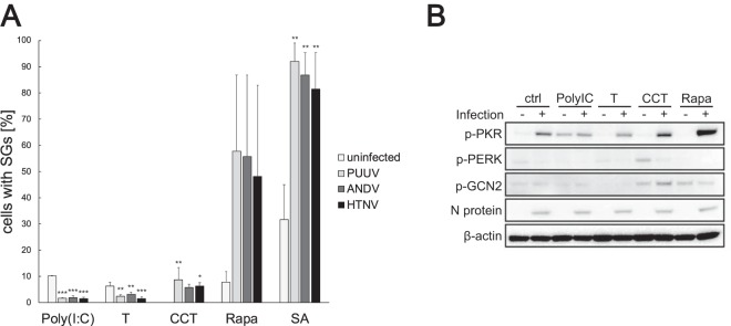
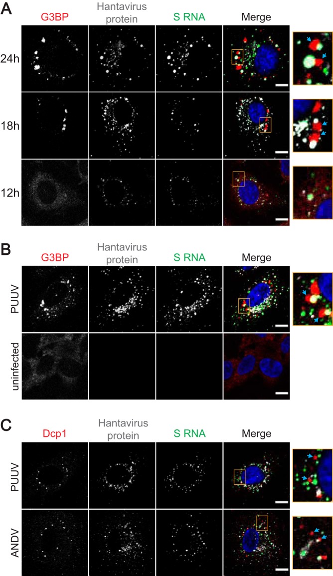
Virus infection frequently triggers host cell stress signaling resulting in translational arrest; as a consequence, many viruses employ means to modulate the host stress response. Hantaviruses are negative-sense, single-stranded RNA viruses known to inhibit host innate immune responses and apoptosis, but their impact on host cell stress signaling remains largely unknown. In this study, we investigated activation of host cell stress responses during hantavirus infection. We show that hantavirus infection causes transient formation of stress granules (SGs) but does so in only a limited proportion of infected cells. Our data indicate some cell type-specific and hantavirus species-specific variability in SG prevalence and show SG formation to be dependent on the activation of protein kinase R (PKR). Hantavirus infection inhibited PKR-dependent SG formation, which could account for the transient nature and low prevalence of SG formation observed during hantavirus infection. In addition, we report only limited colocalization of hantaviral proteins or RNA with SGs and show evidence indicating hantavirus-mediated inhibition of PKR-like endoplasmic reticulum (ER) kinase (PERK).IMPORTANCE Our work presents the first report on stress granule formation during hantavirus infection. We show that hantavirus infection actively inhibits stress granule formation, thereby escaping the detrimental effects on global translation imposed by host stress signaling. Our results highlight a previously uncharacterized aspect of hantavirus-host interactions with possible implications for how hantaviruses are able to cause persistent infection in natural hosts and for pathogenesis.

Keywords: PKR; hantavirus; stress granule.


Figures

FIG 1
FIG 1
PUUV and ANDV infection causes transient formation of SGs in HUVECs. (A) SG formation in hantavirus-infected HUVECs. HUVECs infected with different hantaviruses were fixed 24 h after the infection. Uninfected cells were used as a control. The cells were stained with antibodies against hantavirus proteins (red) and G3BP (green) and with DAPI (4′,6-diamidino-2-phenylindole) (blue). Arrows show SGs. Imaging was performed with confocal microscopy (60×). Scale bars = 10 μm. (B) Time kinetics of SG formation in hantavirus-infected HUVECs. HUVECs infected with different hantaviruses were fixed 12, 18, 24, 48, or 72 h after the infection. Uninfected cells were used as a control. The cells were stained for viral proteins and G3BP. During the investigation using fluorescence microscopy, several pictures of a total of >150 cells were taken at random positions and the number of infected cells showing SGs was determined. n = ≥3. (C) Colocalization of viral proteins with SGs in PUUV-infected cells. HUVECs were fixed 24 h after the infection with PUUV and stained with antibodies against hantavirus proteins (red) and G3BP (green) and with DAPI (blue). Arrows show colocalization of viral proteins with SGs. Imaging was performed with confocal microscopy (60×). Scale bars = 10 μm. (D) Number of infected cells showing colocalization of viral proteins with SGs. HUVECs infected with PUUV and ANDV were fixed 24 h after the infection. The cells were stained for viral proteins and G3BP. During the investigation using fluorescence microscopy, pictures of ≥100 cells with SGs were taken and the number of cells where viral proteins colocalized with SGs was determined. n = 3.
FIG 2
FIG 2
Characterization of infection-induced SGs regarding SG-associated proteins, cell type, and virus pathogenicity. (A) Localization of different SG-associated proteins. HUVECs were infected with PUUV and fixed at 24 h after the infection. In addition, uninfected cells were treated with thapsigargin as described in Materials and Methods. The cells were stained with antibodies against hantavirus proteins (magenta), G3BP (red), and eIF4G, PABP, or HuR (green) and with DAPI (blue). The images show representative cells with stress granules. Imaging was performed with confocal microscopy (60×). Scale bars = 10 μm. (B) Treatment with cycloheximide dissolves infection-induced SGs. HUVECs infected with PUUV were incubated with 20 μg/ml of CHX for 1 h and fixed at 24 h after the infection. The cells were stained for viral proteins and G3BP and analyzed for SG formation under the microscope. During this investigation, several pictures of a total of >100 cells were taken at random positions and the number of infected cells showing SGs was determined. n = 4. (C) Time kinetics of SG formation in hantavirus-infected A549 cells. A549 cells infected with different hantaviruses were fixed 24, 48, or 72 h after the infection. Uninfected cells were used as a control. The cells were stained for viral proteins and G3BP. During the investigation using fluorescence microscopy, several pictures of a total of >150 cells were taken at random positions and the number of infected cells showing SGs was determined. n = 3. (D) Time kinetics of SG formation in HUVECs infected with the nonpathogenic hantaviruses Prospect Hill virus (PHV) and Tula orthohantavirus (TULV). HUVECs were infected with PHV and TULV and fixed 12, 18, 24, 48, or 72 h after the infection. The cells were stained for viral proteins and G3BP, and the number of cells harboring SGs was determined under the microscope. Several pictures were taken at random positions and showed a total of >100 cells. n = 3. **, P < 0.01.
FIG 3
FIG 3
Infection-induced SG formation leads to inhibition of cellular translation. HUVECs infected with PUUV or ANDV were fixed 24 h after the infection. Uninfected untreated cells and uninfected cells treated with 1 μg poly(I·C) (see Materials and Methods) were used as controls. Before fixation, all samples were treated with 10 μg/ml puromycin for 5 min. The cells were stained with antibodies against hantavirus protein (magenta), G3BP (red), or puromycin (green) and DAPI (blue). Arrows show cells with SGs. Imaging was performed with confocal microscopy (60×). Scale bars = 10 μm.
FIG 4
FIG 4
Inhibition and knockdown of PKR reduce infection-induced SG formation. (A) Treatment with the PKR inhibitor C16 reduces infection-induced SG formation. HUVECs infected with PUUV or ANDV were treated with neat or 5× PKR inhibitor C16, PERK inhibitor GSK2606414, or GCN2 inhibitor indirubin-3′-monoxime (I-3′-m) (see Materials and Methods) and fixed at 24 h after the infection. The cells were stained for viral proteins and G3BP. During the investigation using fluorescence microscopy, several pictures of a total of >100 cells were taken at random positions and the number of infected cells showing SGs was determined. Ctrl, control. n ≥ 3. (B) Transfection with PKR siRNA decreases PKR expression. HUVECs were transfected with PKR siRNA or control siRNA as described in Materials and Methods and infected with ANDV after 24 h. Untreated ANDV-infected cells and ANDV-infected cells treated with C16 were used as controls. Cell lysates were collected at 24 h after the infection. In addition, cell lysates of untransfected or PKR siRNA transfected uninfected cells treated with poly(I·C) were collected. The lysates were then analyzed for the expression of PKR and N protein. β-actin was used as a loading control. (C) PKR knockdown reduces infection-induced SG formation in ANDV-infected HUVECs. HUVECs on coverslips were treated as described for panel B, fixed, and stained for viral proteins and G3BP and analyzed for SG formation under the microscope. During this investigation, several pictures of a total of >100 cells were taken at random positions and the number of infected cells showing SGs was determined. n ≥ 3. (D) PKR knockdown increases the production of N protein in ANDV-infected HUVECs. HUVECs were treated as described for panel B. N protein levels were normalized to cellular levels of β-actin. The data are presented as the fold change in relation to the N protein levels of untreated cells. n ≥ 3. (E) HUVECs were treated with control siRNA or PKR siRNA and infected with ANDV after 24 h. The supernatant was collected at 4 days postinfection (p.i.), and the virus titer was determined by endpoint dilution assay. Results shown as 50% tissue culture infectious doses (TCID50) per milliliter. n = 3. *, P < 0.05; **, P < 0.01.
FIG 5
FIG 5
Hantaviruses inhibit PKR-mediated SG formation. (A) Hantavirus infection reduces SG formation induced by poly(I·C) and thapsigargin. HUVECs were infected with different hantaviruses and treated with poly(I·C), thapsigargin, CCT020312 (CCT), rapamycin (Rapa), or sodium arsenite (SA) (see Materials and Methods). Uninfected cells were used as controls. All samples were collected at 72 h after the infection. The cells were stained with antibodies against viral proteins and G3BP and analyzed for SG formation under the microscope. During this investigation, several pictures of a total of >100 cells were taken at random positions, and the number of infected cells showing SGs was determined. n ≥ 3. (B) Phosphorylation of eIF2α kinases. Uninfected (-) and PUUV-infected (+) HUVECs were treated as described for panel A, and cell lysates were collected at 72 h after the infection. The lysates were then analyzed to determine the amount of phospho-PKR, phospho-PERK, or phospho-GCN2. N protein was used to show infection, and β-actin was used as a loading control. *, P < 0.05; **, P < 0.01.
FIG 6
FIG 6
Viral RNA is excluded from SGs and P-bodies. (A) FISH analysis of the localization of ANDV S RNA and SGs at different time points. HUVECs were infected with ANDV and fixed at 12, 18, or 24 h after the infection. Hybridization and staining with antibodies against viral protein (magenta), G3BP (red), and digoxigenin (green) and with DAPI was performed as described in Materials and Methods. Arrows show SGs. (B) FISH analysis of the localization of PUUV S RNA and SGs at 24 h. HUVECs were infected with PUUV and fixed at 24 h after the infection. Hybridization and staining with antibodies against viral protein (magenta), G3BP (red), and digoxigenin (green) and with DAPI was performed as described in Materials and Methods. Arrows show SGs. Uninfected cells were used as a control. (C) FISH analysis of PUUV and ANDV S RNA and P-bodies at 24 h. HUVECs were infected with PUUV or ANDV and fixed at 24 h after the infection. Hybridization and staining with antibodies against viral protein (magenta), Dcp1 (red), and digoxigenin (green) and with DAPI was performed as described in Materials and Methods. Arrows show P-bodies. Imaging for all panels was performed with confocal microscopy (60×). Scale bars = 10 μm.

Similar articles

Cited by

References

    1. Shi M, Lin XD, Chen X, Tian JH, Chen LJ, Li K, Wang W, Eden JS, Shen JJ, Liu L, Holmes EC, Zhang YZ. 2018. The evolutionary history of vertebrate RNA viruses. Nature 556:197–202. doi:10.1038/s41586-018-0012-7. - DOI - PubMed
    1. Jonsson CB, Figueiredo LT, Vapalahti O. 2010. A global perspective on hantavirus ecology, epidemiology, and disease. Clin Microbiol Rev 23:412–441. doi:10.1128/CMR.00062-09. - DOI - PMC - PubMed
    1. Vaheri A, Strandin T, Hepojoki J, Sironen T, Henttonen H, Makela S, Mustonen J. 2013. Uncovering the mysteries of hantavirus infections. Nat Rev Microbiol 11:539–550. doi:10.1038/nrmicro3066. - DOI - PubMed
    1. Klingström J, Smed-Sörensen A, Maleki KT, Solà-Riera C, Ahlm C, Björkström NK, Ljunggren HG. 2019. Innate and adaptive immune responses against human Puumala virus infection: immunopathogenesis and suggestions for novel treatment strategies for severe hantavirus-associated syndromes. J Intern Med 285:510–523. doi:10.1111/joim.12876. - DOI - PMC - PubMed
    1. Pakos-Zebrucka K, Koryga I, Mnich K, Ljujic M, Samali A, Gorman AM. 2016. The integrated stress response. EMBO Rep 17:1374–1395. doi:10.15252/embr.201642195. - DOI - PMC - PubMed

Publication types