. 2019 Nov 13;18(1):158.
doi: 10.1186/s12943-019-1094-z.

Circ-HuR suppresses HuR expression and gastric cancer progression by inhibiting CNBP transactivation

Affiliations

Circ-HuR suppresses HuR expression and gastric cancer progression by inhibiting CNBP transactivation

Feng Yang et al. Mol Cancer. .

Erratum in

Abstract

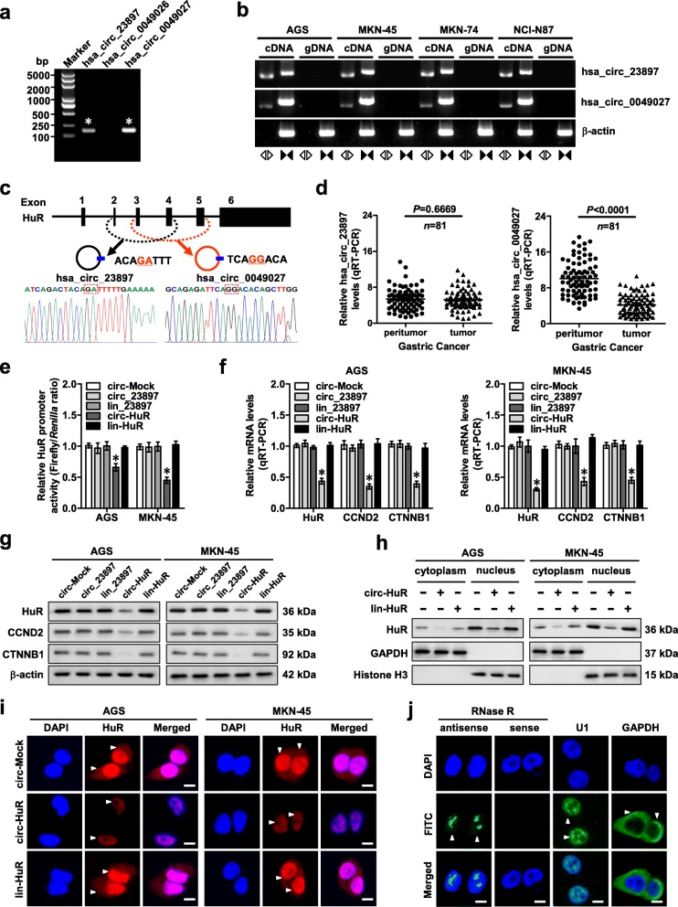
Background: Circular RNAs (circRNAs), a subclass of non-coding RNAs, play essential roles in tumorigenesis and aggressiveness. Our previous study has identified that circAGO2 drives gastric cancer progression through activating human antigen R (HuR), a protein stabilizing AU-rich element-containing mRNAs. However, the functions and underlying mechanisms of circRNAs derived from HuR in gastric cancer progression remain elusive.

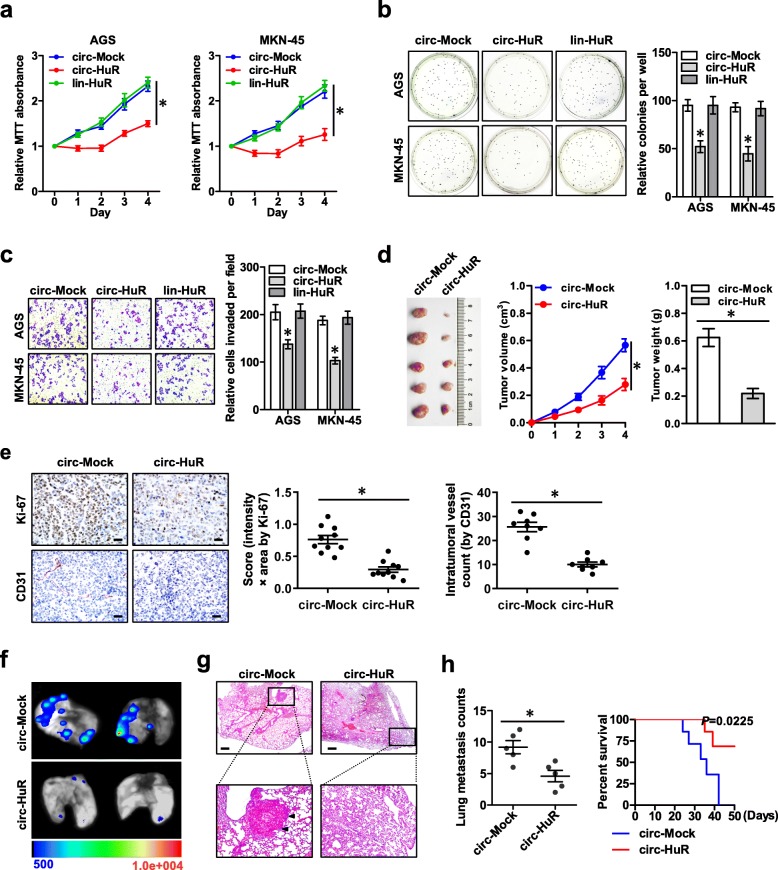
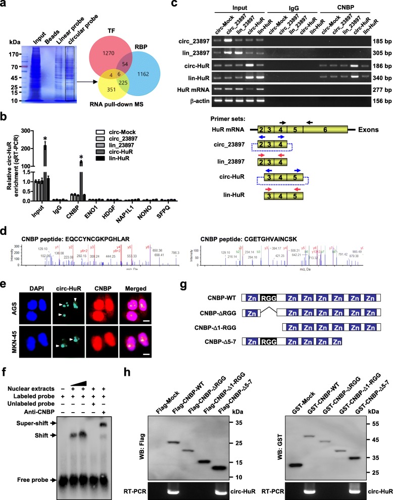
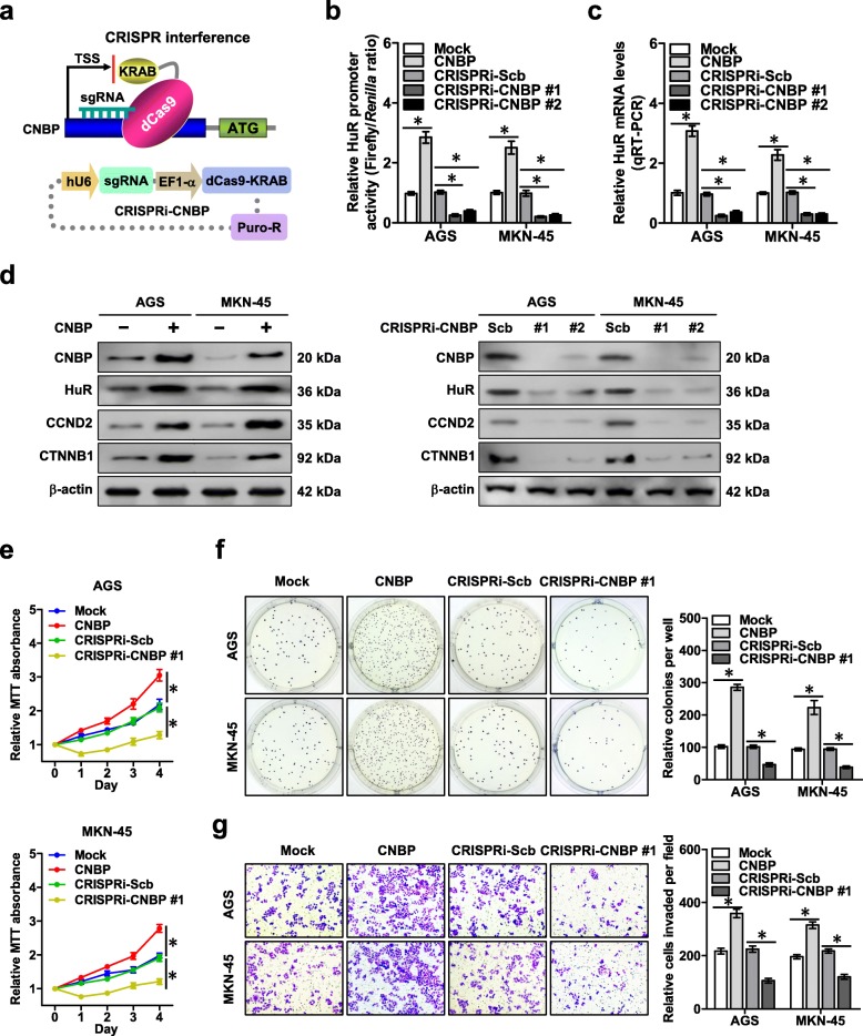
Methods: CircRNAs derived from HuR were detected by real-time quantitative RT-PCR and validated by Sanger sequencing. Biotin-labeled RNA pull-down, mass spectrometry, RNA immunoprecipitation, RNA electrophoretic mobility shift, and in vitro binding assays were applied to identify proteins interacting with circRNA. Gene expression regulation was observed by chromatin immunoprecipitation, dual-luciferase assay, real-time quantitative RT-PCR, and western blot assays. Gain- and loss-of-function studies were performed to observe the impacts of circRNA and its protein partner on the growth, invasion, and metastasis of gastric cancer cells in vitro and in vivo.

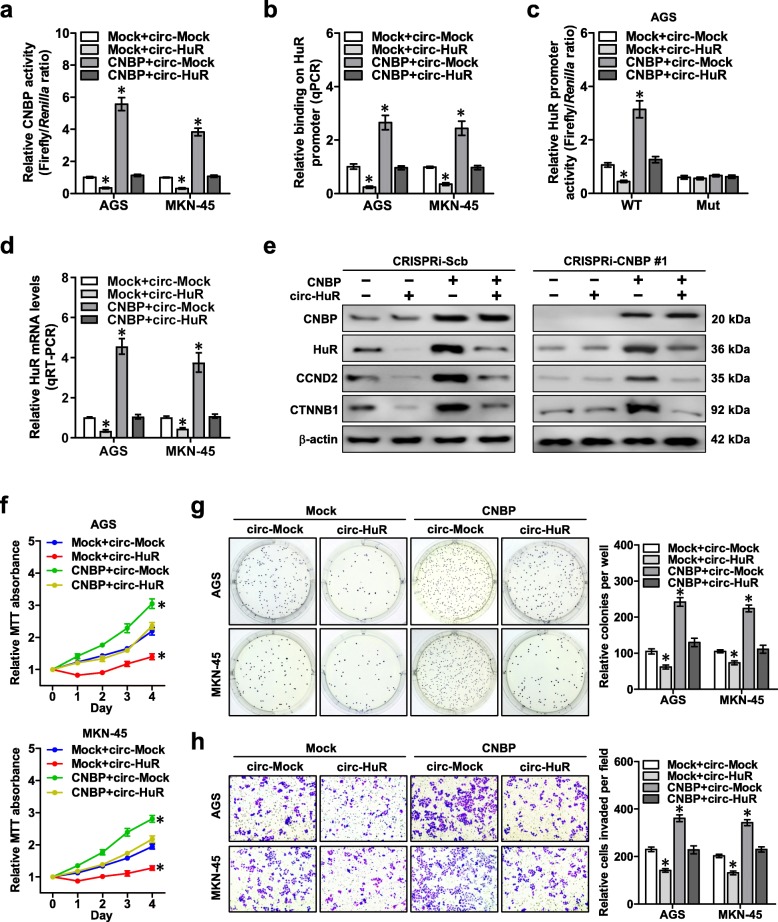
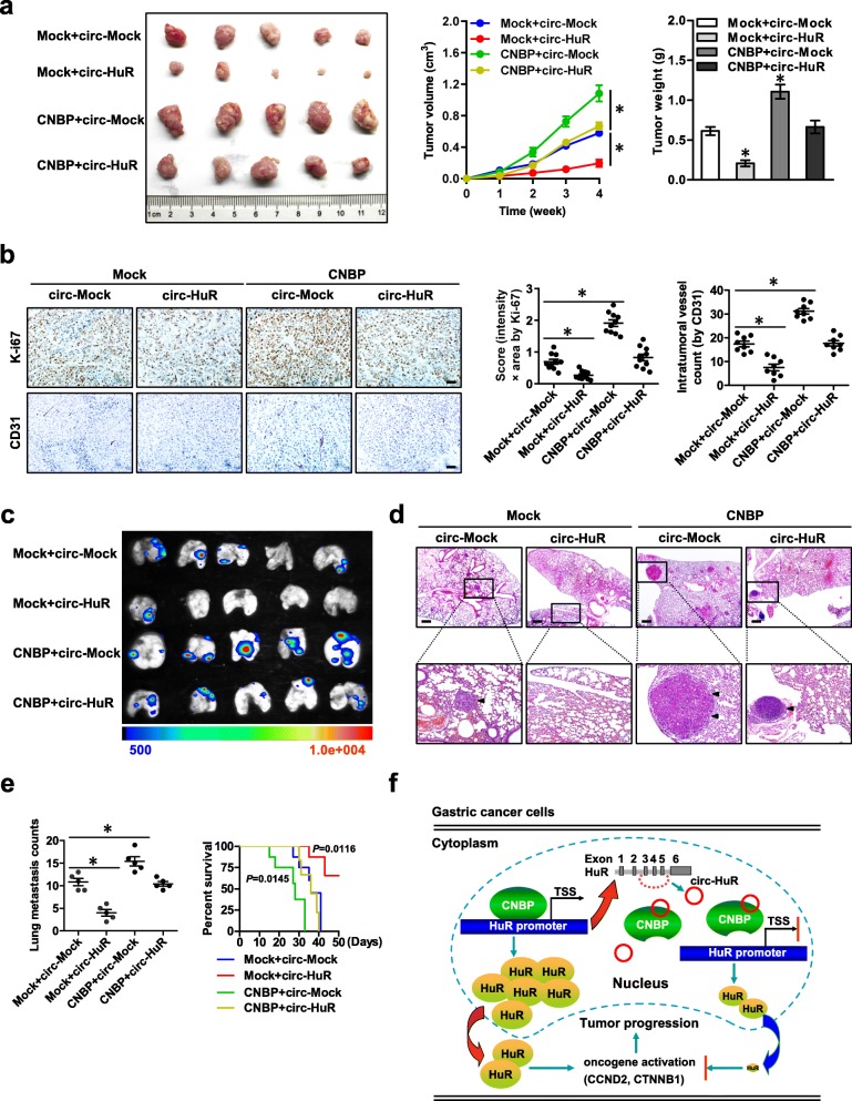
Results: Circ-HuR (hsa_circ_0049027) was predominantly detected in the nucleus, and was down-regulated in gastric cancer tissues and cell lines. Ectopic expression of circ-HuR suppressed the growth, invasion, and metastasis of gastric cancer cells in vitro and in vivo. Mechanistically, circ-HuR interacted with CCHC-type zinc finger nucleic acid binding protein (CNBP), and subsequently restrained its binding to HuR promoter, resulting in down-regulation of HuR and repression of tumor progression.

Conclusions: Circ-HuR serves as a tumor suppressor to inhibit CNBP-facilitated HuR expression and gastric cancer progression, indicating a potential therapeutic target for gastric cancer.

Keywords: CCHC-type zinc finger nucleic acid binding protein; Circular RNAs; Gastric cancer; Human antigen R.


Conflict of interest statement

The authors declare that they have no competing interests.

Figures

Fig. 1
Fig. 1
Circ-HuR is down-regulated and decreases HuR expression in gastric cancer. a RT-PCR assay with divergent primers indicating the detection of three circRNAs derived from HuR in AGS cells. b PCR assay with divergent and convergent primers showing the amplification of circRNAs from cDNA or genomic DNA (gDNA) of gastric cancer cell lines, while β-actin was used as a negative control. c Schematic illustration indicating the generation of hsa_circ_23897 and hsa_circ_0049027 from its host gene, and validation by Sanger sequencing. d Real-time qRT-PCR assay showing the relative levels (normalized to β-actin) of hsa_circ_23897 or hsa_circ_0049027 in the peritumor and tumor tissues of gastric cancer (n = 81). e Dual-luciferase assay indicating the promoter activity of HuR in AGS and MKN-45 cells stably transfected with empty vector (circ-Mock), hsa_circ-23897 (circ_23897), linear circ-23897 (lin_23897), circ-HuR, or linear circ-HuR (lin-HuR). f and g Real-time qRT-PCR (f, normalized to β-actin, n = 5) and western blot (g) assays revealing the transcript and protein levels of HuR and its downstream target genes in AGS and MKN-45 cells stably transfected with circ-Mock, circ-23897, lin_23897, circ-HuR, or lin-HuR. h and i Western blot (h) and immunofluorescence (i) assays showing the cytoplasmic and nuclear accumulation of HuR in AGS and MKN-45 cells stably transfected with circ-Mock, circ-HuR, or lin-HuR. Nuclei were stained by DAPI (blue). Scale bar, 10 μm. j RNA-FISH assay indicating the nuclear localization of circ-HuR in AGS cells using an antisense probe (green), while sense probe was used as a negative control. U1 and GAPDH were applied as positive controls. Scale bar, 10 μm. Student’s t-test and ANOVA analyzed the difference in (d-f). *P < 0.01 vs. circ-Mock. Data are shown as mean ± SEM (error bars) and representative of three independent experiments in (a-c) and (e-j)
Fig. 2
Fig. 2
Over-expression of circ-HuR suppresses the growth and aggressiveness of gastric cancer. a MTT colorimetric assay showing the viability of AGS and MKN-45 cells stably transfected with empty vector (circ-Mock), circ-HuR, or lin-HuR. b and c Soft agar (b) and matrigel invasion (c) assays indicating the in vitro growth and invasion of AGS and MKN-45 cells stably transfected with circ-Mock, circ-HuR, or lin-HuR. d Representative (left panel), in vivo growth curve (middle panel), and weight at the end points (right panel) of xenograft tumors formed by subcutaneous injection of AGS cells stably transfected with circ-Mock or circ-HuR into the dorsal flanks of nude mice (n = 5 for each group). e Representative images (left panel) and quantification (right panel) of immunohistochemical staining showing the expression of Ki-67 and CD31 within xenograft tumors formed by hypodermic injection of AGS cells stably transfected with circ-Mock or circ-HuR (n = 5 for each group). Scale bars: 50 μm. f-h Representative images (f), H&E staining (g, arrowheads), and quantification (h, left panel) of lung metastatic colonization and Kaplan-Meier curves (h, right panel) of nude mice treated with tail vein injection of AGS cells stably transfected with mock or circ-HuR (n = 5 for each group). Scale bar: 100 μm. ANOVA and Student’s t-test analyzed the difference in a-e and h. Log-rank test for survival comparison in (h). *P < 0.01 vs. circ-Mock. Data are shown as mean ± SEM (error bars) and representative of three independent experiments in (a-c)
Fig. 3
Fig. 3
Circ-HuR interacts with CNBP protein in gastric cancer. a Coomassie bright blue staining (left panel), mass spectrometry (MS) assay, and overlapping analysis (Venn diagram, right panel) with established RBP and TF databases revealing the proteins pulled down by biotin-labeled linear or circular forms of circ-HuR from the lysates of AGS cells. b RIP and real-time qRT-PCR assays showing the relative interaction between circ-HuR and six proteins in AGS cells stably transfected with empty vector (circ-Mock), hsa_circ-23897 (circ_23897), linear circ-23897 (lin_23897), circ-HuR, or linear circ-HuR (lin-HuR), with normalization to input of cells transfected with circ-Mock. c RIP assay with primer sets (lower panel) indicating the interaction between circ-HuR and CNBP in AGS cells stably transfected with circ-Mock, circ_23897, lin_23897, circ-HuR, or lin-HuR (upper panel). d MS assay depicting the identified CNBP peptides pulled down by circ-HuR. e Dual RNA-FISH and immunofluorescence staining assay indicating the co-localization of circ-HuR (green) and CNBP (red) in AGS and MKN-45 cells, with nuclei staining with DAPI (blue). Scale bar, 5 μm. f RNA EMSA determining the interaction between endogenous CNBP protein and biotin-labeled circular probe of circ-HuR (arrowheads), with CNBP antibody incubation or competition using an excess of unlabeled circular probe of circ-HuR. g Schematic diagram revealing the domains of CNBP truncations. h In vitro binding assay showing the enriched circ-HuR levels detected by RT-PCR (lower panel) after incubation with full-length or truncations of Flag-tagged or GST-tagged recombinant CNBP protein validated by western blot (upper panel). ANOVA analyzed the difference in (b). *P < 0.01 vs. circ-Mock. Data are shown as mean ± SEM (error bars) and representative of three independent experiments in (b, c, e, f, and h)
Fig. 4
Fig. 4
CNBP promotes HuR expression, growth, and aggressiveness of gastric cancer. a Schematic illustration of dCas9-based CRISPRi for CNBP and small guide RNA (sgRNA) targeting region. b Dual-luciferase assay revealing the promoter activity of HuR in AGS and MKN-45 cells stably transfected with empty vector (mock), CNBP, CRISPRi-Scb, CRISPRi-CNBP #1, or CRISPRi-CNBP #2. (c) and (d) Real-time qRT-PCR (c, normalized to β-actin, n = 5) and western blot (d) assays showing the transcript and protein levels of HuR in AGS and MKN-45 cells stably transfected with mock, CNBP, CRISPRi-Scb, CRISPRi-CNBP #1, or CRISPRi-CNBP #2. e MTT colorimetric assay indicating the viability of AGS and MKN-45 cells stably transfected with mock, CNBP, CRISPRi-Scb, or CRISPRi-CNBP #1. f and g Soft agar (f) and matrigel invasion (g) assays showing the in vitro growth and invasion of AGS and MKN-45 cells stably transfected with mock, CNBP, CRISPRi-Scb, or CRISPRi-CNBP #1. ANOVA analyzed the difference in (b, c and e-g). *P < 0.01 vs. mock or CRISPRi-Scb. Data are shown as mean ± SEM (error bars) and representative of three independent experiments in (b-g)
Fig. 5
Fig. 5
Circ-HuR suppresses HuR expression, growth, and invasion of gastric cancer cells via repressing CNBP transactivation. a Dual-luciferase assay revealing the transactivation of CNBP in AGS and MKN-45 cells stably transfected with empty vector (mock) or CNBP, and those co-transfected with circ-Mock or circ-HuR. b ChIP and qPCR assays showing the changes in binding of CNBP to HuR promoter in AGS and MKN-45 cells stably transfected with mock or CNBP, and those co-transfected with circ-Mock or circ-HuR. c and d Dual-luciferase (c) and real-time qRT-PCR (d) assays indicating the activity of HuR promoter with wild type (WT) or mutant (Mut) CNBP binding site and transcript levels (normalized to β-actin, n = 4) of HuR in AGS and MKN-45 cells stably transfected with mock or CNBP, and those co-transfected with circ-Mock or circ-HuR. e Western blot assay showing the expression of CNBP, HuR, CCND2, and CTNNB1 in AGS cells stably transfected with CRISPRi-Scb or CRISPRi-CNBP #1, and those co-transfected with mock, CNBP, circ-Mock, or circ-HuR. f MTT colorimetric assay indicating the viability of AGS and MKN-45 cells stably transfected with mock or CNBP, and those co-transfected with circ-Mock or circ-HuR. g and h Soft agar (g) and matrigel invasion (h) assays showing in vitro growth and invasion of AGS and MKN-45 cells stably transfected with mock or CNBP, and those co-transfected with circ-Mock or circ-HuR. ANOVA analyzed the difference in (a-d and f-h). *P < 0.01 vs. mock+ circ-Mock. Data are shown as mean ± SEM (error bars) and representative of three independent experiments in (a-h)
Fig. 6
Fig. 6
Circ-HuR suppresses gastric cancer progression by inhibiting CNBP transactivation in vivo. a Representative (left panel), in vivo growth curve (middle panel), and weight at the end points (right panel) of xenograft tumors formed by subcutaneous injection of AGS cells stably transfected with empty vector (mock) or CNBP, and those co-transfected with circ-Mock or circ-HuR into the dorsal flanks of nude mice (n = 5 for each group). b Representative images (left panel) and quantification (right panel) of immunohistochemical staining showing the expression of Ki-67 and CD31 within xenograft tumors formed by hypodermic injection of AGS cells stably transfected with mock or CNBP, and those co-transfected with circ-Mock or circ-HuR (n = 5 for each group). Scale bars: 50 μm. c-e Representative images (c), H&E staining (d, arrowheads), and quantification (e, left panel) of lung metastatic colonization and Kaplan-Meier curves (e, right panel) of nude mice treated with tail vein injection of AGS cells stably transfected with mock or CNBP, and those co-transfected with circ-Mock or circ-HuR (n = 5 for each group). Scale bar: 100 μm. f The mechanisms underlying circ-HuR-suppressed tumor progression: as a nuclear circRNA, circ-HuR interacts with CNBP to inhibit its binding to HuR promoter, resulting in down-regulation of HuR and suppression of gastric cancer progression. ANOVA analyzed the difference in (a, b, e). Log-rank test for survival comparison in (e). *P < 0.01 vs. mock+ circ-Mock. Data are shown as mean ± SEM (error bars)

Similar articles

Cited by

References

    1. Van Cutsem E, Sagaert X, Topal B, Haustermans K, Prenen H. Gastric cancer. Lancet. 2016;388:2654–2664. doi: 10.1016/S0140-6736(16)30354-3. - DOI - PubMed
    1. Zucal C, D'Agostino V, Loffredo R, Mantelli B, NatthakanThongon LP, Latorre E, Provenzani A. Targeting the multifaceted HuR protein, benefits and caveats. Curr Drug Targets. 2015;16:499–515. doi: 10.2174/1389450116666150223163632. - DOI - PubMed
    1. Doller A, el-S A, Huwiler A, Muller R, Radeke HH, Pfeilschifter J, Eberhardt W. Posttranslational modification of the AU-rich element binding protein HuR by protein kinase Cdelta elicits angiotensin II-induced stabilization and nuclear export of cyclooxygenase 2 mRNA. Mol Cell Biol. 2008;28:2608–2625. doi: 10.1128/MCB.01530-07. - DOI - PMC - PubMed
    1. Vazquez-Chantada M, Fernandez-Ramos D, Embade N, Martinez-Lopez N, Varela-Rey M, Woodhoo A, Luka Z, Wagner C, Anglim PP, Finnell RH, et al. HuR/methyl-HuR and AUF1 regulate the MAT expressed during liver proliferation, differentiation, and carcinogenesis. Gastroenterology. 2010;138:1943–1953. doi: 10.1053/j.gastro.2010.01.032. - DOI - PMC - PubMed
    1. Kotta-Loizou I, Giaginis C, Theocharis S. Clinical significance of HuR expression in human malignancy. Med Oncol. 2014;31:161. doi: 10.1007/s12032-014-0161-y. - DOI - PubMed

Publication types