. 2019 Nov 11;20(22):5644.
doi: 10.3390/ijms20225644.

Insights into the Effects of Cancer Associated Mutations at the UPF2 and ATP-Binding Sites of NMD Master Regulator: UPF1

Affiliations

Insights into the Effects of Cancer Associated Mutations at the UPF2 and ATP-Binding Sites of NMD Master Regulator: UPF1

Umesh Kalathiya et al. Int J Mol Sci. .

Abstract

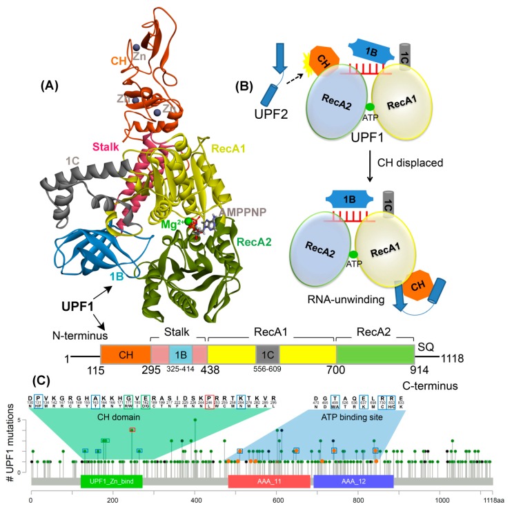
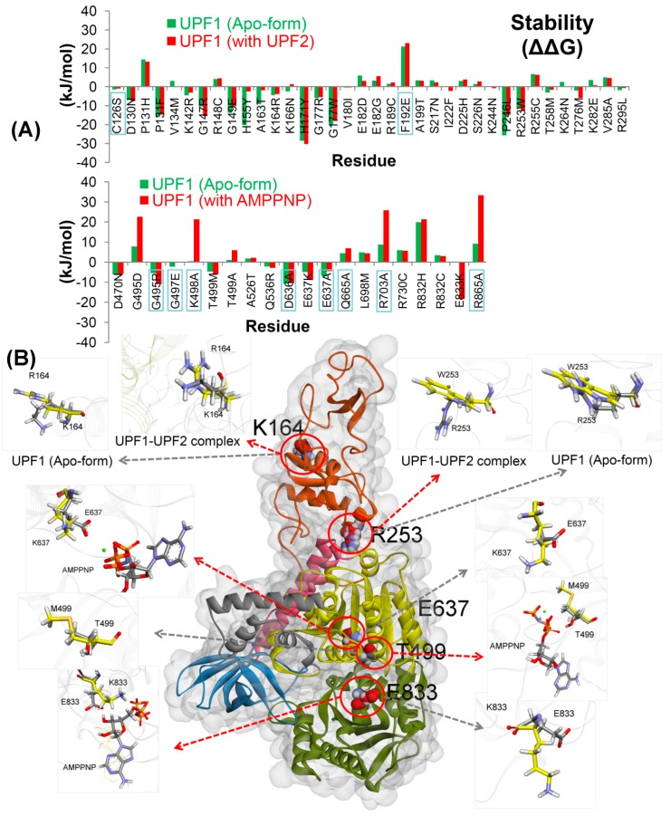
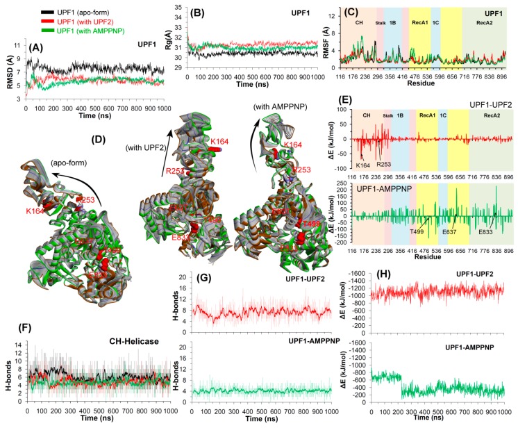
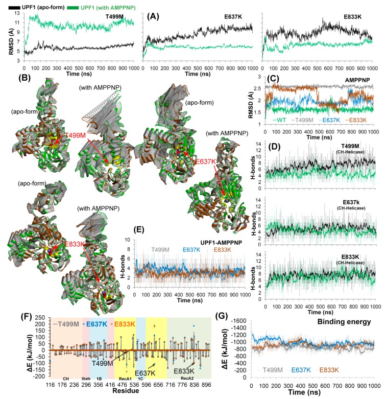
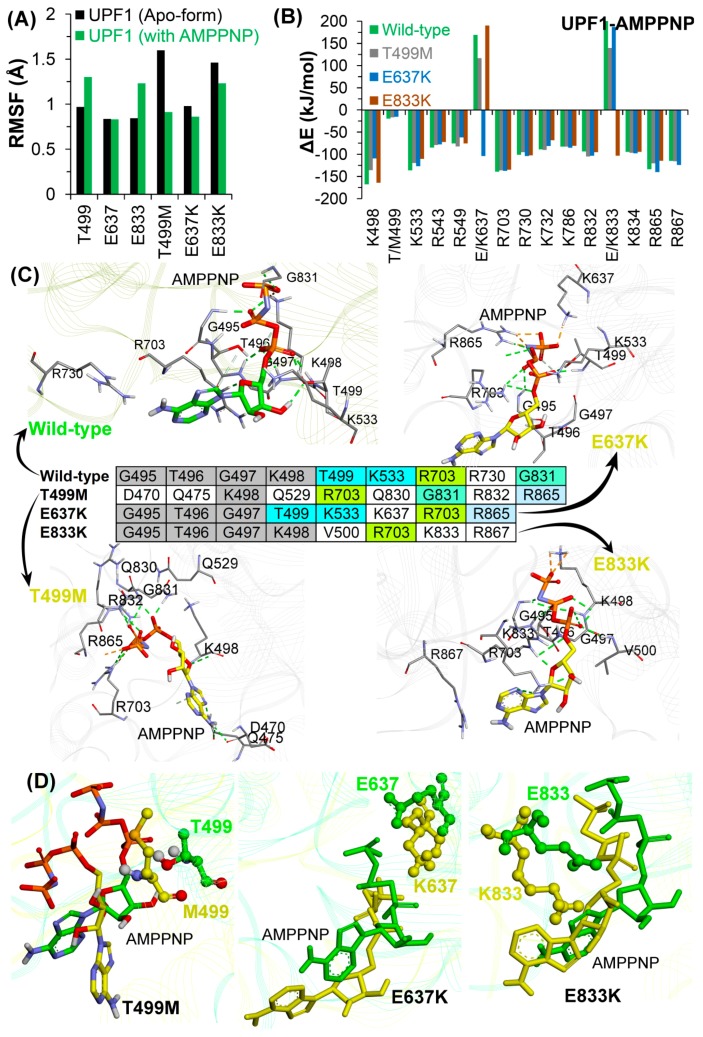
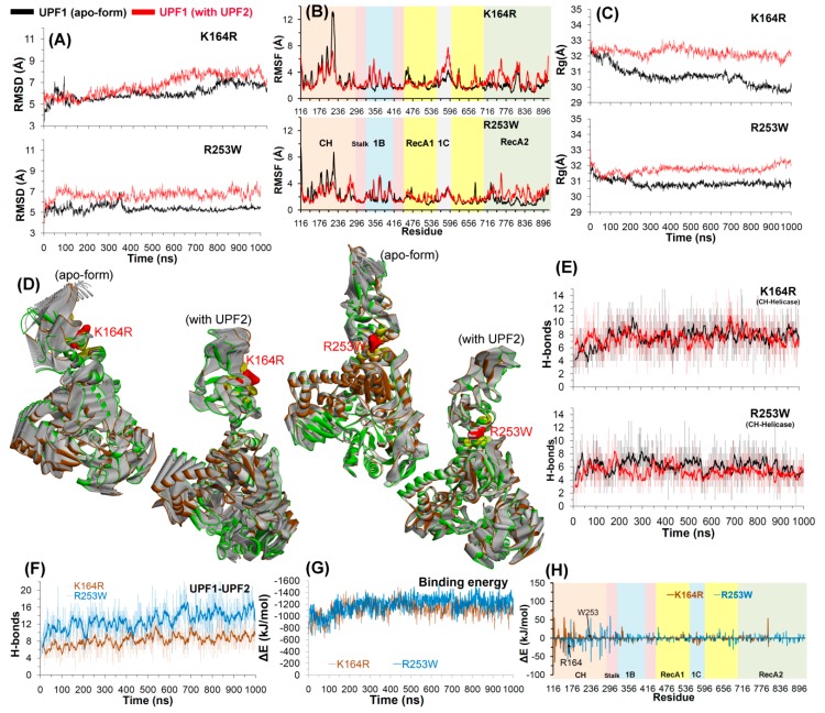
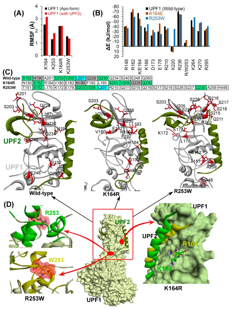
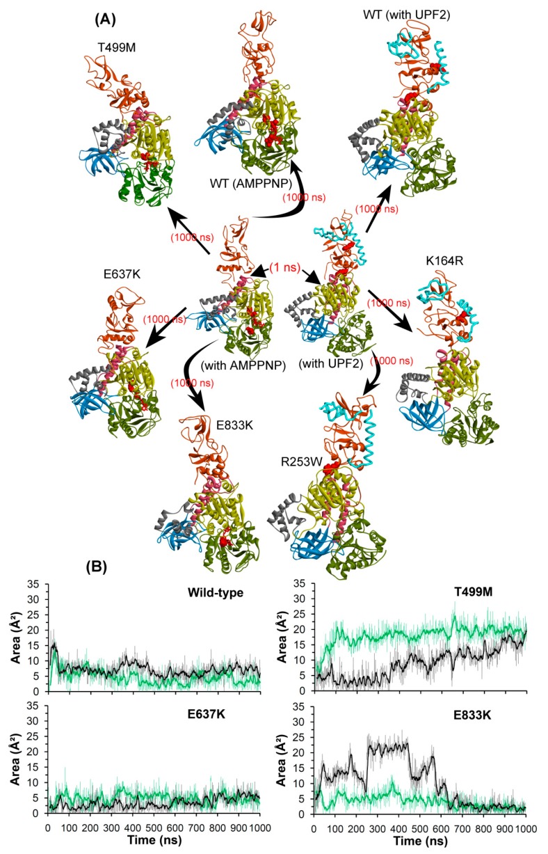
Nonsense-mediated mRNA decay (NMD) is a quality control mechanism that recognizes post-transcriptionally abnormal transcripts and mediates their degradation. The master regulator of NMD is UPF1, an enzyme with intrinsic ATPase and helicase activities. The cancer genomic sequencing data has identified frequently mutated residues in the CH-domain and ATP-binding site of UPF1. In silico screening of UPF1 stability change as a function over 41 cancer mutations has identified five variants with significant effects: K164R, R253W, T499M, E637K, and E833K. To explore the effects of these mutations on the associated energy landscape of UPF1, molecular dynamics simulations (MDS) were performed. MDS identified stable H-bonds between residues S152, S203, S205, Q230/R703, and UPF2/AMPPNP, and suggest that phosphorylation of Serine residues may control UPF1-UPF2 binding. Moreover, the alleles K164R and R253W in the CH-domain improved UPF1-UPF2 binding. In addition, E637K and E833K alleles exhibited improved UPF1-AMPPNP binding compared to the T499M variant; the lower binding is predicted from hindrance caused by the side-chain of T499M to the docking of the tri-phosphate moiety (AMPPNP) into the substrate site. The dynamics of wild-type/mutant systems highlights the flexible nature of the ATP-binding region in UPF1. These insights can facilitate the development of drug discovery strategies for manipulating NMD signaling in cell systems using chemical tools.

Keywords: ATP-binding site; UPF1; UPF2; cancer mutations; molecular dynamics simulations; structural stability.


Conflict of interest statement

The authors declare no competing interests.

Figures

Figure 1
Figure 1
UPF1 structure and mutations from different cancer types. (A) Crystal structure of UPF1 (PDB: 2WJY [10]) representing different domains, ATP analogue (i.e., AMPPNP), and Zn/Mg2+ ions. (B) Schematic model of UPF1 activation mechanism showing how the RNA binding and ATPase/unwinding activity are modulated by UPF2 in an NMD cycle. In the absence of UPF2, the UPF1 has extended RNA interactions via the RecA1, RecA2, 1B, and 1C domains and low RNA-unwinding properties. The CH-domain is displaced upon UPF2 binding (black dotted arrow) and domain 1B changes to a conformation where it does not clamp on the RNA 3′ end and as a result, the RNA-unwinding activity is increased [11]. (C) UPF1 gene mutated in cancer, data retrieved from the cBioPortal database [29]. The frequency of a residue mutation equal to 2, 3, and 4 are marked in blue, green, and red boxes respectively. The mutations from the ATP-binding site are shown in orange. Color scheme: Zn and carbon in grey, Mg in green, oxygen in red, hydrogen in silver, nitrogen in blue, and sulfur in orange.
Figure 2
Figure 2
Effects of mutations on the UPF1 protein. (A) UPF1 structural stability change (∆∆G; kJ/mol) upon mutation. In addition, the mutations from previous studies [4,7,11,13,19,21,32,33] are indicated by labels with the blue box. (B) The UPF1 structure showing mutations studied by MDS. The red arrows represent UPF1 mutations in the complex with UPF2/AMPPNP and the grey arrows show mutations in the apo-form of UPF1. Cα atoms of the wild-type and mutated residues are shown in grey and yellow, respectively (different domains of UPF1 are colored as per Figure 1). Color scheme: oxygen in red, hydrogen in silver, nitrogen in blue, and sulfur in orange.
Figure 3
Figure 3
Structural analysis of wild-type UPF1 in the MD simulations. (AC) RMSD, radius of gyration profile, and RMSF representing the structural changes and fluctuations of residues inthe UPF1 protein. (D) The protein motion corresponding to the first eigenvector defined on the basis of the combined trajectories (green is from the beginning, blue from 400 ns (where the system stabilized), and brown from the end of the MD). (E,H) Energy contribution of each UPF1 residue to the binding with UPF2/AMPPNP and the total binding energy for UPF1-UPF2/AMPPNP, calculated using MM-PBSA. Contribution of residues selected for mutation analysis are labelled and shown with black arrows in (E). (F,G) Number of hydrogen bonds formed between the CH-helicase domains (intramolecular UPF1) and between UPF1-UPF2/AMPPNP (intermolecular). The dark lines represent the moving average of H-bonds formed with a period of 10 ns (i.e., number of H-bonds averaged every 10 ns).
Figure 4
Figure 4
Structural analysis of T499M, E637K, and E833K mutants in the UPF1 protein. (A) RMSD for all atoms (excluding hydrogens) of mutant UPF1. (B) The principal motion projected along the first eigenvector defined on the basis of the combined trajectories (green marks the beginning and brown is the end of MDS; yellow are residues within 5 Å of the mutated residues). (C) RMSD for all atoms (excluding hydrogens) of AMPPNP from the WT and mutant UPF1-AMPPNP systems. (D,E) Number of hydrogen bonds formed between CH-helicase domains (UPF1 intramolecular) and between UPF1-AMPPNP (intermolecular). The dark lines represent trends with a moving average of H-bonds formed with a period of 10 ns (i.e., number of H-bonds averaged every 10 ns). (F,G) Energy contribution (computed using MM-PBSA) of each residue of UPF1 to the binding with AMPPNP and total binding energy for UPF1-AMPPNP complex, respectively. Contribution of residues selected for mutational analysis are labelled and shown with arrows in (F).
Figure 5
Figure 5
Molecular properties of the UPF1-AMPPNP model systems. (A) Substrate binding site residues of the UPF1 protein and studied mutations. RMSFs for wild-type UPF1, mutant models; apo-form and AMPPNP bound UPF1. (B) Individual residues of UPF1 contributing to the binding energy for AMPPNP, calculated by MM-PBSA. (C) Binding mode of AMPPNP to the UPF1 protein in different model systems, and UPF1 residues forming stable H-bonds with AMPPNP having occupancy ≥10%. (D) Binding conformation of AMPPNP with respect to the wild-type and mutated UPF1 (T499M, E637K, and E833K) obtained from the end of MDS.
Figure 6
Figure 6
Structural analysis of UPF1 mutants K164R and R253W. (AC) RMSD, RMSF, and radius of gyration profiles representing structural changes and fluctuations of UPF1 residues. (D) The motion corresponding to the first eigenvector defined on the basis of the combined trajectories. (E,F) Number of hydrogen bonds formed between CH-helicase domains (UPF1 intramolecular interactions) and between UPF1-UPF2 (intermolecular interactions). The dark lines represent trend with a moving average of H-bonds formed with a period of 10 ns (i.e., number of H-bonds averaged every 10 ns). (G,H) Energy of each residue (of the mutant UPF1) contributing to the binding with UPF2 and total binding energy for UPF1-UPF2, calculated by MM-PBSA.
Figure 7
Figure 7
Detailed analysis of UPF1-UPF2 model systems. (A) UPF1 residues involved in binding with UPF2 and their mutations. RMSF values were calculated in native or wild-type UPF1, mutant models, apo-form, and UPF1 bound with UPF2. (B) Individual UPF1 residues contributing to the binding energy with UPF2, were computed using MM-PBSA. (C) Interactions of UPF1 with UPF2 in different model systems, and residues of UPF1 forming stable H-bonds with UPF2 having occupancy ≥10%. (D) Binding mode or conformation of the UPF1 residues (K164 and R253 in native or mutated form R164 and W253) with UPF2 (Color scheme: green represents wild-type and yellow mutated systems, carbon in green/yellow, oxygen in red, nitrogen in blue, and hydrogen in silver).
Figure 8
Figure 8
Structural/conformational changes of UPF1 protein when complexed with UPF2 or AMPPNP. (A) Wild-type and mutant UPF1 protein in complex with AMPPNP or UPF2. (B) Area analysis of structural changes in the ATP-binding region of the UPF1 protein, triangle selected for area calculation was based on Cα atoms coordinates of T499, E637, and E833 residues. The dark lines represent the trend with a moving average of area with a period of 10 ns. Different domains of UPF1 are colored as per Figure 1.

Similar articles

Cited by

References

    1. Serdar L.D., Whiteside D.L., Baker K.E. ATP hydrolysis by UPF1 is required for efficient translation termination at premature stop codons. Nat. Commun. 2016;7:14021. doi: 10.1038/ncomms14021. - DOI - PMC - PubMed
    1. Karamyshev A.L., Karamysheva Z.N. Lost in translation: Ribosome-associated mRNA and protein quality controls. Front. Genet. 2018;9:431. doi: 10.3389/fgene.2018.00431. - DOI - PMC - PubMed
    1. Gleghorn M., Maquat L. UPF1 learns to relax and unwind. Mol. Cell. 2011;41:621–623. doi: 10.1016/j.molcel.2011.03.001. - DOI - PubMed
    1. Kashima I., Yamashita A., Izumi N., Kataoka N., Morishita R., Hoshino S., Ohno M., Dreyfuss G., Ohno S. Binding of a novel SMG-1-Upf1-eRF1-eRF3 complex (SURF) to the exon junction complex triggers Upf1 phosphorylation and nonsense-mediated mRNA decay. Genes Dev. 2006;20:355–367. doi: 10.1101/gad.1389006. - DOI - PMC - PubMed
    1. Hug N., Cáceres J.F. The RNA helicase DHX34 activates NMD by promoting a transition from the surveillance to the decay-inducing complex. Cell Rep. 2014;8:1845–1856. doi: 10.1016/j.celrep.2014.08.020. - DOI - PMC - PubMed