Renal Injury in DENV-4 Fatal Cases: Viremia, Immune Response and Cytokine Profile
- PMID: 31703246
- PMCID: PMC6963280
- DOI: 10.3390/pathogens8040223
Renal Injury in DENV-4 Fatal Cases: Viremia, Immune Response and Cytokine Profile
Abstract
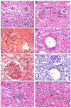
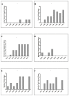
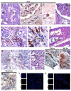
Dengue virus (DENV) infections may result in asymptomatic cases or evolve into a severe disease, which involves multiple organ failure. Renal involvement in dengue can be potentially related to an increased mortality. Aiming to better understand the role of DENV in renal injury observed in human fatal cases, post-mortem investigations were performed in four DENV-4 renal autopsies during dengue epidemics in Brazil. Tissues were submitted to histopathology, immunohistochemistry, viral quantification, and characterization of cytokines and inflammatory mediators. Probably due the high viral load, several lesions were observed in the renal tissue, such as diffuse mononuclear infiltration around the glomerulus in the cortical region and in the medullary vessels, hyalinosis arteriolar, lymphocytic infiltrate, increased capsular fibrosis, proximal convoluted tubule (PCT) damage, edema, PCT debris formation, and thickening of the basal vessel membrane. These changes were associated with DENV-4 infection, as confirmed by the presence of DENV-specific NS3 protein, indicative of viral replication. The exacerbated presence of mononuclear cells at several renal tissue sites culminated in the secretion of proinflammatory cytokines and chemokines. Moreover, it can be suggested that the renal tissue injury observed here may have been due to the combination of both high viral load and exacerbated host immune response.
Keywords: cytokines; dengue 4; fatal case; histopathology; inflammatory mediators; viremia.
Conflict of interest statement
The authors declare no conflict of interest.
Figures


Similar articles
-
Immunopathology of Renal Tissue in Fatal Cases of Dengue in Children.Pathogens. 2022 Dec 15;11(12):1543. doi: 10.3390/pathogens11121543. Pathogens. 2022. PMID: 36558877 Free PMC article.
-
Peripheral Organs of Dengue Fatal Cases Present Strong Pro-Inflammatory Response with Participation of IFN-Gamma-, TNF-Alpha- and RANTES-Producing Cells.PLoS One. 2016 Dec 22;11(12):e0168973. doi: 10.1371/journal.pone.0168973. eCollection 2016. PLoS One. 2016. PMID: 28006034 Free PMC article.
-
Dengue type 4 in Rio de Janeiro, Brazil: case characterization following its introduction in an endemic region.BMC Infect Dis. 2017 Jun 9;17(1):410. doi: 10.1186/s12879-017-2488-4. BMC Infect Dis. 2017. PMID: 28599640 Free PMC article.
-
Early dengue virus interactions: the role of dendritic cells during infection.Virus Res. 2016 Sep 2;223:88-98. doi: 10.1016/j.virusres.2016.07.001. Epub 2016 Jul 2. Virus Res. 2016. PMID: 27381061 Review.
-
Role of Monocytes in the Pathogenesis of Dengue.Arch Immunol Ther Exp (Warsz). 2019 Feb;67(1):27-40. doi: 10.1007/s00005-018-0525-7. Epub 2018 Sep 20. Arch Immunol Ther Exp (Warsz). 2019. PMID: 30238127 Review.
Cited by
-
Prevalence, Characteristics, and Outcomes Associated with Acute Kidney Injury among Adult Patients with Severe Dengue in Mainland China.Am J Trop Med Hyg. 2023 Jun 26;109(2):404-412. doi: 10.4269/ajtmh.22-0803. Print 2023 Aug 2. Am J Trop Med Hyg. 2023. PMID: 37364862 Free PMC article.
-
Immunocompetent Mice Infected by Two Lineages of Dengue Virus Type 2: Observations on the Pathology of the Lung, Heart and Skeletal Muscle.Microorganisms. 2021 Dec 8;9(12):2536. doi: 10.3390/microorganisms9122536. Microorganisms. 2021. PMID: 34946137 Free PMC article.
-
Different Profiles of Cytokines, Chemokines and Coagulation Mediators Associated with Severity in Brazilian Patients Infected with Dengue Virus.Viruses. 2021 Sep 8;13(9):1789. doi: 10.3390/v13091789. Viruses. 2021. PMID: 34578370 Free PMC article.
-
Severe dengue in the intensive care unit.J Intensive Med. 2023 Sep 28;4(1):16-33. doi: 10.1016/j.jointm.2023.07.007. eCollection 2024 Jan. J Intensive Med. 2023. PMID: 38263966 Free PMC article. Review.
-
Brazilian Dengue Virus Type 2-Associated Renal Involvement in a Murine Model: Outcomes after Infection by Two Lineages of the Asian/American Genotype.Pathogens. 2021 Aug 26;10(9):1084. doi: 10.3390/pathogens10091084. Pathogens. 2021. PMID: 34578117 Free PMC article.
References
-
- Kuhn R.J., Zhang W., Rossmann M.G., Pletnev S.V., Corver J., Lenches E., Jones C.T., Mukhopadhyay S., Chipman P.R., Strauss E.G., et al. Structure of dengue virus: Implications for flavivirus organization, maturation, and fusion. Cell. 2002;108:717–725. doi: 10.1016/S0092-8674(02)00660-8. - DOI - PMC - PubMed
-
- Acosta E.G., Kumar A., Bartenschlager R. Revisiting dengue virus-host cell interaction: New insights into molecular and cellular virology. Adv. Virus Res. 2014;88:1–109. - PubMed

