. 2020 Feb;39(7):1600-1616.
doi: 10.1038/s41388-019-1087-9. Epub 2019 Nov 7.

The exosome-mediated autocrine and paracrine actions of plasma gelsolin in ovarian cancer chemoresistance

Affiliations

The exosome-mediated autocrine and paracrine actions of plasma gelsolin in ovarian cancer chemoresistance

Meshach Asare-Werehene et al. Oncogene. 2020 Feb.

Abstract

Ovarian cancer (OVCA) is the most lethal gynecological cancer, due predominantly to late presentation, high recurrence rate and common chemoresistance development. The expression of the actin-associated protein cytosolic gelsolin (GSN) regulates the gynecological cancer cell fate resulting in dysregulation in chemosensitivity. In this study, we report that elevated expression of plasma gelsolin (pGSN), a secreted isoform of GSN and expressed from the same GSN gene, correlates with poorer overall survival and relapse-free survival in patients with OVCA. In addition, it is highly expressed and secreted in chemoresistant OVCA cells than its chemosensitive counterparts. pGSN, secreted and transported via exosomes (Ex-pGSN), upregulates HIF1α-mediated pGSN expression in chemoresistant OVCA cells in an autocrine manner as well as confers cisplatin resistance in otherwise chemosensitive OVCA cells. These findings support our hypothesis that exosomal pGSN promotes OVCA cell survival through both autocrine and paracrine mechanisms that transform chemosensitive cells to resistant counterparts. Specifically, pGSN transported via exosomes is a determinant of chemoresistance in OVCA.


Conflict of interest statement

The authors declare that they have no conflict of interest.

Figures
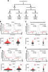
Fig. 1
Fig. 1
High pGSN expression is associated with tumor recurrence in patients with ovarian cancer. a Ovarian cancer public datasets were stratified using histological subtype (serous and endometroid), chemotherapeutic agents, and suboptimal surgical debulking. Kaplan–Meier survival analysis and beeswarm plots with optimal cutoff values of pGSN expression were performed on b only serous patients, c serous and endometroid patients, d serous patients with suboptimal surgical debulking, and e serous and endometroid patients with suboptimal surgical debulking with treatments containing either platinum or platinum + taxol. P values were calculated by the log-rank test
Fig. 2
Fig. 2
pGSN regulates CDDP-induced apoptosis in OVCA cells. a, b CDDP decreased pGSN content and induced apoptosis in chemosensitive (OV2295, OV443, and A2780s) but not chemoresistant (OV90, OV866(2), and A2780cp) OVCA cells. OVCA cells were cultured with or without CDDP (10 µM; 24 h). c, d Silencing pGSN in OV866(2) and A2780cp cells sensitized them to CDDP-induced apoptosis. OV8669(2) and A2780cp cells were transfected with pGSN siRNA (50 nM, 24 h; which specifically knocked down pGSN but not cGSN), and then treated with or without CDDP (10 µM; 24 h). e, f Overexpression of pGSN cDNA attenuated CDDP-induced apoptosis in OV2295 and A2780s cells. OV2295 and A2780s cells were transfected with pGSN cDNA (2 µg; 24 h) and cultured with or without CDDP (10 µM; 24 h). g A2780cp cells (with total GSN knocked down) were cultured with rhpGSN (10 µM; 24 h) before treatment with CDDP (0 and 10 µM; 24 h). pGSN, cGSN, and β-tubulin (loading control) contents were assessed by western blotting (WB) and apoptosis determined morphologically by Hoechst 33258 DNA staining. [a (a; ***p < 0.001 vs b, c, and d); b (a; ***p < 0.001 vs b); c (a; ***p < 0.001 vs b and c); d (a; ***p < 0.001 vs b); e (a; ***p < 0.001 vs b and c); f (a; ***p < 0.001 vs b and c); g (a; ***p < 0.001 vs b and c);]. N = 3
Fig. 3
Fig. 3
Extracellular vesicle characterization and CDDP effect on vesicle size distribution. HGS chemosensitive and resistant cells secrete exosomes (a) and microparticles (b) as confirmed by nanoparticle tracking. c A2780s and A2780cp cells secrete both exosomes and microparticles. OVCA cells were cultured with or without CDDP (10 µM; 24 h). Exosomes and microparticles were isolated from their conditioned media by ultracentrifugation and characterized by Nanoparticle Tracking Analyser. d, e The exosome-to-microparticle ratio is higher in chemoresistant cells compared with chemosensitive cells; their concentrations were not affected by CDDP treatment. f pGSN is predominantly identified in the exosomes compared with microparticles of A2780s and A2780cp; however, pGSN content is higher in the A2780cp cells compared with A2780s cells. Exosomes and microparticles were isolated as described above and pGSN and CD63 (exosome marker) contents assessed by WB. g Electron micrograph showing pGSN in microparticles (mp) and multivesicular bodies/exosomes (mb); white and black arrows showing pGSN in mp and mb, respectively. pGSN in fixed A2780cp cells was immunostained with 18-nm colloidal gold particles, observed and photographed with a Jeol JEM 1230 transmission electron microscope. Scale bars, 500 nm (a), 100 nm (b–e) and 20 nm (f). [d (a; **p < 0.01 vs b); e (a; **p < 0.01 vs b)]. N = 3
Fig. 4
Fig. 4
The integrin signaling pathway is involved in the autocrine upregulation of pGSN content by pGSN. a, b The α5β1 integrin receptor blocker ATN 161 attenuates the upregulation of pGSN by Ex-pGSN (a) and rhpGSN (b). A2780s cells were treated with anti-α5β1 integrin (40 µM; 3 h) followed by A2780cp-derived Ex-pGSN (40 µg/400,000 cells; 24 h) or rhpGSN (10 µM; 24 h). c Knockdown of FAK resulted in the downregulation of HIF1α and pGSN contents. A2780s cells were transfected with FAK siRNA1 and siRNA2 (0–20 pmol; 24 h) before culture with A2780cp-derived Ex-pGSN (40 µg/400,000 cells; 24 h). d pGSN and HIF1-α contents were higher in A2780cp cells compared with A2780s cells; CDDP reduced their content in A2780s but not in A2780cp cells. CDDP-induced apoptosis in A2780cp cells was higher than that in A2780s cells. A2780s and A2780cp cells were cultured with or without CDDP (10 µM; 24 h). e HIF1-α silencing reduced the content of pGSN and sensitized A2780cp cells to CDDP-induced apoptosis. A2780cp cells were transfected with HIF1α siRNA1 and siRNA2 (200 pmol; 24 h) before culture with or without CDDP (10 µM; 24 h). pGSN, FAK, HIF1α, and β-tubulin (loading control) contents were assessed by WB and apoptosis determined morphologically by Hoechst 33258 DNA staining. pGSN levels in the conditioned media were assessed by the sandwich ELISA. [a (a; ***p < 0.001 vs b, c, d); b (a; ***p < 0.001 vs b, c, d); c (a; ***p < 0.001 vs b, c, d); d (a; ***p < 0.001 vs b); e (a; ***p < 0.001 vs b)]. N = 3
Fig. 5
Fig. 5
pGSN-mediated OVCA chemoresistance involves HIF1α modulation by Akt. a Activation of Akt in chemosensitive cells increases the contents of pGSN and HIF1α and renders them resistant to CDDP-induced apoptosis. A2780s cells constitutively expressing an activated Akt (A2780s-A-AKT) and its scrambled control cells (A2780s-CTL) were cultured with or without CDDP (10 µM; 24 h). b pGSN and HIF1α contents are decreased and CDDP-induced apoptosis enhanced in chemoresistant cells when Akt function is downregulated. A2780cp cells constitutively expressing triple-mutant dominant Akt (A2780cp-DN-AKT) or scrambled control (A2780cp-CTL) cultured with or without CDDP (10 µM; 24 h). c Inhibition of proteasomal degradation of HIF1α in chemosensitive cells increases the content of pGSN and attenuates CDDP-induced apoptosis. A2780s cells were pretreated with epoxomycin (10 nM; 3 h) and cultured with or without CDDP (10 µM; 24 h) in the presence of the inhibitor. d Forced expression of a proteasomal nondegradable mutant of HIF1α increases pGSN content and inhibits CDDP-induced apoptosis in chemosensitive cells. A2780s cells were transfected with ΔHIF1α cDNA (1 µg; 24 h) and treated with or without CDDP (10 µM; 24 h). e Induction of proteasomal HIF1α degradation in chemoresistant cells decreases pGSN content and renders them sensitive to CDDP-induced apoptosis. A2780cp cells were pretreated with the proteasome activator 1-methyl PA (10 µM; 3 h), and then cultured with or without CDDP (10 µM; 24 h) in the presence of the activator. pGSN, HIF1α, and β-tubulin (loading control) contents were assessed by WB and apoptosis determined morphologically by Hoechst 33258 DNA staining. [a (a; ***p < 0.001 vs b); b (a; ***p < 0.001 vs b); c (a; ***p < 0.001 vs b); d (a; ***p < 0.001 vs b); e (a; ***p < 0.001 vs b)]. N = 3
Fig. 6
Fig. 6
Chemoresistant cells-derived exosomes enhance HIF1α binding to pGSN promoter region and induces CDDP resistance in chemosensitive OVCA cells. a Chemosensitive OVCA cells (target cells; OV4453, OV2295) were co-cultured with chemoresistant OVCA cells (OV90, OV866(2)), chemosensitive OVCA cells (OV2295), and pGSN-knocked down OV866(2) cells followed by CDDP treatment (10 µm; 24 h). Chemoresistant (OV90 and OV866(2)) but not the chemosensitive OVCA cells conferred CDDP resistance to chemosensitive OVCA cells. OV866(2) cells whose pGSN was knocked down failed to protect OV4453 and OV2295 against CDDP-induced apoptosis. b, c Conditioned media and exosomes from chemoresistant OVCA cells but not the chemosensitive cells increased pGSN content and conferred CDDP resistance to chemosensitive cells. A2780s cells were treated with conditioned media (B, 3 ml; 24 h) or exosomes (cg, 40 µg/400,000 cells; 24 h) derived from cultures of A2780s, PA-1, Hey, OV2295, OV866(2), OV90, and A2780cp cells, and then cultured with or without CDDP (10 µM; 24 h). Exosomes were tagged with pCT-CD63-GFP (1 µg; 24 h) and their uptake by recipient cells (A2780s, labeled with PKH26 red fluorescent dyes) was assessed by IF. e, f Exosomes from chemoresistant cells depleted of pGSN failed to upregulate pGSN content and facilitated CDDP-induced apoptosis compared with exosomes with pGSN. Exosomal pGSN from chemoresistant OVCA cells confer resistance in OV2295 and OV4453 cells. g A2780s cells were cultured with exosomes (40 µg/400,000 cells; 24 h) derived from A2780s, A2780cp, and A2780cp following pGSN knockdown (A2780cp-pGSN-KD) after which they were treated with or without CDDP (10 µM; 24 h). pGSN and β-tubulin contents (loading control) were examined by WB. h HIF1α-pGSN promoter binding is higher in A2780cp than A2780s cells. A2780s and A2780cp cells were cultured with or without CDDP (10 µM; 24 h) and HIF1α-pGSN promoter binding was assessed by the CHIP assay. i Chemoresistant cells-derived exosomes increase HIF1α-pGSN promoter binding and attenuate CDDP-induced apoptosis in chemosensitive cells. A2780s cells were cultured with A2780cp cells-derived exosomes (40 µg/400,000 cells; 24 h), and then cultured with or without CDDP (10 µM; 24 h). HIF1α-pGSN promoter binding was assessed by ChIP assay. [a (a; ***p < 0.001 vs b); b (a; ***p < 0.001 vs b and c); d (a; ***p < 0.001 vs b and c); e (a; ***p < 0.001 vs b); f (a; ***p < 0.001 vs b); g (a; ***p < 0.001 vs b); h (a; **p < 0.01 vs b); i (a; **p < 0.01 vs b)]. N = 3
Fig. 7
Fig. 7
Hypothetical models illustrating the autocrine and paracrine mechanisms of Ex-pGSN in OVCA chemoresistance. a Chemoresistant cells (CR)-derived Ex-pGSN autoregulates its own gene expression and induces CDDP resistance in chemosensitive OVCA cells (CS) in a paracrine manner by activating the α5β1/FAK/Akt/HIF1α/pGSN signaling pathway. b Aside the direct activation of the α5β1/FAK/Akt/HIF1α/pGSN signaling pathway, it is also possible that exosomal pGSN-α5β1 integrin could be internalized (1) leading to the release of pGSN which further activates (2) signaling cascades resulting in chemoresistance. Upon internalization, exosomal pGSN could be released from the endosomes and secreted via transcytosis (3); a phenomenon that could propel the autocrine and paracrine mechanisms described in a. There is also the possibility that exosomal pGSN could be uptaken and pGSN released to activate (4) signaling cascades resulting in chemoresistance

Similar articles

Cited by

References

    1. Ferlay J, Soerjomataram I, Dikshit R, Eser S, Mathers C, Rebelo M, et al. Cancer incidence and mortality worldwide: sources, methods and major patterns in GLOBOCAN 2012. Int J Cancer. 2015;136:E359–86. - PubMed
    1. Armstrong DK, Bundy B, Wenzel L, Huang HQ, Baergen R, Lele S, et al. Intraperitoneal cisplatin and paclitaxel in ovarian cancer. N Engl J Med. 2006;354:34–43. - PubMed
    1. Stefanou DT, Bamias A, Episkopou H, Kyrtopoulos SA, Likka M, Kalampokas T, et al. Aberrant DNA damage response pathways may predict the outcome of platinum chemotherapy in ovarian cancer. PLoS ONE. 2015;10:e0117654. - PMC - PubMed
    1. Fu LJ, Wang B. Investigation of the hub genes and related mechanism in ovarian cancer via bioinformatics analysis. J Ovarian Res. 2013;6:92. - PMC - PubMed
    1. Yang X, Fraser M, Moll UM, Basak A, Tsang BK. Akt-mediated cisplatin resistance in ovarian cancer: modulation of p53 action on caspase-dependent mitochondrial death pathway. Cancer Res. 2006;66:3126–36. - PubMed

Publication types

MeSH terms