. 2019 Sep 15:2019:4957878.
doi: 10.1155/2019/4957878. eCollection 2019.

Bovine Herpesvirus 1 Productive Infection Led to Inactivation of Nrf2 Signaling through Diverse Approaches

Affiliations

Bovine Herpesvirus 1 Productive Infection Led to Inactivation of Nrf2 Signaling through Diverse Approaches

Xiaotian Fu et al. Oxid Med Cell Longev. .

Abstract

Bovine herpesvirus type 1 (BoHV-1) is a significant cofactor for bovine respiratory disease complex (BRDC), the most important inflammatory disease in cattle. BoHV-1 infection in cell cultures induces overproduction of pathogenic reactive oxygen species (ROS) and the depletion of nuclear factor erythroid 2 p45-related factor 2 (Nrf2), a master transcriptional factor regulating a panel of antioxidant and cellular defense genes in response to oxidative stress. In this study, we reported that the virus productive infection in MDBK cells at the later stage significantly decreased the expression levels of heme oxygenase-1 (HO-1) and NAD(P)H quinone oxidoreductase-1 (NQO1) proteins, the canonical downstream targets regulated by Nrf2, inhibited Nrf2 acetylation, reduced the accumulation of Nrf2 proteins in the nucleus, and relocalized nuclear Nrf2 proteins to form dot-like staining patterns in confocal microscope assay. The differential expression of Kelch-like ECH associated protein 1 (KEAP1) and DJ-1 proteins as well as the decreased association between KEAP1 and DJ-1 promoted Nrf2 degradation through the ubiquitin proteasome pathway. These data indicated that the BoHV-1 infection may significantly suppress the Nrf2 signaling pathway. Moreover, we found that there was an association between Nrf2 and LaminA/C, H3K9ac, and H3K18ac, and the binding ratios were altered following the virus infection. Taken together, for the first time, we provided evidence showing that BoHV-1 infection inhibited the Nrf2 signaling pathway by complicated mechanisms including promoting Nrf2 degradation, relocalization of nuclear Nrf2, and inhibition of Nrf2 acetylation.


Conflict of interest statement

The authors declare that they have no conflict of interest.

Figures
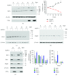
Figure 1
Figure 1
The effects of BoHV-1 infection on the expression of Nrf2-regulated downstream targets. (a, c, d, and e) MDBK cells in 60 mm dishes were mock infected or infected with BoHV-1 at an MOI of 0.1 for 4, 8, 16, 24, 36, and 48 h. The cell lysates were then prepared for Western blots to detect the expression of Nrf2, HO-1, NQO1, and VP16 using the Nrf2 antibody (Abcam, cat# ab137550, 1 : 500), HO-1 monoclonal antibody (mouse) (Enzo Life Sciences, cat# ADI-OSA-110-D, 1 : 1000), NQO1 polyclonal antibody (ABclonal, cat# A0047, 1 : 1000), and VP16 antibody (a gift from Prof. Vikram Misra at the University of Saskatchewan, 1 : 2000). (b) MDBK cells were seeded into 24-well plates. After overnight incubation, the cells were infected with BoHV-1 at an MOI of 0.1. After infection for 4, 8, 16, 24, 36, and 48 h, the cells were collected and cell numbers were counted using a Trypan-blue exclusion test. Data shown are representative of three independent experiments. (f) The band intensity was analyzed with software ImageJ. Each analysis was compared with that of uninfected control at each time point, which was arbitrarily set as 100%. These images are representative of those from three independent experiments. (g) Total RNA was prepared at 16 and 24 hpi in MDBK cells, and the mRNA levels of Nrf2, HO-1, and NQO1 were measured by qRT-PCR. Each analysis was compared with that of uninfected control, which was arbitrarily set as 100%. Data represent three independent experiments. The significance was assessed with Student's t-test (P < 0.05).
Figure 2
Figure 2
The effects of Trolox on the expression of Nrf2 and its downstream targets. (a, b, and c) MDBK cells in 60 mm dishes pretreated with Trolox (1 mM) or DMSO control for 1 h were infected with BoHV-1 (MOI = 0.1); in the presence of Trolox or DMSO control for 24 h, the cell lysates were prepared for Western blots to detect the expression of Nrf2 (a), HO-1 (b), and NQO1 (c). (e, f, and g) MDBK cells in 60 mm dishes pretreated with Trolox (1 mM) or DMSO control for 1 h were exposed to tBHP in the presence of Trolox or DMSO control for 2 h; the cell lysates were prepared for Western blots to detect the expression of Nrf2 (e), HO-1 (f), and NQO1 (g). (d and h) The relative band intensity was analyzed with software ImageJ, and each analysis was compared with that of uninfected control at each time point, which was arbitrarily set as 100%. Data shown are representative of three independent experiments. (i) MDBK cells in 24-well plates pretreated with Trolox at indicated concentrations or MDSO control were infected with BoHV-1 (MOI = 0.1) for 24 hours in the presence of an inhibitor or DMSO. The cell cultures were subjected to frozen-thawing twice, and viral yield was determined with the results being expressed as TCID50/mL. (j) The cytotoxicity of Trolox in MDBK cells for 24 h was analyzed by Trypan-blue exclusion. The significance was assessed with Student's t-test (P < 0.05).
Figure 3
Figure 3
BoHV-1 infection promoted Nrf2 depletion through a proteasome pathway. (a, c, and i) MDBK cells in 60 mm dishes were infected with BoHV-1 at an MOI of 0.1 in the presence of MG132 (1 μM) or DMSO control. After infection for 24 h, the cell lysates were prepared for Western blotting to detect the expression of Nrf2 (a) or were prepared for IP using antibodies against Nrf2 (c), DJ-1 (d), and unrelated IgG (k). The IP samples were subjected to immunoblots using antibodies against ubiquitin (Cell Signaling Technology, cat# 3936, 1 : 1000) and Nrf2 (Abcam, cat# ab137550, 1 : 500), respectively. (d and e) MDBK cells in 60 mm dishes were mock infected or infected with BoHV-1 (MOI = 0.1) for an indicated time length. The cell lysates were then prepared for Western blots to detect the expression of KEAP1 and DJ-1 using antibodies against KEAP1 mAb (Cell Signaling Technology, cat# 8047, 1 : 1000) and DJ-1 (ABclonal, cat# A0201, 1 : 1000), respectively. Data shown are representative of three independent experiments. (f, h, and j) The band intensity was analyzed with software ImageJ. Each analysis was compared with that of control, which was arbitrarily set as 100%. These images are representative of those from three independent experiments. The significance was assessed with Student's t-test (P < 0.05). (g) MDBK cells in 60 mm dishes were mock infected or infected with BoHV-1 (MOI = 0.1) for 24 hpi. Then, the nuclear proteins were prepared using a commercial kit, and the levels of protein DJ-1 were detected by immunoblots. Data shown are representative of three independent experiments. WCE: whole cell extract.
Figure 4
Figure 4
BoHV-1 infection altered the localization of nuclear Nrf2 protein. MDBK cells in 2-well chamber slides were mock infected (a) or infected with BoHV-1 (MOI = 0.1) (b) for 24 hours. After three washings with PBS, cells were fixed with 4% formaldehyde, and Nrf2 was detected by IFA using the Nrf2 antibody (1 : 500) and LaminA/C antibody (1 : 500). DAPI staining was used to stain nuclear DNA. Images were obtained by performing confocal microscopy (Leica). These images are representative of three independent experiments. (c) Zoom-in cells showing typical dot-like staining. (d) The percentage of dot-like staining-positive cells among ~400 cells was estimated from photos derived from three independent experiments. Scale bar: 200 μM.
Figure 5
Figure 5
BoHV-1 infection altered the accumulation of Nrf2 in the nucleus. (a) MDBK cells in 60 mm dishes were mock infected or infected with BoHV-1 (MOI = 0.1) for 24 h, the cell cultures were collected for extracting nuclear proteins using commercial nuclear protein purification kit (Beyotime Biotechnology, cat# P0027). Nrf2 was detected by Western blot using the antibody against Nrf2 (Abcam, cat# ab137550). LaminA/C was detected and used as protein loading control. (b) Protein fractions of both the cytosol and nucleus were subjected to Western blots using the antibody against β-tubulin. (c) MDBK cells in 60 mm dishes were mock treated or treated with Trolox (1 mM) for 2 h; the cell lysates were collected for the purification of nuclear proteins and cytosol proteins using commercial nuclear protein purification kit (Beyotime Biotechnology, cat# P0027). Nrf2 was detected by Western blot using the antibody against Nrf2 (Abcam, cat# ab137550). LaminA/C and β-tubulin were detected to characterize whether each fraction was contaminated. These images are representative of three independent experiments.
Figure 6
Figure 6
The posttranslational modification of Nrf2 stimulated by virus infection. MDBK cells in 60 mm dishes were mock infected or infected with BoHV-1 (MOI = 0.1) for 24 h; the cell cultures were collected to produce whole cell lysates (a and d) or to extract nuclear proteins using commercial nuclear protein purification kit (Beyotime Biotechnology, cat# P0027) (b and c). IP was performed using the antibody against Nrf2 (Abcam, cat# ab137550). Then, levels of phospho-Nrf2 in whole cell extracts (WCE) (a) and the nucleus (b) and acetylation levels of Nrf2 in the nucleus (c) and WCE (d) were detected by immunoblots using the antibody against the Nrf2 antibody (Abcam, cat# ab1375501:500), acetyl Lysine antibody (ABclonal, cat# A2391, 1 : 1000), and phospho-(Ser/Thr) Phe antibody (Cell Signaling Technology, cat# 9631, 1 : 1000). Data shown are representative of three independent experiments. The significance was assessed with Student's t-test (P < 0.05).
Figure 7
Figure 7
BoHV-1 affected the association between Nrf2 and LaminA/C. MDBK cells in 60 mm dishes were mock infected or infected with BoHV-1 for 24 h. Nucleus proteins were purified using commercial nuclear protein purification kit (Beyotime Biotechnology, cat# P0027). IP was performed using antibodies against Nrf2 (a), LaminA/C (b), Nrf2 and unrelated IgG (c), and LaminA/C and IgG (d) (R: rabbit; M: mouse). Then, Western blots were performed using corresponding antibodies. (e) MDBK cells in 60 mm dishes were mock treated or treated with Trolox (1 mM) for 2 h. Nucleus proteins were purified using commercial nuclear protein purification kit (Beyotime Biotechnology, cat# P0027), and LaminA/C was detected by Western blots.
Figure 8
Figure 8
BoHV-1 affected the association between Nrf2 and H3K9ac/H3K18ac. MDBK cells in 60 mm dishes were mock infected or infected for 24 h. The cell lysates were prepared to perform IP using antibodies against Nrf2 (a), H3K9ac (b), H3K18ac (c), H3K9ac and rabbit IgG (d), and H3K18ac and rabbit IgG (e). The precipitated proteins were subjected to Western blots using the following antibodies: Nrf2 (Abcam, cat# ab137550, 1 : 1000), acetyl-Histone H3 (Lys9) rabbit mAb (Cell Signaling Technology, cat# 9649, 1 : 1000), and acetyl-Histone H3 (Lys18) rabbit mAb (Cell Signaling Technology, cat# 13998, 1 : 1000). Data shown are representative of three independent experiments. The significance was assessed with Student's t-test (P < 0.05).

Similar articles

Cited by

References

    1. Hodgson P. D., Aich P., Manuja A., et al. Effect of stress on viral–bacterial synergy in bovine respiratory disease: novel mechanisms to regulate inflammation. Comparative and Functional Genomics. 2005;6(4):244–250. doi: 10.1002/cfg.474. - DOI - PMC - PubMed
    1. Rice J. A., Carrasco-Medina L., Hodgins D. C., Shewen P. E. Mannheimia haemolytica and bovine respiratory disease. Animal Health Research Reviews. 2007;8(2):117–128. doi: 10.1017/S1466252307001375. - DOI - PubMed
    1. Jones C., Chowdhury S. A review of the biology of bovine herpesvirus type 1 (BHV-1), its role as a cofactor in the bovine respiratory disease complex and development of improved vaccines. Animal Health Research Reviews. 2007;8(2):187–205. doi: 10.1017/S146625230700134X. - DOI - PubMed
    1. Cardoso T. C., Rosa A. C. G., Ferreira H. L., et al. Bovine herpesviruses induce different cell death forms in neuronal and glial-derived tumor cell cultures. Journal of Neurovirology. 2016;22(6):725–735. doi: 10.1007/s13365-016-0444-5. - DOI - PubMed
    1. Sena L. A., Chandel N. S. Physiological roles of mitochondrial reactive oxygen species. Molecular Cell. 2012;48(2):158–167. doi: 10.1016/j.molcel.2012.09.025. - DOI - PMC - PubMed

MeSH terms