. 2019;1(1):48-60.
Epub 2019 Aug 25.

RNA-dependent Amplification of Mammalian mRNA Encoding Extracellullar Matrix Proteins: Identification of Chimeric RNA Intermediates for α1, β1, and γ1 Chains of Laminin

Affiliations

RNA-dependent Amplification of Mammalian mRNA Encoding Extracellullar Matrix Proteins: Identification of Chimeric RNA Intermediates for α1, β1, and γ1 Chains of Laminin

Vladimir Volloch et al. Ann Integr Mol Med. 2019.

Abstract

De novo production of RNA on RNA template, a process known as RNA-dependent RNA synthesis, RdRs, and the enzymatic activity conducting it, RNA-dependent RNA polymerase, RdRp, were initially considered to be exclusively virus-specific. Eventually, however, the occurrence of RdRs and the ubiquitous presence of conventional RdRp were demonstrated in numerous eukaryotic organisms. The evidence that the enzymatic machinery capable of RdRs is present in mammalian cells was derived from studies of viruses, such as hepatitis delta virus, HDV, that do not encode RdRp yet undergo a robust RNA replication once inside the mammalian host; thus firmly establishing its occurrence and functionality. Moreover, it became clear that RdRp activity, apparently in a non-conventional form, is constitutively present in most, if not in all, mammalian cells. Because such activity was shown to produce short transcripts, given its apparent involvement in RNA interference phenomena, and because double-stranded RNA is known to trigger cellular responses leading to its degradation, it was generally assumed that its role in mammalian cells is restricted to a regulatory function. However, at the same time, an enzymatic activity capable of generating complete antisense RNA complements of mRNAs was discovered in mammalian cells undergoing terminal differentiation. Moreover, observations of widespread synthesis of antisense RNAs initiating at the 3'poly(A) of mRNAs in human cells suggested an extensive cellular utilization of mammalian RdRp. These results led to the development of a model of RdRp-facilitated and antisense RNA-mediated amplification of mammalian mRNA. Recent detection of the major model-predicted identifiers, chimeric RNA intermediates containing both sense and antisense RNA strands covalently joined in a rigorously predicted and uniquely defined manner, as well as the identification of a putative chimeric RNA end product of this process, validated the proposed model. The results corroborating mammalian RNA-dependent mRNA amplification were obtained in vivo with cells undergoing terminal erythroid differentiation and programmed for only a short survival span. This raises a question of whether mammalian RNA-dependent mRNA amplification is a specialized occurrence limited to extreme circumstances of terminal differentiation or a general physiological phenomenon. The present study addresses this question by testing for the occurrence of RNA-dependent amplification of mRNA encoding extracellular matrix proteins abundantly produced throughout the tissue and organ development and homeostasis, an exceptionally revealing indicator of the range and scope of this phenomenon. We report here the detection of major identifiers of RNA-dependent amplification of mRNA encoding α1, β1, and γ1 chains of laminin in mouse tissues producing large quantities of extracellular matrix proteins. The results obtained warrant reinterpretation of the mechanisms involved in ubiquitous and abundant production and deposition of extracellular matrix proteins, confirm the occurrence of mammalian RNA-dependent mRNA amplification as a new mode of genomic protein-encoding information transfer, and establish it as a general physiological phenomenon.

Keywords: Antisense-strand RNA; Chimeric RNA intermediate; RNA-dependent RNA polymerase; RNA-dependent amplification of mammalian mRNA; Self-priming; Sensestrand RNA.


Figures

Figure 1:
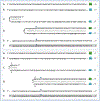
Figure 1:. Sequence of a chimeric junction containing antisense and sense segments of laminin α1 mRNA and the projected pathway of its generation.
Uppercase letters – nucleotide sequence of the sense strand; lowercase letters – nucleotide sequence of the antisense strand. Highlighted in green – “AUG” translation initiation codon on the sense strand; highlighted in blue – “uac” complement of translation initiation codon on the antisense strand. In italics and highlighted in grey – detected chimeric fragments. Blue arrows: Position of antisense/sense junction. The top and bottom panels depict amplification of mRNA molecules originated from different TSSs; note that self-priming positions and, consequently, chimeric junction sequences in two panels are different. A: 5′ terminal region of laminin al mRNA. B: Antisense complement of the 5’ terminal region of α1 laminin mRNA. C: Projected folding of the antisense strand into self-priming configuration. D: Extension of selfprimed antisense strand into sense-oriented sequence. E: Projected chimeric junction sequence. F: Detected chimeric junction sequence. Complete sequences of the chimeric junctions shown in this Figure are provided in the main text above. Note that the priming occurs within the segment of antisense strand corresponding to the 5′UTR of mRNA, thus preserving the coding capacity of amplified mRNA.
Figure 2:
Figure 2:. Sequence of a chimeric junction containing antisense and sense segments of laminin β1 mRNA and the projected pathway of its generation.
Uppercase letters – nucleotide sequence of the sense strand; lowercase letters – nucleotide sequence of the antisense strand. Highlighted in green – “AUG” translation initiation codon on the sense strand; highlighted in blue – “uac” complement of translation initiation codon on the antisense strand. In italics and highlighted in grey – detected chimeric fragments. Blue arrows: Position of antisense/sense RNA junction. The top and bottom panels depict amplification of mRNA molecules originated from different TSSs; note that self-priming positions and, consequently, chimeric junction sequences shown in two panels are different. A: 5’ terminal region of laminin pi mRNA. B: Antisense complement of the 5’ terminal region of pi laminin mRNA. C: Folding of the antisense strand into self-priming configuration. D: Extension of self-primed antisense strand into sense-oriented sequence. E: Projected chimeric junction sequence. F: Detected chimeric junction sequence. Complete sequences of the chimeric junctions shown in this Figure are provided in the main text above. Note that the priming occurs within the segment of antisense strand corresponding to the 5′UTR of mRNA, thus preserving the coding capacity of amplified mRNA.
Figure 3:
Figure 3:. Sequence of a chimeric junction containing antisense and sense segments of laminin γ1 mRNA and the projected pathway of its generation.
Uppercase letters – nucleotide sequence of the sense strand; lowercase letters – nucleotide sequence of the antisense strand. Highlighted in green – “AUG” translation initiation codon on the sense strand; highlighted in blue – “uac” complement of translation initiation codon on the antisense strand. In italics and highlighted in grey – detected chimeric fragments. Blue arrows: Position of antisense/sense RNA junction. The top and bottom panels depict amplification of mRNA molecules originated from different TSSs; note that self-priming positions and, consequently, chimeric junction sequences shown in two panels are different. A: 5’ terminal region of laminin γ1 mRNA. B: Antisense complement of the 5’ terminal region of γ1 laminin mRNA. C: Projected folding of the antisense strand into self-priming configuration. D: Extension of self-primed antisense strand into sense-oriented sequence. E: Projected chimeric junction sequence. F: Detected chimeric junction sequence. Complete sequences of the chimeric junctions shown in this Figure are provided in the main text above. Note that the priming occurs within the segment of antisense strand corresponding to the 5′UTR of conventional mRNA, thus preserving the coding capacity of amplified mRNA.
Figure 4:
Figure 4:. Projected stages of of RdRp-directed, antisense RNA-mediated amplification of mammalian mRNA.
Top panel: Conventional, genome-originated mRNA molecule. Bottom panel: Projected stages of antisense RNA-mediated mRNA amplification. Boxed line – sense strand RNA. Single line – antisense strand RNA. “AUG” – functional translation initiation codon (could be other than “AUG”). “TCE”– 3′-terminal complementary element; “ICE”– internal complementary element, both on the antisense RNA strand. Yellow circle -helicase/modifying activity complex. Blue lines (both single and boxed) – RNA strand modified and separated from its complement by a helicase complex. Red arrowhead – position of cleavage of the chimeric intermediate. Step 1: Synthesis of antisense strand; step 2: Strand separation; step 3: Folding of antisense strand into self-priming configuration; step 4: Extension of self-primed antisense RNA; step 5: Strand separation; step 6: Cleavage of the chimeric intermediate; step 7: End-products of amplification. Note that chimeric RNA end product retains the intact coding capacity of conventional mRNA.
Figure 5:
Figure 5:. TSS shift as a potential regulator of the eligibility of an mRNA for the amplification process.
Single line – 3′ terminus of the antisense strand. Filled grey boxes -sense strand. Filled blue boxes – topologically compatible complementary elements on the antisense strand. A: One of the complementary elements is 3’-terminal; folding results in a self-priming structure which is extended into the sense strand. B: Both complementary elements are internal, no self-priming is possible; TSS shift in the downstream direction makes one of the elements 3’-terminal and allows self-priming and extension into the sense strand. C: There are no complementary elements, no self-priming can occur; TSS shift in the upstream direction generates complementary elements one of which is 3’-terminal and thus enables self-priming and extension. Note that processes depicted in panels B and C can occur in reverse resulting in a loss, rather than the acquisition, of the eligibility.
Figure 6:
Figure 6:. RNA-dependent RNA polymerase can transcribe the cap “G” of mRNA.
Data shown is adapted from Figure 1, top panel. Uppercase letters – nucleotide sequence of the sense strand; lowercase letters – nucleotide sequence of the antisense strand. “u” highlighted in blue – 3′-terminal nucleotide of the antisense strand corresponding to the transcription start site of mRNA; “uC” highlighted in blue –the projected junction structure in the absence of the cap “G” transcription. “c” highlighted in green – transcript of the cap”G”; “ucC” highlighted in green – the resulting junction structure when cap “G” is transcribed. A: Projected self-priming configuration of the antisense strand in the absence of the cap “G” transcription. B: Projected nucleotide sequence of the sense/antisense junction in the absence of the cap “G” transcription. C: Detected nucleotide sequence of the sense/ antisense junction. D: Self-priming configuration of the antisense strand as defined by experimental results. Note that the genomic sequence upstream of the TSS cannot account for the additional 3′-terminal “C” in the antisense strand.
Figure 7:
Figure 7:. Novel experimental design: in vitro generation of a chimeric polypeptide containing murine alpha 1 laminin chain, initiated from the antisense RNA and non-contiguously encoded in the genome.
Uppercase letters: sense strand RNA. Lowercase letters: antisense strand RNA. Highlighted in green: AUG translation initiation codon. 5′-CAU-3′ (highlighted in blue) on the sense RNA: Complement of 5’-aug-3’ (highlighted in green) on the antisense RNA. “INS” highlighted in grey: Insert encoding a Tag peptide and lacking initiation codon; “TAG” highlighted in grey: Peptide encoded by “INS”. “ins” highlighted in grey: “INS” complement on the antisense RNA. Amino acid sequence highlighted in blue: Polypeptide encoded by conventional mRNA or by the amplified mRNA. Amino acid sequence highlighted in grey: The N-end extension of conventional polypeptide; underlined portion: Amino acids encoded by the antisense RNA. Highlighted in yellow: Editing changes resulting in replacement of the “aug” by the “aca” on the antisense RNA. Red arrowhead: Position of cleavage of the chimeric intermediate. Top panel: A-conventional mRNA encoding alpha 1 chain of laminin. B-chimeric RNA end product of amplification (adapted from Figure 1) encoding the same polypeptide as “A”. C-amino acid sequence encoded by either “A” or “B”. Middle panel: A-projected edited “dormant” mRNA originated from edited alpha 1 laminin gene. B-antisense complement of edited alpha 1 laminin mRNA. C-folding of the antisense strand into self-priming configuration; 3’terminal “c” is a transcript of the 5’capG of mRNA. D-extension of self-primed antisense strand into sense-oriented sequence followed by strand separation and cleavage of the chimeric intermediate. E-chimeric RNA end product of RNA-dependent amplification of edited alpha 1 laminin mRNA. F-projected translational outcome. Bottom panel: Same as the middle panel with the exception of editing changes resulting in replacement of the “aug” by the “aca” (highlighted in yellow) on the antisense RNA.

Similar articles

Cited by

References

    1. Volloch V. Cytoplasmic synthesis of globin RNA in differentiated murine erythroleukemia cells: possible involvement of RNA-dependent RNA polymerase. Proc Natl Acad Sci U S A. 1986;83(5):1208–12. - PMC - PubMed
    1. Volloch V, Schweitzer B, Rits S. Synthesis of globin RNA in enucleated differentiating murine erythroleukemia cells. J Cell Biol. 1987;105(1):137–43. - PMC - PubMed
    1. Volloch VZ, Schweitzer B, Rits S. Evolutionarily conserved elements in the 5’ untranslated region of beta globin mRNA mediate site-specific priming of a unique hairpin structure during cDNA synthesis. Nucleic Acids Res. 1994;22(24):5302–9. - PMC - PubMed
    1. Volloch V, Schweitzer B, Rits S. Antisense globin RNA in mouse erythroid tissues: structure, origin, and possible function. Proc Natl Acad Sci U S A. 1996;93(6):2476–81. - PMC - PubMed
    1. Rits S, Olsen B, Volloch V. RNA-dependent synthesis of mammalian mRNA: Identification of chimeric intermediate and putative end-product. BioRxiv. 2016.

LinkOut - more resources