. 2019 Oct 25;9(1):15296.
doi: 10.1038/s41598-019-51773-x.

Thioredoxin-related transmembrane protein 2 (TMX2) regulates the Ran protein gradient and importin-β-dependent nuclear cargo transport

Affiliations

Thioredoxin-related transmembrane protein 2 (TMX2) regulates the Ran protein gradient and importin-β-dependent nuclear cargo transport

Ami Oguro et al. Sci Rep. .

Abstract

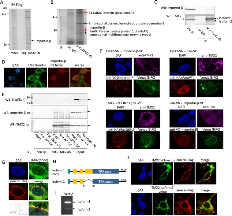
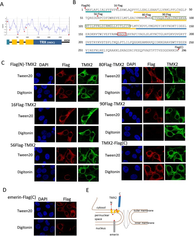
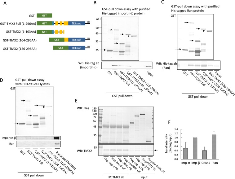
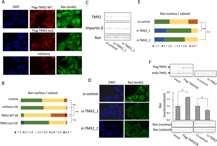
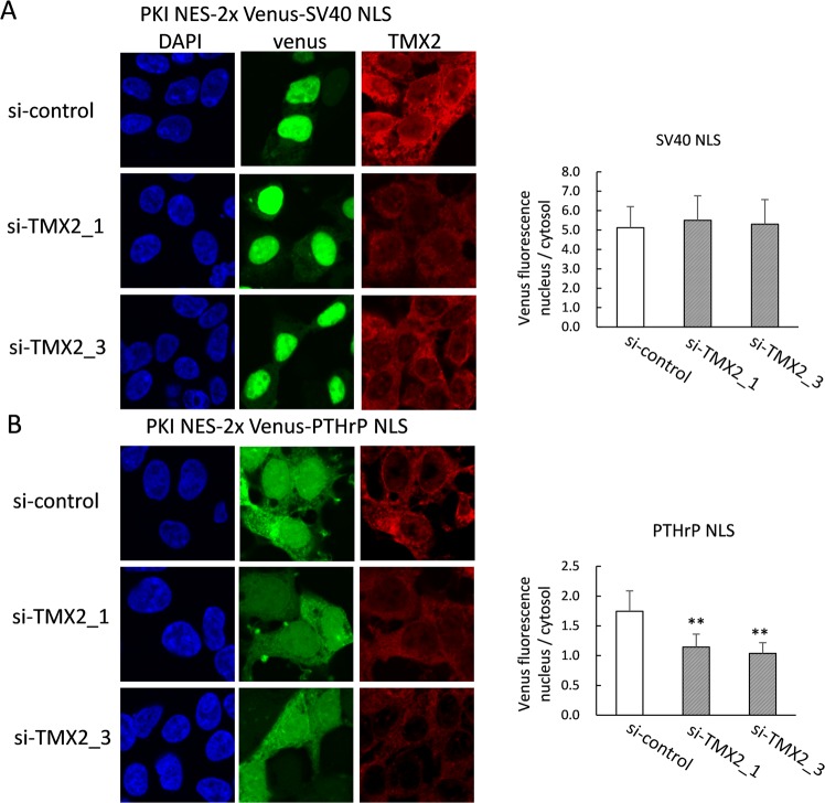
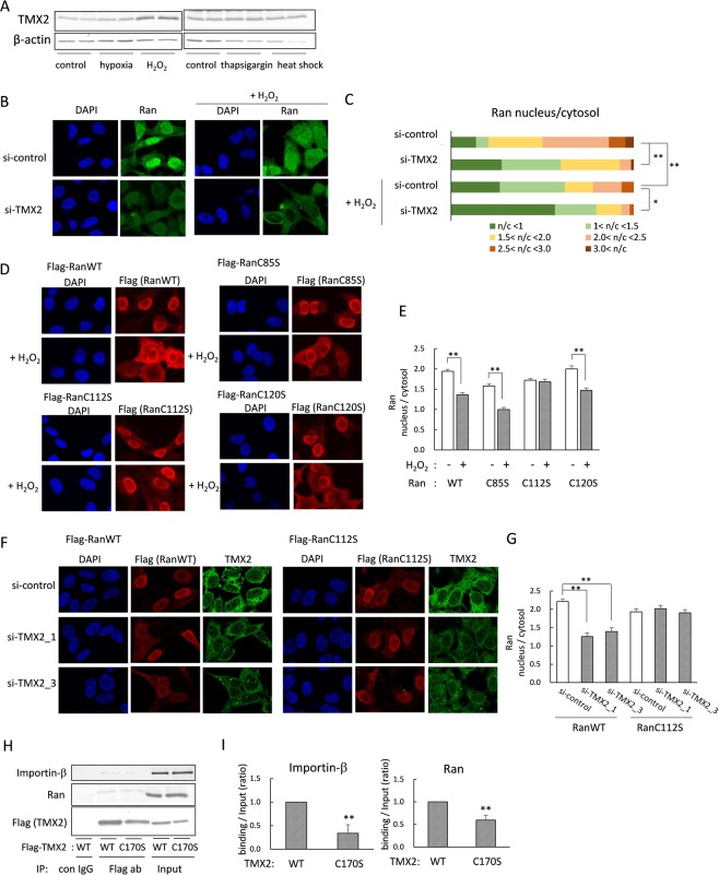
TMX2 is a thioredoxin family protein, but its functions have not been clarified. To elucidate the function of TMX2, we explored TMX2-interacting proteins by LC-MS. As a result, importin-β, Ran GTPase (Ran), RanGAP, and RanBP2 were identified. Importin-β is an adaptor protein which imports cargoes from cytosol to the nucleus, and is exported into the cytosol by interaction with RanGTP. At the cytoplasmic nuclear pore, RanGAP and RanBP2 facilitate hydrolysis of RanGTP to RanGDP and the disassembly of the Ran-importin-β complex, which allows the recycling of importin-β and reentry of Ran into the nucleus. Despite its interaction of TMX2 with importin-β, we showed that TMX2 is not a transport cargo. We found that TMX2 localizes in the outer nuclear membrane with its N-terminus and C-terminus facing the cytoplasm, where it co-localizes with importin-β and Ran. Ran is predominantly distributed in the nucleus, but TMX2 knockdown disrupted the nucleocytoplasmic Ran gradient, and the cysteine 112 residue of Ran was important in its regulation by TMX2. In addition, knockdown of TMX2 suppressed importin-β-mediated transport of protein. These results suggest that TMX2 works as a regulator of protein nuclear transport, and that TMX2 facilitates the nucleocytoplasmic Ran cycle by interaction with nuclear pore proteins.


Conflict of interest statement

The authors declare no competing interests.

Figures

Figure 1
Figure 1
TMX2 interacts with nuclear import protein. (A) Flag-TMX2 was overexpressed in HEK293 cells and immunoprecipitated with anti-Flag antibody. The band indicated by the arrow was analyzed by LC-MS. (B) Endogenous TMX2-intereacting proteins in HEK293 cells were immunoprecipitated with anti-TMX2 antibody and analyzed by LC-MS. (C) HEK293 cell lysates were immunoprecipitated with anti-TMX2 antibody and analyzed by western blot with anti-importin-β antibody. (D) mCherry-importin-β was expressed in HEK293 cells and immunostained with anti-TMX2 antibody. (E) Flag-Ran WT or -Ran Q69L mutant was overexpressed in cells and immunoprecipitated with anti-TMX2 antibody. (F) TMX2-VN was co-expressed with importin-β-VC, HA-RanWT-VC, or HA-RanQ69L-VC in HEK293 cells, and the overexpression of these proteins were confirmed by anti-TMX2, anti-VC, or anti-HA antibody. Venus fluorescence as BiFC signal indicates the interaction of these fusion proteins. BiFC signal was also detected by co-expression of Ran-VN and importin-β-VC as a positive control. (G) Co-localization of endogenous TMX2 and Flag-emerin was analyzed by immunofluorescence. (H) A diagram of TMX2 WT and isoform 2, which lacks part of the transmembrane region. (I) TMX2 WT and isoform 2 cDNA were amplified by PCR from HEK293 cDNA with the primer sets at 191–507th of TMX2 WT nucleotide. (J) Venus fused with TMX2 WT or isoform 2 was expressed in HEK293 cells, and co-localization with emerin-Flag was analyzed.
Figure 2
Figure 2
Topology of TMX2 in the nuclear envelope. (A) A hydrophobic profile of the TMX2 protein was obtained by the SOSUI program, and predicted its transmembrane regions. (B) Protein sequences of TMX2. The predicted transmembrane regions are indicated by yellow bars, and thioredoxin-like domain was by blue bar. TMX2 isoform 2 lacks part of the transmembrane region as indicated by green box. The conserved S-X-X-C motif was indicated by red box. (C) Flag tag was inserted into TMX2 at the N- or C- terminus, or the amino acid position 16, 56, 80, or 90 as indicated by red arrowheads (A) or red circles (B), and overexpressed in HEK293 cells. These cells were fixed with PFA, and permeabilized by Tween20 or digitonin. The localizations of these proteins were analyzed with anti-Flag antibody or polyclonal anti-TMX2 antibody, which recognizes an epitope in full length TMX2. (D) Flag fused-emerin at the C terminus was expressed in cells, and permeabilized by Tween20 or digitonin after fixation. (E) The speculated topology of TMX2 in the nuclear outer membrane.
Figure 3
Figure 3
Binding between TMX2 and importin-β or Ran. (A) Scheme of GST-tagged TMX2 full-length and deletion mutants. (B,C) GST-tagged TMX2 proteins were immobilized on glutathione-Sepharose beads and incubated with purified His-tagged importin-β or Ran protein. The precipitated importin-β or Ran was analyzed by immunoblot with anti-His tag antibody. (D) TMX2-packed glutathione-Sepharose was incubated with HEK293 cell lysates, and the precipitant with TMX2 was analyzed with anti-importin-β or Ran antibody. (E) Flag-importin-α, -importin-β, -CRM1, or -RanQ69L was expressed in HEK293 cells, and cell lysates were immunoprecipitated with anti-TMX2 antibody. The precipitates were detected with anti-Flag antibody. (F) The ratio of band intensity of binding/input was quantitated. Values are the means ± S.D. for three separate experiments. The value of importin-β was set at 1.0.
Figure 4
Figure 4
Regulation of the Ran protein gradient by TMX2. (A) Flag-TMX2 WT, Flag-isoform 2, or mCherry was overexpressed in HEK293 cells, and the distribution of endogenous Ran was analyzed by immunofluorescence microcopy. (B) The ratio of Ran levels in the nucleus/cytosol of each kind of cell (n = 60) was quantified. (C) si-RNA targeting for the negative control or TMX2 was lipofected into HEK293 cells, and TMX2, importin-β, or Ran protein levels were analyzed by western blotting. (D) Endogenous Ran distribution in TMX2 knockdown cells was analyzed by immunofluorescence microcopy. (E) The ratio of Ran levels in the nucleus/cytosol of each kind of cell (n = 60) was quantified. (F) The nuclear and cytosolic fractions of TMX2-overexpressing or -knockdown cells were extracted, and analyzed by western blotting with anti-Ran antibody. Values are the means ± S.D. for three experiments. The Ran nucleus/cytosol ratio of control cells (mock or si-control) was set at 1.0. *p < 0.05, **p < 0.01.
Figure 5
Figure 5
TMX2 knockdown decreased importin-β-dependent nuclear transport of protein. The expression plasmid for NES-2xVenus-SV40 NLS (A) or NES-2xVenus-PTHrP NLS (B) was co-transfected with control si-RNA or si-TMX2 into HEK293 cells. After 48 h, cells were fixed and incubated with anti-TMX2 antibody, followed by secondary antibody labeling with Dylight 647. The nucleus/cytosol ratio of Venus fluorescence was measured in about 30 cells. Values are the means ± S.D. **p < 0.01 compared with the si-control.
Figure 6
Figure 6
The cysteine 112 of Ran was involved in the regulation of Ran gradient by TMX2. (A) HEK293 cells were cultured under 1% oxygen concentration for 6 h or heat stress condition at 42 °C for 3 h, or in the presence of 200 μM H2O2 for 4 h or 200 nM thapsigargin for 4 h, and TMX2 protein induction was investigated. (B) Hela cells were lipofected with si-control or si-TMX2, and treated with 200 μM H2O2 for 20 min. After fixing with PFA, the endogenous Ran distribution was observed by immunofluorescence microscopy. (C) The Ran nucleus/cytosol signal-intensity ratios of about 40 cells were quantitated. (D) Flag-Ran WT, C85S, C112S, or C120S was expressed in Hela cells, and the cells were treated with 200 μM H2O2 for 30 min. The localization of Ran was analyzed by immunostaining with anti-Flag antibody. (E) The nucleus/cytosol ratio of Ran was quantified in about 45 cells. Values are the means ± S.E. (F) Ran WT or C112S/Flag pcDNA was co-transfected with si-TMX2 into cells. After 48 h, the localization of Ran was analyzed by anti-Flag antibody. Knockdown of TMX2 was confirmed by anti-TMX2 antibody. (G) The nucleus/cytosol ratio of Ran was quantified in about 40 cells. (H) HEK293 cells were overexpressed with Flag-tagged TMX2 WT or C170S mutant, and the cell lysates were immunoprecipitated with anti-Flag antibody. The precipitant was analyzed by anti-importin-β or Ran antibody. (I) The band intensity of importin-β or Ran binding/Input was quantified. Values are the means ± S.D. for three separate experiments. The control value was set at 1.0. *p < 0.05, **p < 0.01.

Similar articles

Cited by

References

    1. Matsuo Y, et al. Identification of a novel thioredoxin-related transmembrane protein. J Biol Chem. 2001;276:10032–10038. doi: 10.1074/jbc.M011037200. - DOI - PubMed
    1. Lynes EM, et al. Palmitoylated TMX and calnexin target to the mitochondria-associated membrane. EMBO J. 2012;31:457–470. doi: 10.1038/emboj.2011.384. - DOI - PMC - PubMed
    1. Matsuo Y, et al. TMX, a human transmembrane oxidoreductase of the thioredoxin family: the possible role in disulfide-linked protein folding in the endoplasmic reticulum. Arch Biochem Biophys. 2004;423:81–87. doi: 10.1016/j.abb.2003.11.003. - DOI - PubMed
    1. Haugstetter J, Blicher T, Ellgaard L. Identification and characterization of a novel thioredoxin-related transmembrane protein of the endoplasmic reticulum. J Biol Chem. 2005;280:8371–8380. doi: 10.1074/jbc.M413924200. - DOI - PubMed
    1. Sugiura Y, et al. Novel thioredoxin-related transmembrane protein TMX4 has reductase activity. J Biol Chem. 2010;285:7135–7142. doi: 10.1074/jbc.M109.082545. - DOI - PMC - PubMed

Publication types