. 2020 Jan 2;77(1):26-38.e7.
doi: 10.1016/j.molcel.2019.09.024. Epub 2019 Oct 22.

53BP1 Enforces Distinct Pre- and Post-resection Blocks on Homologous Recombination

Affiliations

53BP1 Enforces Distinct Pre- and Post-resection Blocks on Homologous Recombination

Elsa Callen et al. Mol Cell. .

Abstract

53BP1 activity drives genome instability and lethality in BRCA1-deficient mice by inhibiting homologous recombination (HR). The anti-recombinogenic functions of 53BP1 require phosphorylation-dependent interactions with PTIP and RIF1/shieldin effector complexes. While RIF1/shieldin blocks 5'-3' nucleolytic processing of DNA ends, it remains unclear how PTIP antagonizes HR. Here, we show that mutation of the PTIP interaction site in 53BP1 (S25A) allows sufficient DNA2-dependent end resection to rescue the lethality of BRCA1Δ11 mice, despite increasing RIF1 "end-blocking" at DNA damage sites. However, double-mutant cells fail to complete HR, as excessive shieldin activity also inhibits RNF168-mediated loading of PALB2/RAD51. As a result, BRCA1Δ1153BP1S25A mice exhibit hallmark features of HR insufficiency, including premature aging and hypersensitivity to PARPi. Disruption of shieldin or forced targeting of PALB2 to ssDNA in BRCA1D1153BP1S25A cells restores RNF168 recruitment, RAD51 nucleofilament formation, and PARPi resistance. Our study therefore reveals a critical function of shieldin post-resection that limits the loading of RAD51.

Keywords: 53BP1; BRCA1; PARPi; aging; cancer; homologous recombination; resection; shieldin.


Conflict of interest statement

DECLARATION OF INTERESTS

The authors declare no competing interests.

Figures
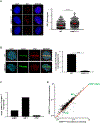
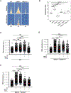
Figure 1.
Figure 1.. Serine 25 to Alanine mutation in 53BP1 compromises PTIP binding while increasing RIF1 association at DSBs.
(A) Left: WT, 53BP1−/− and 53BP1S25A MEFs were assayed for RIF1 (red) IRIF (10 Gy, 1 hour recovery). Cells were counterstained with DAPI (blue). Scale bar represents 10 μm. Right: quantification of RIF1 foci per nucleus in WT and 53BP1S25A MEFs, normalized by nuclear area (per 100 μm2). A minimum of 300 nuclei were quantified per condition using the Gen5 spot analysis software. A representative experiment (n=3) is shown. Statistical significance was determined by Mann-Whitney t-test. (B) Left: PTIP (red) recruitment to laser microirradiation damage in WT and 53BP1S25A MEFs. Damaged cells are indicated by γ-H2AX tracks (green). Scale bar represents 10 μm. Right: quantification of cells with PTIP/γ-H2AX colocalization. Statistical significance was determined by Mann-Whitney t-test. (C, D) Graphs depicting the relative ion intensities for 53BP1, RIF1 and PTIP peptides associated with the 53BP1 complex in HeLa-S cells expressing FLAG-HA-53BP1S25A or FLAG-HA-53BP1WT, as determined by mass spectrometry.
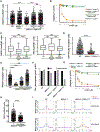
Figure 2.
Figure 2.. 53BP1S25A mutation reverts lethality of BRCA1Δ11 mice but promotes accelerated aging and an elevated rate of senescence
(A) Table showing the expected and observed frequency of breeding outcomes from BRCA1+/Δ1153BP1+/S25A x BRCA1+/Δ1153BP1+/S25A intercrosses. (B) Kaplan-Meier survival analysis of WT and BRCA1Δ1153BP1S25A mice (n=14). A significantly shorter lifespan was observed in BRCA1Δ1153BP1S25A mice compared to WT (p < 0.0001). Log-rank (Mantel-Cox) test was used to determine statistical significance. (C) Representative X-ray CT projection image showing increased kyphosis in a 4 month old BRCA1Δ1153BP1S25A mouse compared with a WT littermate. (D) Representative image of E13.5 WT, BRCA1Δ11, 53BP1S25A and BRCA1Δ1153BP1S25A embryos showing the decrease in size of BRCA1Δ11 and BRCA1Δ1153BP1S25A animals. (E) In vitro growth of primary WT, BRCA1Δ11, 53BP1S25A and BRCA1Δ1153BP1S25ä MEFs at passage 2.
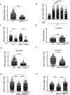
Figure 3.
Figure 3.. BRCA1Δ1153BP1S25A mice are sensitive to PARPi and defective in RAD51 filament assembly
(A) Representative histological images of the small intestine of BRCA1Δ1153BP1S25A and WT littermate controls upon treatment with PARPi by daily oral gavage. Image was taken 9 days after treatment. In BRCA1Δ1153BP1S25A mice, small intestinal villi are blunted and fused and the lamina propria is expanded by increased inflammatory cells; intestinal crypts are unorganized and cells display atypia. (B) Genomic instability (chromosome breaks and radials) measured in metaphase spreads from B lymphocytes derived from WT, BRCA1Δ11, 53BP1S25A and BRCA1Δ1153BP1S25A mice after PARPi treatment. Cells were stimulated for 2 days and then, treated for 16 hours with 1 μM PARPi. At least 50 cells were scored per condition. Experiment was repeated 5 times. Statistical significance was determined by Mann-Whitney t-test. (C) Colony formation in WT, BRCA1Δ11, 53BP1S25A and BRCA1Δ1153BP1S25A MEFs measured 9 days after continual treatment with 1 μM PARPi. Data are plotted relative to the plating efficiency of untreated controls of the same genotype. Statistical significance was determined by Mann-Whitney t-test. (D) RAD51 foci per EdU+ nucleus in WT, BRCA1Δ1153BP1S25A and BRCA1Δ1153BP1−/− MEFs measured 4 hours after 10 Gy IR, normalized by nuclear area (per 100 μm2). Statistical significance was determined by Mann-Whitney t-test. A minimum of 300 nuclei were quantified using the Gen5 spot analysis software. A representative experiment (n=2) is shown. Statistical significance was determined by Mann-Whitney t-test.
Figure 4.
Figure 4.. BRCA 1Δ1153BP1S25A cells exhibit normal levels of end resection catalyzed mainly by DNA2
(A) Top panel: Heat map of END-seq signals across individual AsiSI sites in WT, 53BP1−/− and BRCA1Δ11 MEFs measured 5 hours after AsiSI induction. Lower panel: ChIP-seq for ssDNA bound by RPA in the same cells. Heat maps are ordered by END-seq signal intensity in WT cells. (B) Box plots showing quantification of resection end points in the top 10% resected breaks in WT, 53BP1−/−, BRCA1Δ11, 53BP1S25A, BRCA1An53BP1−/− and BRCA1Δ1153BP1S25A MEFs at AsiSI cleaved DSB sites. Welch’s t-test was used to determine statistical significance. (C) Quantification of the intensity of chromatin bound RPA in individual EdU-positive nuclei from WT and EXO1-depleted MEFs, either pre-treated or not with 1 μM DNA2i prior to 10 Gy IR. Cells were analyzed 4 hours post-IR. (D) Quantification of the intensity of chromatin bound RPA in individual EdU-positive nuclei from EXO1-proficient and EXO1-depleted BRCA1Δ1153BP1S25A MEFs, either pre-treated or not with 1 μM DNA2i prior to 10 Gy IR. Cells were analyzed 4 hours post-IR. (E) Quantification of the intensity of chromatin bound RPA in individual EdU-positive nuclei from EXO1-proficient and EXO1-depleted BRCA1Δ11SHLD3−/− MEFs, either pre-treated or not with 1 μM DNA2i prior to 10 Gy IR. Cells were analyzed 4 hours post-IR. In panels C, D, and E a minimum of 300 nuclei per condition were quantified using the Gen5 spot analysis software. A representative experiment (n=2) is shown. Statistical significance was determined by Mann-Whitney t-test.
Figure 5.
Figure 5.. RAD51 loading defect in BRCA1Δ1153BP1S25A cells is corrected by Shld3 depletion or forced PALB2 chromatin binding
(A) RAD51 foci per EdU+ nucleus in WT, BRCA1Δ1153BP1−/−, BRCA1Δ1153BP1S25A and BRCA1Δ1153BP1S25ä MEFs deficient in SHLD3 measured 4 hours after 5 Gy IR. Two independent BRCA1Δ1153BP1S25ASHLD3−/− clones (#1 and #2) were used. Foci numbers are normalized by nuclear area (per 100 μm2). (B) Viability of WT, BRCA1Δ1153BP1S25A and BRCA1Δ1153BPIS25ASHLD3−/− MEFs measured by CellTiter-Glo 10 days after PARPi treatment. (C) RAD51 (left panel) and RPA (right panel) foci formation 4 hours after 10 Gy IR in WT cells expressing FHA-SHLD2 containing (SHLD2C) or lacking (SHLD2Cm1) the OB fold (Noordermeer et al., 2018). (D) GFP-PALB2 foci per EdU+ nucleus in WT and BRCA1Δ1153BP1S25A MEFs overexpressing PALB2 measured 4 hours after 5 Gy IR. (E) RAD51 foci per EdU+ nucleus in WT, BRCA1Δ1153BP1S25A, and BRCA1Δ1153BP1S25A MEFs overexpressing PALB2 or PALB2 fused with the FHA domain of RNF8, measured 4 hours after 10 Gy IR. Foci numbers are normalized by nuclear area (per 100 μm). (F) Chromosomal aberrations in WT, BRCA1Δ11, BRCA1Δ1153BP1−/− and BRCA1Δ1153BP1S25A expressing empty vector or FHA-PALB2. (G) Viability of BRCA1Δ1153BP1S25A and BRCA1Δ1153BPΓ MEFs with or without FHA-PALB2 protein measured by CellTiter-Glo 10 days after PARPi treatment. (H) RIF1 foci per EdU+-nucleus in BRCA1Δ1153BP1S25A and BRCA1Δ1153BP1S25A MEFs expressing FHA-PALB2 measured 4 hours after 10 Gy IR. (I) Aggregate plot for ssDNA bound by RAD51 as measured by ChIP-seq at AsiSI sites, separated into sense and antisense strands. WT, BRCA1Δ11 and BRCA1Δ1153BP1S25A MEFs (top panels) are compared with their respective counterparts expressing FHA-PALB2 (bottom panels). In panels A, D, E and H, a minimum of 300 nuclei per condition were quantified using the Gen5 spot analysis software. In panel C, a minimum of 130 nuclei per condition were quantified. A representative experiment (n=2) is shown. Statistical significance was determined by Mann-Whitney t-test in all the indicated panels.
Figure 6.
Figure 6.. Shieldin blocks RNF168 recruitment post-resection in BRCA1Δ1153BP1S25A cells
(A) Quantification of RNF168 foci in individual EdU+ nuclei from WT and BRCA1Δ11 MEFs. Cells were irradiated with 5 Gy and analyzed 4 hours post-IR. (B) Quantification of RNF168 foci in individual EdU+ nuclei from WT and EXO1-depleted MEFs, pretreated or not with DNA2i (1 μM). Cells were either un-irradiated or irradiated with 10 Gy and analyzed 4 hours post-IR. (C) Quantification of RNF168 foci in individual EdU+ nuclei from WT and BRCA1Δ1153BP1−/− MEFs pretreated with ATRi (AZ20, 10 μM). Cells were either un-irradiated or irradiated with 10 Gy and analyzed 4 hours post-IR. (D) Quantification of RNF168 foci in WT and BRCA1Δ1153BP1S25A MEFs 1 hour after 10 Gy IR. Statistical significance was determined by Wilcoxon Rank Sum test. (E) Quantification of RNF168 foci in individual EdU+ nuclei from WT and BRCA1Δ1153BP1S25A MEFs 4 hours post IR (5 Gy). Statistical significance was determined by Wilcoxon Rank Sum test. (F) Quantification of RAD 18 foci in individual EdU+ nuclei from WT and BRCA1Δ1153BP1S25A MEFs. Cells were irradiated with 5 Gy and analyzed 4 hours post-IR. (G) Quantification of RNF168 foci in individual EdU+ nuclei from WT, BRCA1Δ1153BP1S25A and BRCA1Δ1153BP1S25ASHLD3−/− MEFs. Cells were irradiated with 5 Gy and analyzed 4 hours post IR. (H) Quantification of RAD 18 foci in individual EdU+ nuclei from WT, BRCA1Δ1153BP1S25A MEFs and BRCA1Δ1153BP1S25ASHLD3−/− MEFs. Cells were irradiated with 5 Gy and analyzed 4 hours post-IR. In panels A-H, a minimum of 300 nuclei per condition were quantified using the Gen5 spot analysis software. A representative experiment (n=2) is shown. Unless otherwise noted, statistical significance was determined by Mann-Whitney t-test in all the panels.
Figure 7.
Figure 7.. RING-less BRCA1 supports end resection but not RAD51 filament assembly
Left: IR-induced RPA foci per EdU+nuclei in BRCA1F2/F2 MEFs with adenoviral Cre infection which deletes BRCA1 exon 2 (BRCA2Δ2/Δ2) and in BRCA1Δ11 cells. Right: IR-induced RAD51 foci per EdU+ nucleus in BRCA2Δ2/Δ2 and BRCA1Δ11 cells. A minimum of 300 nuclei per condition were quantified using the Gen5 spot analysis software. A representative experiment (n=2) is shown. Statistical significance was determined by Mann-Whitney t-test.

Similar articles

Cited by

References

    1. Alver RC, Chadha GS, Gillespie PJ, and Blow JJ (2017). Reversal of DDK-Mediated MCM Phosphorylation by Rif1-PP1 Regulates Replication Initiation and Replisome Stability Independently of ATR/Chk1. Cell Rep 18, 2508–2520. - PMC - PubMed
    1. Ameziane N, May P, Haitjema A, van de Vrugt HJ, van Rossum-Fikkert SE, Ristic D, Williams GJ, Balk J, Rockx D, Li H, et al. (2015). A novel Fanconi anaemia subtype associated with a dominant-negative mutation in RAD51. Nat Commun 6, 8829. - PMC - PubMed
    1. Aymard F, Bugler B, Schmidt CK, Guillou E, Caron P, Briois S, Iacovoni JS, Daburon V, Miller KM, Jackson SP, et al. (2014). Transcriptionally active chromatin recruits homologous recombination at DNA double-strand breaks. Nat Struct Mol Biol 21, 366–374. - PMC - PubMed
    1. Baer R, and Ludwig T (2002). The BRCA1/BARD1 heterodimer, a tumor suppressor complex with ubiquitin E3 ligase activity. Curr Opin Genet Dev 12, 86–91. - PubMed
    1. Bouwman P, Aly A, Escandell JM, Pieterse M, Bartkova J, van der Gulden H, Hiddingh S, Thanasoula M, Kulkarni A, Yang Q, et al. (2010). 53BP1 loss rescues BRCA1 deficiency and is associated with triple-negative and BRCA-mutated breast cancers. Nat Struct Mol Biol 17, 688–695. - PMC - PubMed

Publication types

MeSH terms

Substances