. 2019 Dec;213(4):1271-1288.
doi: 10.1534/genetics.119.302713. Epub 2019 Oct 23.

Tel1 Activation by the MRX Complex Is Sufficient for Telomere Length Regulation but Not for the DNA Damage Response in Saccharomyces cerevisiae

Affiliations

Tel1 Activation by the MRX Complex Is Sufficient for Telomere Length Regulation but Not for the DNA Damage Response in Saccharomyces cerevisiae

Rebecca Keener et al. Genetics. 2019 Dec.

Abstract

Previous models suggested that regulation of telomere length in Saccharomyces cerevisiae by Tel1(ATM) and Mec1(ATR) would parallel the established pathways regulating the DNA damage response. Here, we provide evidence that telomere length regulation differs from the DNA damage response in both the Tel1 and Mec1 pathways. We found that Rad53 mediates a Mec1 telomere length regulation pathway but is dispensable for Tel1 telomere length regulation, whereas in the DNA damage response, Rad53 is regulated by both Mec1 and Tel1 Using epistasis analysis with a Tel1 hypermorphic allele, Tel1-hy909, we found that the MRX complex is not required downstream of Tel1 for telomere elongation but is required downstream of Tel1 for the DNA damage response. Our data suggest that nucleolytic telomere end processing is not a required step for telomerase to elongate telomeres.

Keywords: DNA damage response; MRX complex; Tel1; epistasis; telomere.


Figures

Figure 1
Figure 1
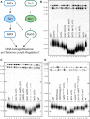
Rad53 is in the Mec1 telomere length regulation pathway. (A) Diagram representing a simplified, current understanding of Tel1/Mec1 pathways in the DNA damage response. (B–D) Southern blot analysis of telomeres from segregants with the indicated genotype. Two independent, haploid segregants are shown for each genotype. Median telomere length is quantitated in Figure S1, A–C. (B) Haploid cells were passaged on solid media for ∼120 population doublings to decrease telomere length heterogeneity. Segregants are from JHUy937-1. Two biological replicates were assayed after 120 population doublings for each genotype. (C) Segregants are from yRK6002 and yRK6003. (D) Both Rad53 and rad531−4/9−12AQ are epitope tagged with a 3xFLAG tag. Haploids were passaged for ∼100 population doublings. Segregants are yRK6008-1, yRK6008-2, yRK6009-1, yRK6009-2, yRK6010-1, yRK6010-2, yRK6011-1, yRK6011-2, yRK6012-1, yRK6012-2, yRK6013-1, yRK6013-2, yRK6014-1, yRK6014-2, yRK6015-1, and yRK6015-2.
Figure 2
Figure 2
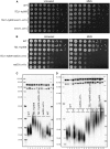
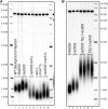
Tel1-hy909 requires Rad53 for the DNA damage response but not telomere elongation. (A and B) Yeast dilution series of untreated cells or cells cultured in 0.02% MMS for 1 hr. The genotype is indicated to the left of the panels. (A) Segregants are from yRK5126 and yRK5127. (B) Segregants are from yRK5028 and yRK5059. (C) Southern blot analysis of telomeres from segregants with the indicated genotype. Two independent, haploid segregants are shown for each genotype. Additional biological replicates were assayed for each genotype: WT, n = 35; TEL1-hy909, n = 42; TEL1-hy909 rad53Δ crt1Δ, n = 6; rad53Δ crt1Δ, n = 5. Because the TEL1-hy909 hypermorph elongates telomeres in the parental diploid (Figure S4B), we observed increased telomere length heterogeneity across all genotypes in the haploid segregants and observe the wild-type segregant telomeres were longer compared to other Southern blots. Segregants are from yRK5028 and yRK5059. (D) Southern blot analysis of telomeres from segregants with the indicated genotype. Segregants were passaged on solid media for ∼120 population doublings. Passage number is indicated: 1 = first passage or 5 = fifth passage. Segregants are from yRK5028. Additional biological replicates examined for P1 samples, see Figure 2C legend. Two biological replicates were assayed for each genotype at P5.
Figure 3
Figure 3
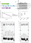
The mrx-18A S/T-Q mutant does not affect the DNA damage response, NHEJ, or telomere length. (A) Domain structure of the MRX complex indicating location of S/T-Q motifs with data from Lee et al. 2013; Shima et al. 2005; Becker et al. 2006 and are consistent with NCBI annotation (Mre11: BAA02017.1, Rad50: CAA65494, Xrs2: AAA35220.(1). S/T-Q motifs are indicated with a bar and the corresponding S or T residue number. (B) Western blots examining stability of the MRX complex in MRX-tag and mrx-18A strains. Quantitation was performed relative to Pgk1 loading control and normalized to the second lane of the Western blot. The average relative protein level in MRX-tag cells was 1.11 for Mre11-3HA, 1.07 for Rad50-G6-V5, and 0.88 for Xrs2-13myc. The average protein level in mrx-18A cells was 1.01 for mre11-4A-3HA, 1.02 for rad50-10A-G6-V5, and 0.69 for xrs2-4A-13myc. By unpaired two-tailed Student t-test there was no significant difference between the tagged and mutant-tagged protein for any MRX complex component. Strains used in the Western blot are derived from yRK79, yRK80, yRK81, and yRK83. (C) Proportion of colonies on cells treated with 0.01% MMS over 120 min (see Materials and Methods). Proportion is calculated as the number of colonies at a given time point relative to the number of colonies for that genotype at t = 0. The average and SE of the mean of six technical replicates is plotted for each genotype with error bars only going upward for clarity. Strains included are yRK114, yRK128, yRK104, and yRK92. (D) Plasmid end-joining assay results with three technical replicates for each of two biological replicates (see Materials and Methods). Black circles correspond to the first biological replicate and pink triangles correspond to the second biological replicate. An unpaired two-tailed Student’s t-test comparing MRX-tag to mrx-18A had a P-value = 0.068 and was not significant (n.s.). Comparison of mrx-18A to mre11Δ had a P-value < 0.0001 (***). Strains included are segregants from yRK79, yRK80, yRK81, yRK83, and yRK5064. (E) Southern blot analysis of telomeres from strains with the indicated genotype. Two independent, haploid segregants are shown for each genotype. The rad50Δ haploid was yRK2024 and was passaged for 200 generations. All other genotypes were segregants of yRK3018, yRK35, or yRK36 and were not passaged. Additional biological replicates were assayed for each genotype: WT, n = 12; MRE11-3HA, n = 6; mre11-4A-3HA, n = 6; RAD50-G6-V5, n = 6; rad50-10A-G6-V5, n = 6; XRS2-13myc, n = 6; xrs2-4A-13myc, n = 4. (F) Southern blot analysis of telomeres from strains with the indicated genotype. The median telomere lengths are reported in Figure S7A. The mre11Δ haploids were yRK1018 and yRK1019 and were passaged for ∼200 population doublings. WT, MRX-tag, and mrx-18A haploids were segregants from yRK79, yRK80, yRK81, and yRK83. Additional biological replicates were assayed for each genotype: WT, n = 12; MRX-tag, n = 6; mrx-18A, n = 8.
Figure 4
Figure 4
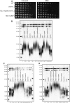
Tel1-hy909 requires the MRX complex for the DNA damage response but not for telomere elongation. (A) Yeast dilution series of untreated cells or cells treated with 0.02% MMS for 1 hr. The genotype is indicated to the left of the panels. To account for growth differences between the genotypes different amounts of cells were collected for the initial dilution. A total of 0.5 OD of cells were collected for WT and TEL1-hy909, 1.5 OD of cells were collected for mre11Δ, and 8.0 OD of cells were collected for TEL1-hy909 mre11Δ. Strains used in this assay were yRK114, yRK126, yRK128, yRK104, yRK141, yRK92, yRK93, and yRK122. (B–D) Southern blot analysis of telomeres from strains with the indicated genotype. Two independent, haploid segregants are shown for each genotype. (B) Segregants are from JHUy816, yRK79, yRK80, yRK81, and yRK83. Cells underwent minimal propagation before genomic DNA was prepared. Additional biological replicates were assayed for each genotype: WT, n = 35; TEL1-hy909, n = 42; TEL1-hy909 mre11Δ, n = 21; mre11Δ, n = 18; TEL1-hy909 rad50Δ, n = 18; rad50Δ, n = 15; TEL1-hy909 xrs2Δ, n = 4; xrs2Δ, n = 4. (C) Segregants are from yRK5150 and yRK5151. Cells underwent minimal propagation before genomic DNA was prepared. Additional biological replicates were assayed for each genotype: WT, n = 35; exo1Δ, n = 4; TEL1-hy909 exo1Δ, n = 4; TEL1-hy909 exo1Δ mre11Δ, n = 4; TEL1-hy909 mre11Δ, n = 21; TEL1-hy909, n = 42; mre11Δ exo1Δ, n = 2; mre11Δ, n = 18. (D) Segregants are from yRK5152 and yRK5153. Cells underwent minimal propagation before genomic DNA was prepared. Additional biological replicates were assayed for each genotype: WT, n = 25; sae2Δ, n = 6; TEL1-hy909 sae2Δ, n = 6; TEL1-hy909 mre11Δ sae2Δ, n = 4; TEL1-hy909 mre11Δ, n = 21; TEL1-hy909, n = 42; mre11Δ sae2Δ, n = 2; mre11Δ, n = 18.
Figure 5
Figure 5
rad50S telomere elongation is dependent on Tel1. (A and B) CRISPR/Cas9 was used to knock-in the rad50S allele into a wild-type haploid strain (yRK114). A transformant that was not edited at the RAD50 locus but was transformed with the Cas9 plasmid was used as a control and is referred to as RAD50 (A, lane 2). Both RAD50 and rad50S transformants were passaged on solid media for ∼120 population doublings (see A, lanes 2–4, yRK2112-5, yRK2113-5, and yRK2116-5). rad50S or RAD50 cells were transformed to introduce tel1Δ, mre11Δ (A, lanes 5–10), or TEL1-hy909 (B, lanes 4–7). Cells were passaged on solid media for ∼120 population doublings. The strains used were yRK2118-5, yRK2120-5, yRK2121-5, yRK2122-5, yRK2123-5, yRK2124-5, yRK2125-5, yRK2126-5, yRK2127-5, and yRK2128-5. Biological replicates were assayed for each genotype: rad50S, n = 5; rad50S tel1Δ, n = 2; tel1Δ, n = 3; mre11Δ, n = 1; rad50S mre11Δ, n = 2; rad50S TEL1-hy909, n = 2; TEL1-hy909, n = 2.
Figure 6
Figure 6
Tel1 regulates telomere length in a pathway distinct from the DNA damage response. Diagram demonstrating the distinctions between Tel1 pathways in the DNA damage response and telomere length regulation. (A) The DNA damage response is most strongly regulated by Mec1 and Rad53 as indicated with the bold arrows, although Tel1 signaling through Rad53 and MRX plays a role. The MRX complex is both upstream and downstream of Tel1 in the DNA damage response. (B) For telomere length regulation, Tel1 does not require MRX after activation and Rad53 does not plan a role in the Tel1 telomere length regulation pathway. The Tel1/MRX pathway plays the major role in telomere length compared to a minor role of Mec1/Rad53 pathway.

Similar articles

Cited by

References

    1. Alani E., Padmore R., and Kleckner N., 1990. Analysis of wild-type and rad50 mutants of yeast suggests an intimate relationship between meiotic chromosome synapsis and recombination. Cell 61: 419–436. 10.1016/0092-8674(90)90524-I - DOI - PubMed
    1. Albuquerque C. P., Smolka M. B., Payne S. H., Bafna V., Eng J. et al. , 2008. A multidimensional chromatography technology for in-depth phosphoproteome analysis. Mol. Cell. Proteomics 7: 1389–1396. 10.1074/mcp.M700468-MCP200 - DOI - PMC - PubMed
    1. Anand R., Memisoglu G. and Haber J., 2017. Cas9-mediated gene editing in Saccharomyces cerevisiae. Protoc. Exch. 10.1038/protex.2017.021a - DOI
    1. Armanios M., and Blackburn E. H., 2012. The telomere syndromes. Nat. Rev. Genet. 13: 693–704 [corrigenda: Nat. Rev. Genet. 14: 235 (2013)]. 10.1038/nrg3246 - DOI - PMC - PubMed
    1. Arora S., Deshpande R. A., Budd M., Campbell J., Revere A. et al. , 2017. Genetic separation of Sae2 nuclease activity from Mre11 nuclease functions in budding yeast. Mol. Cell. Biol. 37: e00156-17. 10.1128/MCB.00156-17 - DOI - PMC - PubMed

Publication types

MeSH terms

Substances